Horsetails:
relic plants from prehistory
by
Dave Walker, UK
(Please note: this article celebrates
the attractive features of horsetails from the perspective of a UK amateur
naturalist but does not offer any help on their eradication. The following
Google search will list a wide variety of advice for different areas of the
world www.google.com.)
 If
the hero in H. G. Wells' story 'The Time Machine' travelled back
ca. 350 million years, he couldn't fail to notice horsetails as
he stepped out of the machine into a steaming swamp. The
horsetails would have been a dominant part of the vegetation with
magnificent specimens reaching 30m or more in height and 1m in
diameter.
If
the hero in H. G. Wells' story 'The Time Machine' travelled back
ca. 350 million years, he couldn't fail to notice horsetails as
he stepped out of the machine into a steaming swamp. The
horsetails would have been a dominant part of the vegetation with
magnificent specimens reaching 30m or more in height and 1m in
diameter.
Unfortunately the common
horsetail plants of today are much smaller than their prehistoric
relatives and it's only the fossil record that provides a glimpse
of their heyday.
Horsetails don't have flowers to
attract the attention or showy leaves like ferns, but
nevertheless they are attractive plants with interesting macro
and microscopic features.
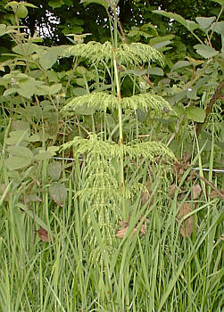
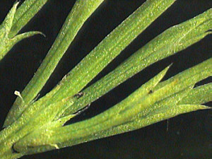
The image right shows a
horsetail (ca. 0.3m tall) in one of its typical habitats i.e. in
the partial shade growing amongst the understorey of a damp wood
edge. (The central plant with pale green stem and whorls of
branches). It's easy to see why they are often overlooked, as
many of the common species are barely taller than the surrounding
grasses. Depending on the species, they grow in habitats such as
roadsides, stream banks and salt flats.
In urban areas away from nature
reserves, the species found are likely to be common, so it's
worth studying them in situ with a 10X hand lens and if possible
bring home the top half of a stem with a whorl of branches for
closer inspection.
The whorls of branches (see left
below) are very attractive in some species, especially those of
the wood horsetail (Equisetum sylvaticum) which
resembles a miniature Christmas tree. The green whorls aren't
actually leaves; the leaves in horsetails are reduced to sheaths
which clasp the stem (see images below).
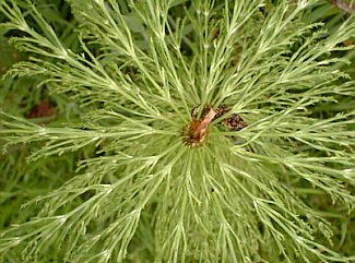
A whorl of branches
|
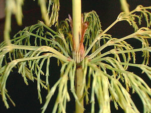
Close up of whorl showing the brown
leaves clasping the stem
|
Detail of base of a clasping leaf |
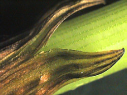
Detail of upper part of a clasping
leaf. |
Detail of the branches which
look like thin leaves.
|
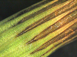
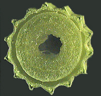 A cross section of the jointed stem is worth
looking at as they are ridged with hollow centres; features which
are useful diagnostic clues to identify the species. Just a crude
cross section 1mm thick when viewed with a hand lens or stereo
'scope will show the detail shown right.
A cross section of the jointed stem is worth
looking at as they are ridged with hollow centres; features which
are useful diagnostic clues to identify the species. Just a crude
cross section 1mm thick when viewed with a hand lens or stereo
'scope will show the detail shown right.
There are only ca. thirty
species of horsetails in the world which are all in the genus
Equisetum (Phylum Sphenophyta). In the UK there are only eight or
so species, but despite this they are not always easy to
identify, as they form hybrids and their characters are variable.
In the UK an excellent guide to identifying horsetails (and
ferns) is reference 1.
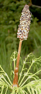 Some species
produce two kinds of shoots, one vegetative and another bearing a
cone called the strobilus (shown left). When ripe this cone
releases tiny spores. These cones don't last very long, but if
you find a horsetail with just ripening spores, collect some
spores and study under the microscope. As they dry they have
hairs wrapped around each spore called elaters which uncoil and
help distribute the spores. This phenomenom seems a neat
subject for the enthusiast of video microscopy, if you've
captured any footage of spores and would like to share it let me know.
Some species
produce two kinds of shoots, one vegetative and another bearing a
cone called the strobilus (shown left). When ripe this cone
releases tiny spores. These cones don't last very long, but if
you find a horsetail with just ripening spores, collect some
spores and study under the microscope. As they dry they have
hairs wrapped around each spore called elaters which uncoil and
help distribute the spores. This phenomenom seems a neat
subject for the enthusiast of video microscopy, if you've
captured any footage of spores and would like to share it let me know.
Horsetails (also known as the
scouring rush) have been used for scouring pans and polishing, as
the epidermal cells in the stem contain silica which make them
abrasive. They are poisonous to livestock although they have been
used in some folk medicines. (Addition:
I had an interesting email from a reader who told me that his
geese loved eating horsetail plants, which suggests they are not
poisonous to all farm animals!)
If you have the facilities you
may like to try preparing your own stained plant sections to
study cellular detail, although their silica content may make
this trickier than for other plants. I'm fortunate to have a set
of sections prepared by an accomplished UK slide maker called
John Nicholls.
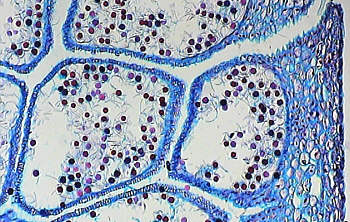
E. arvense. T/S cone showing spore
development (3.5X objective). Alcian Blue & Safranin
stain.
Slide prepared by J. Nicholls 1988.
|
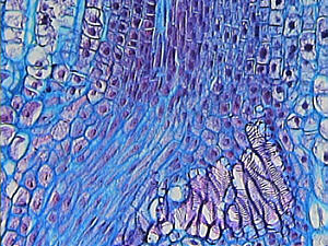
E. arvense. T/S cone showing cell
detail (9X objective). Alcian Blue & Safranin stain.
Slide prepared by J. Nicholls 1988.
|
So if you haven't studied
horsetails before, it's worth keeping an eye out for them, as
they are fascinating plants and in their own way are as
attractive as the more commonly studied and admired flowering
plants.
General comments to the author Dave
Walker welcomed. But please
don't ask me how to eradicate them. Try a keyword search like 'horsetails' with
'eradication' on eg www.google.com.
The best advice may vary depending on which country you are in.
The author is an amateur
naturalist. In-depth information on horsetails can be found via
web search engines or visit brittanica.com where Encyclopaedia Britannica
entries are complemented by links to online resources.
Footnote
There are some exotic horsetails
with heights approaching their prehistoric forebears. E.
giganteum is a South American species 10m tall (2cm
diameter) which is supported by the tall vegetation surrounding
it.
References
1) 'The Fern Guide'. A field
guide to the study of ferns, clubmosses, quillworts and
horsetails of the British Isles. By J. Merryweather and M. Hill.
Field Studies Council, 1995, 2nd Edition.
2) Encyclopaedia Britannica,
15th edition, 1993.
Pictures were all taken by the
author.
© Microscopy UK
or their contributors.
Published in the
June 1999 edition of Micscape Magazine.
Please report any
Web problems or offer general comments to the Micscape Editor,
via the contact on current Micscape Index.
Micscape is the
on-line monthly magazine of the Microscopy UK web
site at Microscopy-UK
WIDTH=1
© Onview.net Ltd, Microscopy-UK, and all contributors 1995 onwards. All rights
reserved. Main site is at www.microscopy-uk.org.uk with full mirror at www.microscopy-uk.net.