I'm sure most of us had our first 'peep'
through a microscope using brightfield illumination, and
thereafter many have accepted this fundamental lighting , as
the 'bread and butter' of microscope illumination techniques. Traditionally, brightfield illumination,
using a substage condenser, has been the mainstay of
illumination together with other variations such as phase
contrast for many years.
Brightfield involves a highly directional, and intense light source, accurately focussed by a condenser to the upper surface of the slide, under control of the substage iris diaphragm. But most of the research and development related to improvement of the compound microscope appears, understandably, to have been directed towards the higher powers, and particularly pushing the upper limits of resolving ability. Manipulation of the instrument under low power has changed little, if at all since the earliest days of microscopy, leaving the microscopist to manage as best they can. True, the advent of variable intensity illumination has helped quench the intense lighting experienced at low powers, and the manufacture of condensers with removable tops does help to broaden the illuminated field.
I remember being intrigued with daylight as the source of light for the first time, and despite opinion to the contrary, was impressed by the 'purity' it imparted to the images.
Later on, in an effort to solve an old problem of providing full field illumination for low powers, which some condensers are incapable of without recourse to removing their top elements, I discovered a very simple method of broadening the condenser's cone of light on the slide. By placing a piece of ground glass, such as a camera focussing screen, in the light path below the substage condenser, a significant increase in the width of the field's illumination occurs at the same condenser setting. The idea of being able to do this in a matter of seconds satisfied my 'convenience at little cost' mentality immensely, and soon thereafter I also noticed that diffuse lighting had some interesting and beneficial properties too. It was as if the qualities of daylight viewing could be created instantly from an electric power source, and soon had me experimenting, trying to improve what I believe to be is an extremely pleasant method of observation at low to medium powers. At last I felt somewhat emancipated from the silhouette inducing and diffraction bordered imagery of brightfield !
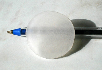
Method : Light diffused before it gets into the condenser as shown below :-
|
Simple trial can be set-up like this using a sheet of ground glass. |
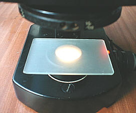
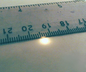
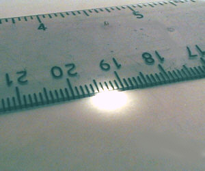
The ground glass scatters the near parallel beam of light before entering the condenser, and after entering the condenser in a multitude of varying angles is 'focussed' into a large diffuse cone upon the slide. The photographs below illustrate this clearly, depicting the results of the light impinging on plain paper resting on the microscope's stage. Notice also show the absence of color around the larger diffused circle of light, presumably caused by the 'remixing' of spectral aberrations of the condenser ?
 |
 |
The notion of a well designed and constructed brightfield condenser being bombarded with diffuse light might raise a few eyebrows, but of course no harm will come of it.
The photo's below illustrate my original intention of using diffuse light to broaden the field's illumination at low power :-
 |
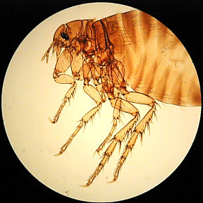 |
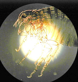
Photo left shows typical lighting appearance using low power with an unsuitably small image from the condenser. Two pieces of ground glass separated by a couple of inches and placed directly below the substage condenser expand the illuminated field considerably, as seen in the photo on the right. The eye fails to detect the slight unevenness of illumination during direct observation, though it can be seen here in the photograph. ( Human Flea. x4 achromatic objective.) |
Features of diffuse lighting
I find this method not only broadens the field illumination by up to 2.5x, and therefore a great bonus for low powers, it also brings about some very useful though subtle changes in the image. The first is that diffusion by the ground glass attenuates the light source, and far from being a hindrance, helps to reduce the bright fields experienced at low powers to the extent that the iris can be left fully open, so the full resolution of the objective can be used to advantage. Importantly too is the fact that if the electric light source is left at full power, it's colour temperature is ideally suited, being whiter than if diminished by a dimmer control, and creating more faithfully coloured imagery. Whilst the dimmer control on many microscopes is a useful aid, the fact that the colour of the light decidedly reddens when used does present problems, especially for photography.
Subjective assessment of diffused lighting with low powers ( up to approx. 100x )
I find the quality of the images from many subjects with diffuse light to be most pleasing, and importantly restful on the eyes. The inherent contrast sometimes experienced with bright diffraction bordered subjects can be tiring, and subtle colour variations are lost. Generally, the images have an airy quality, and appear more natural when observing subjects such as insect structures, and other semi transparent subjects in diffused light.
Last year I acquired some fluorite optics, and looked at some favourite slides with diffuse illumination. With the 10x objective I marvelled at the image of a well mounted proboscis from a blowfly, and could hardly believe the inherent quality of the image, which unfortunately I could never reproduce here, as a simple photo from my digicam would be quite incapable of capturing such detail, subtleties of colour, lighting and 'depth' . Another bonus is that with some delicate subjects the iris diaphragm can be closed carefully, appearing to reduce 'diffraction boundary' effects when using diffuse light, with consequently improved balance between light intensity and subject density and colour rendering.
I must emphasise the careful use of the substage diaphragm during experiments and subsequent observations, if you decide to use diffused lighting. Some subjects and optics will support the use of a full cone of light from the condenser, and benefit from this, whilst more delicate subjects need careful reduction of the iris to the point were the background and subject boundaries appear with least diffraction and yet enough contrast to be pleasing and functional.
Experienced observers who are fully aware of the effects of iris closure on resolution, diffraction and image degradation in standard brightfield, should find that diffuse lighting brings about some interesting and subtle effects.
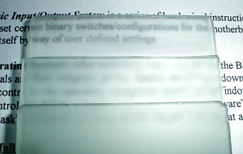
Diffuser materials
|
Three layers of ground glass demonstrate diffusion |
Luckily, quite a number of common materials have properties which suit our purposes of scattering light, though some impart subtle shades of colour which understandably some observers may not like.
Ground glass (frosted), some translucent plastics, opalescent materials, and paper will all to a greater or lesser extent diffuse light . Ground glass is probably the best, and is easily 'frosted' by grinding two pieces together with a pinch of medium grit 'Carborundum' powder ( silicon carbide ) and soapy water until both take on even defragmentation of their surfaces. I made four pieces of ground glass for use as photographic focussing screens , one pair with a coarser grade of grit, and they are perfectly suited for diffusion. The actual grade of grit is not so important so long as it is not too fine, ie not finer than about 180-220. The lower the number, the coarser the grit, and something in the region of about 80-120 or more should be . (Telescope mirror kit suppliers sell abrasive powders, they are expensive but we only need a pinch ? Valve grinding paste may be a cheaper alternative if it is still available ? 'Wet and Dry' emery paper will do the job but care is required to produce an evenly 'etched' surface)
I successfully treated the surfaces of a piece of thickish translucent plastic on both sides with a disposable 'nail file', cut to fit into the filter tray, and whilst not quite as effective as ground glass, would give someone an idea of how effective diffuse lighting can be before venturing further :-
 |
It is quite instructive and enlightening to slide a piece of white paper, preferably thinnish, under the substage, or in the filter tray and see the effects under low power, especially if you have the common problem of uneven field illumination. True, the image will be on the dark side even with the iris open, but an idea of what happens will be self evident. The effect is very similar with ground glass, but this has more efficient dispersal and transmission of light, and most importantly no tainting of source colour.
Placement of diffuser
The position of the diffuser in the optical path is quite important, because in some situations the field may show subtle variations of intensity, commonly dimmer around the edges of the field. This is a property of the distribution of scattered light, but this can be 'eliminated' for all practical purposes by making sure the diffuser is as close to the condenser as possible, and depending on the results, a second diffuser may be required which should be located ideally a couple of inches or more below the first. If there is little room between your condenser and field lens, two or three diffusers can be edge-taped together to do the job. The whole business of finding the best location, and number of diffusing sheets to suit your set-up will take only a few minutes at most.
Summary: Advantages of using Diffuse lighting
Expands the illuminated field of all condensers .
Imparts an 'airy' field and 'natural lighting' qualities to images.
Eye friendly !
Auto-attenuation of full power light source, allows full objective resolution when required at pleasing light levels, faithful colour rendering of subjects, more dilated pupils allow easier observing alignment and more relaxed viewing, and also suppression of floater's and specks in optical train.
Capable of really beautiful renderings of subjects such as largish mounted insects (parts), and even strewn diatoms are rendered more faithfully at low to medium powers with sensitive control of the iris diaphragm, which can convey images rendering their delicate colouring more faithfully.
Couldn't be more convenient to implement.
Cheap, simply technology.
Con's
May not suit some observers, though initial appraisal may change in the course of time especially if compared to standard brightfield in A/B comparisons, with careful use of iris closure.
Some subjects may not be suitable.
Depending on personal preferences, is limited to low and mid power observations and may not suit photography so well since this process reduces contrast, leaving the delicate structures more difficult to capture in print. I believe it to be an observational aid, in the first instance, and therefore not necessarily suited to photomicrography.
Conclusions
If you have never attempted to use your microscope under low power with diffuse lighting, I urge you to do so, even if only to expand the illuminated field if you find this necessary.
Have a look at a number of subjects with and without the diffuser, adjusting the iris diaphragm frequently in both situations, before coming to a conclusion.
I'm confident that a number of readers will take to this gentle yet subtle lighting alternative to 'unhindered' brightfield, and use it as enthusiastically and frequently as I do for low to medium power observations.
(All
photo's taken with Olympus 830L digicam )