Getting
micro-organisms in focus
using
mouse-over images with different optical sections
by Bill Ells & Wim
van Egmond
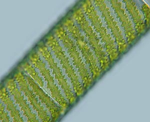
Spirogyra
wait for the second image to load and move your mouse over the image to switch focus
Bill mentioned the problems of getting microscopic organisms with a distinct three
dimensional shape in focus. When you observe these creatures under the microscope you can
change your area of focus very easily by adjusting the height of the sample. This way you
can get an idea of the actual 3D shape of the organism. The only way to capture this in
images is by video or film.
There is one (more limited method), a simple trick I demonstrated in the article an Interactive Rotifer. This time I used the mouse-over trick to show 2 different optical sections through two freshwater algae. On the first page it was a desmid. I could show both the cell wall structure and the cell contents. On this page I photographed the common pond-scum Spirogyra. The first image shows the chloroplasts that run in a helix. When you move your mouse over the image we go deeper inside the cell. There the nucleus can be seen hanging between thin fibres.