During my studies of Australian desmids I am required to make drawings of each cell in three different views; front (face view), end (apical view) and side (lateral view). Some desmids look very similar when viewed in the usual front view but can be closely differentiated from closely allied species when they are viewed either end-on or side-on.Even a front view can reveal different structures when the image is focussed up and down. The two black and white images taken from the same specimen show Euastrum spinulosum Delponte at two different focus levels.
The picture below (left) shows the outline of the cell with its short spines on the corners of each arm (lobe). The picture on the right was taken at a higher focus level showing the prominent ring of central granules together with a secondary ring.
It is apparent that it requires at least two photomicrographs to be taken to illustrate the cell structure (in fact a third photo taken at a focus in between these two would reveal more cell wall granules). By drawing cells such as this one, all of the cell wall structures can be drawn on one drawing by focussing up and down which gives a pseudo 3-D image with any granule, spine or other structure that protrudes up and out of the plane of focus. Although most drawings are not 'pretty' like photomicrographs they can reveal more detail.
These photomicrographs were taken using 35mm black and white film and scanning the negatives using a Nikon Coolscan III scanner.
Comments to the author Mike Dingley are welcomed.
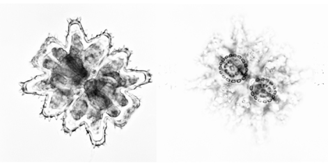
Two views of the desmid. Size ca. 60µm x 45µm.