Editor's note: Demodex is a tiny mite ca. 0.1 - 0.4 mm long that lives on humans. Aaron Messing describes a simple method of collecting and observing them, and shares some images. See also M Halit Umar's Micscape article Demodex: An unusual inhabitant of hair follicles for further details and images of this mite.
Aaron Messing writes:
Take a clean glass slide and holding it at a 45 degree angle to your skin, scrape across your forehead just above your eyebrows. (Please see additional collecting notes and important safety notice in the footnotes.) Gather the fatty substance that collects along the edge of the slide with a small spatula or scalpel and place the material in the middle of a clean slide. Place one small drop of high viscosity immersion oil on top of the specimen and cover with a cover slip. Gently press the cover slip down and move the cover slip slightly and gently from side to side to spread the specimen. Examine under the microscope using phase contrast or DIC for best results. Use 100X to locate the animal and 400X for detail.
The high viscosity immersion oil is Type B. It has the same refractive index as Type A but will not flow to the underside of the slide and make a mess. The immersion oil also dissolves and hides the sebum. The debris seen in the image backgrounds is dead skin cells that normally slough off.
Commments to the author Aaron Messing are welcomed. Larger master images are available from the author if required.Acknowledgement: The technique followed is that described on the Biomedia Associates 'Learning Programs for Biology' web site which has a wealth of resources for teachers and students. This web site's page on Demodex with a neat animated image is here.
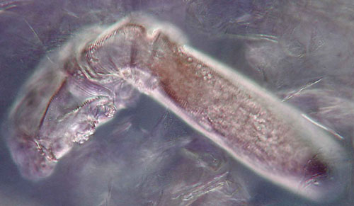
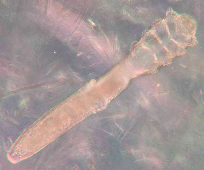
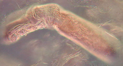
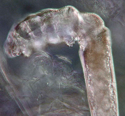
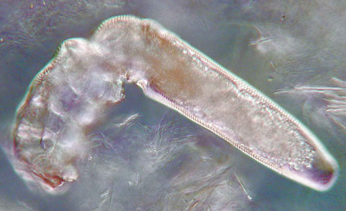
Images
Optical magnification 400x using differential
interference contrast and with compensators.
View of the entire mite.
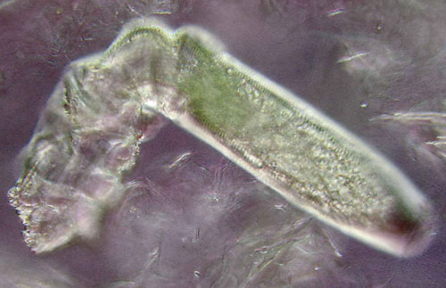
This image and those below show various detail by using different
focus planes as the mite rolls under the slide.
Additional collecting notes by the author:
The slide is drawn across the surface of the skin in a single direction as one would use a broom to sweep a surface to gather dust carefully. Also the scraping is a single pass at moderate pressure. The sharp edge of the glass is not moved across the skin as one would use a knife to cut, as this would risk cutting the skin. A slide used at the correct angle with a sweeping type motion is not likely to cut into the skin unless there is also a lateral cutting action.Micscape safety notice:
The practical tip is given by the author in good faith, but is probably only suitable for adults and not for unsupervised youngsters. There are always finite risks with glass; more so if collecting from the face, so please take great care.Disclaimer: No responsibility is accepted by the author, On.View Ltd who host Microscopy-UK / Micscape, the site administrators or other site contributors for any harm to persons or property incurred by following this collecting method.