For instance, a microscopist cannot follow and measure the fast moving microbes, such as flagellates, ciliates, rotifers, etc. Furthermore, unskilled investigators like me cannot identify and measure a microorganism at the same time. Thus we have to look up a key first and may lose the time needed for measuring it. In another case, photomicrographers like me will concentrate on taking a series of photographs and may forget a measurement.
As many microscopists often
encounter these problems, I offer "a measuring method for microorganisms"
which my teacher taught me if you have a stage micrometer. A size measurement
is accomplished by using your printed photographs.
|
1 |
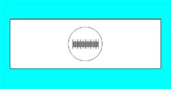 |
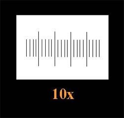
Take photographs of a stage micrometer using different objectives (4x, 10x, 20x and 40x). |
|
2 |
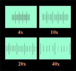 |
Develop that film to get pictures of the scale at different sizes according to the magnifying power of each objective. |
|
3 |
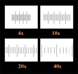 |
Photocopy the scales pictures on transparency sheets. One scales picture or more per one sheet (up to you). |
|
4 |
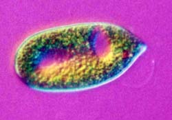 |
When taking a photograph of any organisms, you should also record the magnifying power of the objective used for each picture, such as the 10x objective for this Euglena image. |
|
5 |
 |
Thus, you can measure the organism by using the transparency for the objective it was taken with. (In this case, the 10x objective for the Euglena image.) |
|
6 |
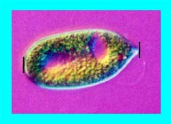 |
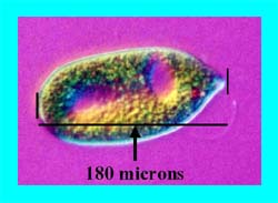
Set two points (|) in the picture in your mind's eye but don't mark the picture, e.g. as shown in the left hand figure. |
|
7 |
 |
After that, put the transparency sheet (10x) on the picture (10x).
On this transparency, 1 space of a stage micrometer = 0.01 millimeter or 10 microns. Thus you count the number of divisions between the two chosen marks. |
|
8 |
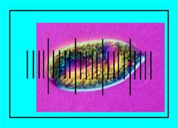 |
For this picture, the number of spaces
= 18 spaces and which = 180 microns. |
|
|
||
|
9 |
 |
In the scanned image displayed on the computer screen, drag a line from the first to the second points chosen. |
|
10 |
 |
If the line is oblique, you can rotate it to a straight line. At the moment, you know that this line equals 180 microns in length. |
|
11 |
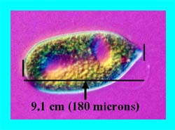 |
You can now determine the length of this line by using e.g. the routine Format Autoshape (for Microsoft Office). In the example on the left, this line is about 9.1 cm in length. |
|
12 |
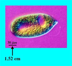 |
To make
a scale bar, reduce the length of this line by a suitable factor e.g. 3,
5 times. For example, I divided this line length by 6, so the line is about
1.52 cm in length (9.1/6). Then the bar length was reduced from 9.1 to
1.52 cm.
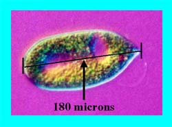
By doing this, you will get the microorganism picture with an appropriate scale bar for 30 microns (180/6). |
|
13 |
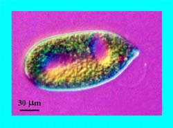 |
Finally, by using all of these steps, microscopists can measure the size
of a microorganism and can make a suitable scale bar as demonstrated in
the left hand Euglena figure.
Note: For this explanation, I used a Euglena picture as an example. In this case all of the steps were made in Microsoft PowerPoint®, i.e. images of the stage micrometer, scale bar, transparency sheet, etc. in order to present the steps easily. |
Although this measuring method is effective and useful from my point of view, it also has some restrictions. A limitation of this technique is that the enlarging and printing process for the image and scale prints must be identical to enable measurements to be made. For example, if you print your image at 4x6 inch in size, you will need to do this for the scale picture as well (4x6 inch).
I hope this method can help a little for the measurement of microorganisms. If any microscopists cannot understand all the steps. Please do not hesitate to contact me. I will try to help in every way that I can.
All comments to the author Chitchai Chantangsi are welcomed.
Acknowledgement:
I
would love to express gratitude to Assistant Professor Dr. Malinee Chutmongkonkul,
my nice teacher. The first person who explained to me "What is a microorganism?"
and taught me to use a microscope and this measuring method. I am particularly
indebted to Dave Walker for his kindness with all of my articles, especially
this one, that cannot be complete without his valuable suggestions.
Microscopy
UK Front Page
Micscape
Magazine
Article
Library
© Microscopy UK or their contributors.
Please report any Web problems or
offer general comments to the
Micscape
Editor,
via the contact on current Micscape
Index.
Micscape is the on-line monthly magazine
of the Microscopy UK web
site at Microscopy-UK
WIDTH=1