 |
In the Summer of 2002, I was introduced to bryophytes (mosses and liverworts) on a nature walk organised by the Friends of Corstorphine Hill and led by an experienced Edinburgh bryologist. After a very interesting and informative day out, I was easily persuaded to sign up for a bryophyte workshop to be held, in the Royal Botanic Garden of Edinburgh, later that year. After attending the workshop and receiving some excellent teaching and demonstrations I had no choice but to add these fascinating plants to my natural history interests. | |
| In November 2002 I heard about the (UK based) Millennium Commission
Awards. The commission supports a scheme that awards generous amounts of
money to individuals, enabling them to carry out their own projects concerned
with the local community, buildings and natural environment.
This seemed like the perfect opportunity to combine my new interest in bryophytes together with my fascination for buildings and produce something that would encourage community appreciation of the environment. I submitted my project proposal and was fortunate enough to be given an award. |
Millennium project image |
|
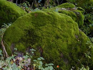
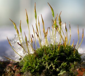
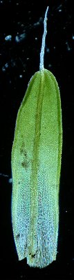
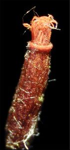
Tortula muralis cushion with mature capsules |
The main "deliverable" item in my project is a final report but, while
on the path to this end, I thought my investigations might be of interest
to others inclined to bryophytes. For this purpose, I offer some of the
images I created while studying my first moss, Tortula muralis.
The standard equipment needed by the aspiring bryologist is a X20 pocket lens, some paper packets for samples and, in the UK, a copy of E. V. Watson's book, "British Mosses and Liverworts". This latter item is aimed at a UK audience but as bryophytes live everywhere on the planet, it is probably useful in many countries. |
|
| The identification of a moss begins with an examination of its general
growth habit and mode of branching, leaf characteristics, colour and shape
of any capsules etc. Referral to a book containing an identification key
leads the user through a series of questions and answers that will (if
you are lucky) arrive at a generic or specific identification
However, until one becomes very familiar with mosses, it is often necessary to subject the specimen to examination under a microscope and use a key such as that found in Watson. For me, this is where the fun begins and will I confess at once that I need very little excuse to use my microscope. |
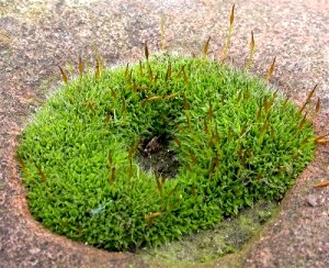
Tortula muralis cushion in and old railing hole |
|
Tortula muralis cushion with young capsules |
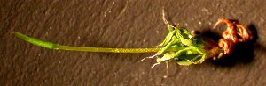
Tortula muralis is a small plant growing in low cushions or sometimes in wide patches about 1 cm tall, commonly on walls. The leaves are usually deep green or bright yellow-green. The cushions consist of many individual plants and it is thought that this communal habit provides some protection against desiccation. | |
|
The leaves and hair points are twisted when dry and the leaves widely spreading when moist. The specimen shown on the right was photographed dry then after applying some water. It is apparent the the leaves absorb moisture and swell. Note that little change is seen in the capsule. |
Dry specimen 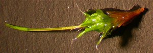
Moist specimen |
|
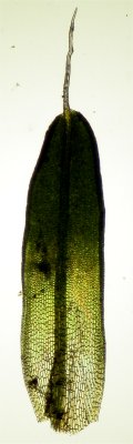  |
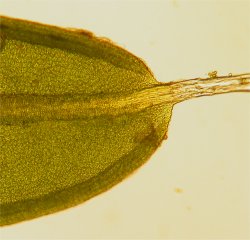
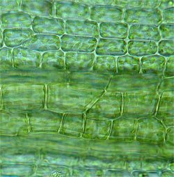
The shape and sizes of the leaf, its upper, mid and basal
cells, vein, leaf margin and tip are used in identification of the species.
The images on the left shows a leaf from the stem mounted in a drop
of water under a cover slip. The leaf was examined under bright and dark-field
illumination.
The leaf is tongue shaped with a long hair point and recurved (folded) margins. |
|
|
Under a higher power objective, the hair point is seen to be devoid of chloroplasts and the cells appear to be empty. When dry, these hair points appear grey or white to the observer. |
Tortula muralis Leaf hair |
|
Tortula muralis. Top of leaf |
The image on the left shows the top of the leaf and it can be seen that the hair tip is a continuation of the central "nerve". The folded or recurved margins of the leaf are also evident. | |
| The cells from the top of the leaf to mid-leaf are described as being "papillose" and have wart like protuberances on their surface. This accounts for their unusual appearance. | 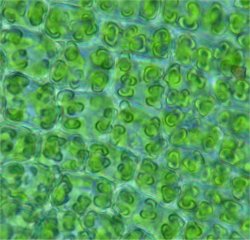
Tortula muralis papillose cells above mid-leaf |
|
Tortula muralis nerve cells and cells below mid-leaf |
Below about the mid-leaf, the cells are regular, rectangular in outline and have lost the papillae of the previous cells. The chloroplasts are clearly seen. Note the nerve cells which appear to be larger than the leaf blade cells. | |
|
Moving nearer the base of the leaf, the cells become elongate, rectangular and have few or no chloroplasts. |
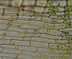
Tortula muralis rectangular outline cells near leaf base |
|
Tortula muralis leaf base |
This image of the leaf base shows another view of the rectangular shaped basal cells which are devoid of chloroplasts. | |
| The young spore capsule (2 mm long) is covered with a thin paper-like
calyptra that serves to protect it during growth.
The calyptra eventually falls off to show the operculum, a lid, which in turns falls off to reveal the peristome that regulates the dispersal of the spores. |
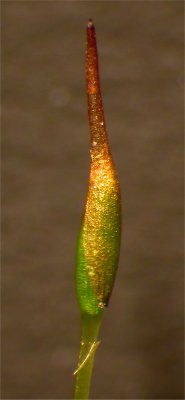
Tortula muralis capsule showing calyptra |
|
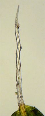
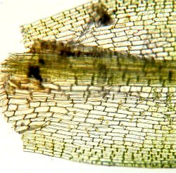
Ripe Tortula muralis capsule showing peristome |
In this image of a mature capsule, the twisted peristome teeth can
be seen.
In dry weather, the teeth untwist and allow spores to be shaken out of the capsule and dispersed by the wind. When moist, the teeth twist together and prevent the spores from being shaken out. The common English name, wall screw-moss, is presumably derived from the twisted appearance of the peristome. |
|
| The image on the right is a top view of the peristome. | 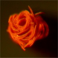
Tortula muralis top view of peristome |
|
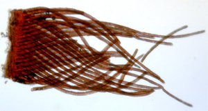
Detached peristome |
With a bit of fiddling, in a drop of water, I was able to detach a peristome from a capsule and mount in in water under a coverslip. The resulting image shows the criss-cross arrangement of the peristome teeth. | |
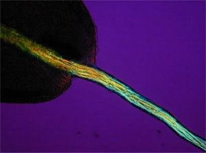
| I wondered how the peristome would look under crossed polars and the image of the teeth is shown on the right. The complex structure of the teeth shows up well and probably provides a clue as to how these teeth are able to twist. | 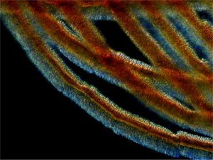
Peristome teeth under crossed polars |
|
Leaf hair under crossed polars |
Having discovered that parts of the moss exhibited birefringence, I took another look at the leaf hair and was pleased to see that it to provided an interesting image. | |
| As a final image, lest this mossy tale becomes a saga, here is a crossed polar image of cells near the base of the leaf. The cell walls show up very clearly and the folded leaf margin is also evident. | 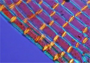
Leaf base cells under crossed polars |
|
| For those interested in the equipment I used, here is a list of my
resources.
Camera - Nikon Coolpix 950 Microscope - Leitz Ortholux II Stereo microscope - Very old Zeiss X10 Hand lens X20 Hand lens |
||
| I am really enjoying my exploration of bryophytes as it involves outdoor
field trips, close-up photography and most of all, an excuse to use my
microscopes.
I do hope that this article has proved of some interest to Micscape
viewers and constructive comment would be most welcome.
|
References:
Watson E. V. (1981) British mosses and Liverworts 3rd Edition. Cambridge University Press. ISBN 0 521 29472 X |