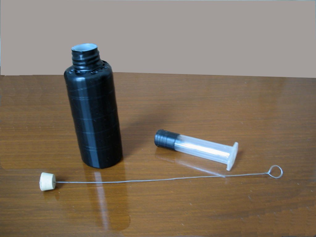
title image
A DETACHABLE-NECK FLASK TECHNIQUE TO SAMPLE MICROINVERTEBRATES FROM FINE GRAINED SUBSTRATA.
WALTER  DIONI                       Cancún (Quintana Roo) Mexico

Key words: New sampler, sampling, micro invertebrates, detritus.

All the technical terms displayed in yellow are explained in the Glossary. It could be a good idea to pay an initial visit to it.


INTRODUCTION


Most microscopists gather their aquatic samples of organic sediments from the bottom (benthos) or from materials adhering to submerged objects (bioderms), or to rooted or floating plants, (periphyton). Normally they are small samples.

The examination of those samples is done in the laboratory, taking drops with pipettes from different parts of the collection. So chance plays a most important part in the amateur’s findings; and the time that it takes to examine each sample causes it to deteriorate and only a few of the many present species can be detected.

The specialists in diverse branches of the zoology of micro invertebrates, and mainly the ecologists, use, whenever they can, harvesting methods that concentrate in small volumes the microfauna of great volumes of the habitat. The typical example is the plankton net which collects in a small container the living beings existing in the water column by straining when it is dragged through.

The specialists in edaphology, for example, when they want to gather soil nematodes, use a Baermann funnel; the soil micro-entomologists use the Berlese-Tullgren funnel, to separate acarii and other groups of micro-arthropods from litter or soil.

The instrument that I will present in a professional version in Appendix I and in an "amateur’s" one here, performs a similar task with micro-invertebrates, which are abundant in fine bottom sediments (ooze) or in filamentous algae floating or fixed to some substrate (plocon and heteroplocon) or in the periphyton or bioderms that surrounds the stems and leaves of rooted plants, submerged or emergent, in the different water bodies (bafon), or in sediments that the floating plants (pleuston) can catch between their roots.

In all those cases the sample is an abundant collection of organic microparticles grouped in floccules, between which the micro-invertebrates move and hide themselves. It is very frustrating while examining at the microscope our drop of sample to see that the micro-invertebrate of interest, after some fast movements in free solution, that whet up our curiosity, subsequently hide up and disappear for long minutes behind a floccule of detritus. This is for example an irritating and common behavior of Gastrotrichs and Catenulida.

Protozoa, rotifers, gastrotrichs, nemertines, turbellaria, entomostracans (copepods, cladocera, and ostracoda), nematodes, microoligochaeta (naidids and enchytreids) and freshwater mites can be represented in the samples. (Try their names in your browser's search engine to find a definition of those terms you don’t know). The sample won't reveal information of the first developmental stages of the larvae of many insects, that also live in the same habitats.

In order to separate the organisms from the sediment, and to concentrate them in a small volume of clean water, which can be easily screened out, I have designed the "Detachable-Neck Sampler”.
 

The instrument

 
The sampler for amateurs (that is the one that most interests me in this article) was constructed with plastic or glass bottles of different sizes, according to the volume of the sample that is to be treated. But it is not practical to work with volumes greater than 200-250 or so milliliters.

extractor 01
bottles and syringes most amateurs can gather from around the home

A tube of transparent plastic of about 7 to 10 cm in length, and a diameter similar to the neck of the bottle is adapted to it so that it fits tightly, without allowing any loss of water. It is possible to complete the adjustment surrounding the end of the tube with one or two turns of plastic electrician's tape. Clear plastic adhesive tape is not as efficient for this task. Hypodermic syringes with their ends cut away can make excellent samplers. You can wipe the printed scale off with thinner.

extractor 02
all the materials used for the prototype

Obtaining a watertight closing is possibly the more difficult step for the amateur. The bottle and the added tube, totally full of water, must not lose liquid over several hours. The tube fit does not have to extend beyond the neck of the bottle, so that the micro-invertebrates can easily find their way up. In addition it must be easily removed to examine its content.

Once this simple system is built and used, the removable tube must be pulled out and a rubber stopper is placed in the interior of the bottle fixed to the end of a rigid wire, preferably stainless steel or copper, that exceeds conveniently the length of the assembled instrument. Choose a shaft of not less than a millimeter in diameter to allow an easy manipulation (the wire I used for the prototype is the only stainless steel one I could find, but is too thin). [David Walker suggested the thinner style of wire clothes hanger when straightened and this should work fine]. The stopper must be of a diameter that exactly closes the lower end of the separable tube, and must be soft, to adapt itself easily.
 
extractor 03
the just finished prototype

The bottle to be used is painted externally a dull black, or one can wrap it (even the bottom) with black electrician’s tape, so that only the removable tube is transparent.

In order to use this instrument, it is filled to approximately one third or one quarter of its volume with detritus, or filamentous algae, or vegetable roots, or any other suitable materials. The cork is placed in the interior and the sampler is assembled sliding the tube down the shaft and fitting it to the neck.

The instrument is now totally filled to near the end of the chimney with water from the sampling place, it is placed in a well lit site (or it is illuminated with a desk lamp in the laboratory) and it is left to rest for 3 or 4 hours.

extractor 04
the assembled extractor and the washing bottle

 The micro-invertebrates will respond to several physiological factors. They will be attracted by the light, (positive phototaxis) they will move away of the bottom towards the surface (negative geotaxis) and, as almost all of them are aerobes (it is to say that they need oxygen) they will flee from carbon dioxide that will be produced and accumulated at the bottom, and will be attracted by oxygen that diffuses from the small surface of the tube.

 After one or two hours a magnifying glass will show the activity of many different micro-invertebrates near the superior end of the tube.  It can be the time to begin to sample.


extractor 05
harvesting the collected micro-fauna

Using the rigid handle, the stopper is raised, and it will close the lower end of the tube. Once ensured that it is closed, and firmly holding the shaft with your thumb and index finger, the tube must be removed with care, and emptied into a container of suitable volume.

Now the tube can be opened and with one washing bottle filled with water from the same source as the sample, the walls must be flooded to release the organisms that could be adherent to the walls.

extractor 06
washing the tube to loosen possibly attached organisms
water coloured for illustration, it must be the same source as the sample water

Reassemble the sampler, fill up the tube with water, and start a new cycle. The procedure can be repeated for 6 to 12 or more hours.

 Due to different response times from different species, there occurs sometimes different dominances in the successive samples, which results in a useful additional sampling, and offers to the amateur additional information on the oxygen exigencies and the taxis of the sampled organisms.

 Of all the groups listed above, the only ones that give a poor response are most of the nematodes, and micro-oligocheta, and perhaps some turbellaria, probably due to positive tigmotaxis (they need to be in contact with sediment particles), their negative phototaxis and positive geotaxis. A Baermann’s funnel suitably modified for these small samples will be more efficient.

 The harvesting must use the smallest possible amount of water to provide one clean and abundant qualitative sample, that shows richness and diversity of the most important faunistic groups and gives the opportunity to select easily (aided by micropipettes) the species of greatest interest.

 The collected organisms can be observed live, the obvious and immediate election, to obtain a clear idea of the aspect, structure, color and behavior of the studied species and to allow the photography of the better specimens. Some organisms can also be used to start mono or polyspecific cultures.

 The remaining ones can be fixed “en masse”, or can be anesthetized beforehand to observe them or to fix them for definitive preparation and mounting.

 Of course the election of which of these methods to use will be dictated by the interest of the microscopists.

 
NOTE: This is one of those cases in which we must decide between economy or ease of work. The professional version works more easily than the amateur’s one. The approximate price of the standard glass version is around 33.oo dollars.

 
APENDIX   I – Professional version

 DETACHABLE-NECK FLASK CONCENTRATOR
 

extractor 07
a laboratory glassware version

 An Erlenmeyer flask of 250 or 125 milliliters with a TS 24/40 ground neck is fitted with an inner TS ground joint of the same size. The tube of the joint must be cut to a length of 70-75 mm up from the ground portion. When it is desired, the chimney thus constructed can be closed by means of a Nº 6 or 7 rubber stopper, fitted to its lower hole. The stopper, minor diameter upwards, is fixed to a copper or stainless steel wire rigid shaft, with a diameter of  2 or 2.5 mm, and a convenient length. 

The sediments and other materials to examine can be pre-concentrated and sorted by size, sieving them through a graded series of ASTM standard sieves. Sieving can be avoided, but this treatment provides in most of the cases a cleaner and more abundant collection.

 An additional advantage of size sorting the sediments, is that it allows a coarse separation of taxonomic groups, which can require different preservation methods, giving the opportunity for a more individualized treatment.

 The instrument was designed in 1976 to help collect the inhabitants of the rhizosphere of tropical floating plants (pleuston) of a great variety of kinds and sizes (Azolla, Pistia, Eicchornia, Salvinia, etc.) but it was also useful in the treatment of ooze samples from an ox-bow lake whose bottom is formed to a large extent by vegetal detritus, and fine clay.

 During the tests made, the abundant material retained by the smaller meshes was divided into 3 aliquots. One of these was immediately fixed with 3% formalin (37% formaldehyde). A duplicate was treated at 80 powers under a stereoscopic microscope with Rousselet anesthetic. When most of the organisms were anesthetized they were fixed with 3% formalin or 2% glutaraldehyde. The third one was placed in a beaker with capacity five or six times the volume of the sample and at least 3 volumes of water heated to 80-90ºC were suddenly added. The fixative was added some minutes later. The sample was allowed to sediment in the same container (covered from dust) for 18 to 24 hours, and the supernatant was carefully discarded. Five percent of glycerol was routinely added to all the fixed samples.

 Although this protocol is a standard routine for samples that would have a deferred examination (time series, or numerous samples taken simultaneously to study distribution patterns) the method is of course very useful also to obtain material to study some groups alive (Bdelloidea for example), or to start cultures.

 All the work needs a minimum of equipment and the routine can be run just as well in the field (its greater utility) as in the laboratory.

 The combination of the three used techniques provides an ample range of well preserved specimens, without unnecessary tests and delays.

 The relation between the sampling scheme (at least several hours) and the short “turnover" of the protozoa and some other invertebrates, can allow perhaps for a non proportional increase of the densities during the period of sampling.

 On the other hand, the ambient conditions in the container change with time. This fact is at the base of the usefulness of the instrument and can selectively affect the mortality of some taxons.

 Therefore the technique is proposed as a qualitative one. Perhaps it can be used quantitatively with the entomostracans and the rotifers, that respond more quickly, but this will have to be verified with additional investigations.


APPENDIX  II


Glossary

a. Taxism: oriented (positive or negative) movement (displacement) of an organism in response to an outer stimulus, light, gravity, etc. i.e. the protozoa show positive quimiotaxis when they are accumulated next to a drop of air under the cover slide.

b. Tropism: direction of growth of a generally sessile organism (a plant by example) in response to an outer stimulus. i.e: the roots show positive geotropism, the stem negative geotropism.

c. Aerobes.- organisms that they need oxygen to breathe. Those that  don’t need it are denominated "anaerobes".

d. Axenic cultures.- they are monospecific cultures using a media designed to provide species with all the chemical nutrients they need, without use of live food. Several species of protozoa (and also other micro-invertebrates) can be cultivated by this technique.

e. Baermann’s funnel. A funnel, almost full of water, with a sieve where the sample is placed, and a rubber tube on the end of its tip, closed by a valve or clamp, very useful to gather nematodes from the edaphon, or finely chopped vegetal tissues. In addition to the soil nematodes it is common to gather also microoligochaeta.

f. Berlese-Tullgren funnel.- A funnel similar to the previous one, but whose tip is placed over a tube or bottle with alcohol, and in whose sieve are placed samples of soil or litter. A lamp warms up swiftly the surface of the sample and as this dries up the micro-invertebrates go down and they finish falling into the alcohol.

g. Benthos.- bottom materials, with the inhabiting organisms.

h. Bioderm. Any set of microscopic live beings, animals, algae, fungi, bacteria, which grow on a live or inanimate firm surface. Almost a synonym of periphyton, but these are a bioderm on plants.

i. Diversity.- A sample composed by a single or a few species is a "uniform", or "little diverse" one. When one sample has many species it is a sample with great "diversity". They has been designed mathematical indices to measure that diversity based on the proportions of the diverse species that compose the sample.  

j. Edaphology.- science that studies the soil. The set of animals, fungi, bacteria and algae that lives in this habitat is denominate "edaphon".

k. Heteroplocon. It is plocon loose, and originally or secondarily floating.

l. Monospecific cultures or polyspecific cultures.  It is possible to make rather easily “polyspecific cultures” of micro-invertebrates, in which  several species grow up together, normally on a mixture of foods, or preying one on the other. Cultures with only a single species ("monospecific") are more difficult to establish. In this type of culture one selected organism is grown mixed with an appropriate prey. For example: one protozoan with food bacteria.

m. Ooze. It is the organic detritus sediment layer, which is deposited on the surface of the bottom of the water bodies. This layer is inhabited by a multitude of organisms that form a chain of decomposers of the organic components of the detritus.

n. Periphyton.- Layer of bioderm, adhered to the submerged parts of plants (stems, leaves).

o. Pleuston.- The free floating plants with the animals, algae and bacteria that inhabit them, be they in their leaves, or especially on and between the pending roots of the same ones.

p. Plocon.- filamentous seaweeds adhered to an alive or inanimate substrate,  with the microscopic beings that live on or among them.

q. Qualitative sample. It is that in which only the kinds of elements that compose it, not its amounts or proportions, are of interest.

r.  Richness. Is the number of species that are present in the sample.

s.  Wash bottle.- laboratory bottle with an extension tube finished in a small orifice, that allows direction of a fine water spurt to wash surfaces. It can be conveniently replaced, in amateur work, with any little “squeeze dropper bottle” leftover from some old medicine.


Comments to the author, Walter Dioni , are welcomed.


Microscopy UK Front Page
Micscape Magazine
Article Library


© Microscopy UK or their contributors.

Published in the March 2005 edition of Micscape.

Please report any Web problems or offer general comments to the Micscape Editor.

Micscape is the on-line monthly magazine of the Microscopy UK web
site at Microscopy-UK


© Onview.net Ltd, Microscopy-UK, and all contributors 1995 onwards. All rights reserved. Main site is at www.microscopy-uk.org.uk with full mirror at www.microscopy-uk.net.