Cryptomonas
|
Changes in Microscopic Pond-Life by Michael
Morgan, UK |
It was interesting to observe, over a relatively short period, the change in the predominant organisms present in the pond.
On 11th January a sample of water when examined microscopically revealed a large number of Cryptomonas.
Cryptomonas
Cryptomonas is a typical Cryptomonad flagellate. Each cell is 10 - 50µm in size.
The Cryptomonads are distinguished by the presence of characteristic extrusomes. These, latter, also referred to as ejectisomes, consist of two connected spiral ribbons held under tension.
If the cell is stressed by an external stimulus the ejectisomes will discharge and propel the cell in a zig-zag direction away from the stimulus.
Cryptomonads have been considered both algae, in the order Cryptomonadida and protozoans in the class Cryptophyceae.
On 31st January the water sample was very noticeably greener than the previous sample on 11th of the month.
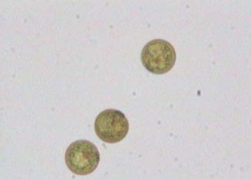
Microscopic examination showed the sample to be teeming with the alga Chlamydomonas.
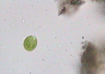
Chlamydomonas
Chlamydomonas is a unicellular, motile, biflagellate green alga, oval to rounded in shape, depending on the species. Some possess an outer, gelatinous envelope. The shape of the chloroplast and number and position of pyrenoids vary according to species. The cells can be from 5 - 20µm in length.
Together with the microscopic examination of the samples, various chemical tests were also routinely performed. It was interesting to observe the pH measurements with the changes in algal population of the pond.
With the appearance of the large numbers of Chlamydomonas, the morning pH registered 6.71 units. The afternoon pH was pushed to 8.24. This being a reflection on the photosynthetic activity.
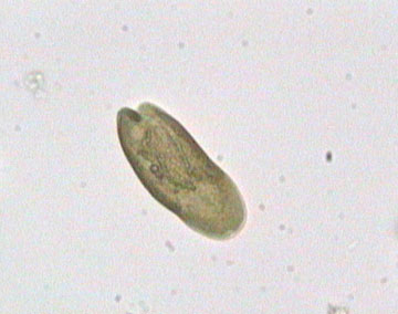
On 12th February the water sample appeared much less green than seen of late. Microscopy now showed the presence of large numbers of Trachelomonas.
Trachelomonas
Trachelomonas is a free-swimming loricated euglenid. The green chloroplasts may be seen, but the cells are usually brownish in colour, which is due to the absorption of metal salts by the test.
On 20th February, microscopy now appears to show a balancing out of the population present and is represented by individual members of all the above species.
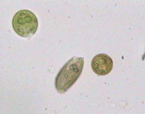
All the above species described
Comments to the
author
Mike
Morgan
are welcomed.
All images
by the author
Published in the March 2006 edition of Micscape.
Please report any Web problems or offer general comments to the Micscape Editor .
Micscape is the on-line monthly magazine of the Microscopy UK web site at Microscopy-UK
© Onview.net Ltd, Microscopy-UK, and all contributors 1995
onwards. All rights reserved.
Main site is
at www.microscopy-uk.org.uk
with full mirror
at www.microscopy-uk.net
.