|
|
A Gallery of Beta - Naphthyl Acetate Photomicrographs (using
a variety of illumination techniques) |
|
|
A Gallery of Beta - Naphthyl Acetate Photomicrographs (using
a variety of illumination techniques) |
Beta-naphthyl acetate seems to be a
compound that is not commonly used outside of biological
research. There is some indication that it is an intermediate in
the production of some scent producing substances for the fragrance
industry.
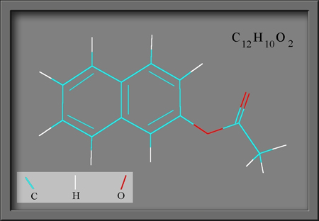
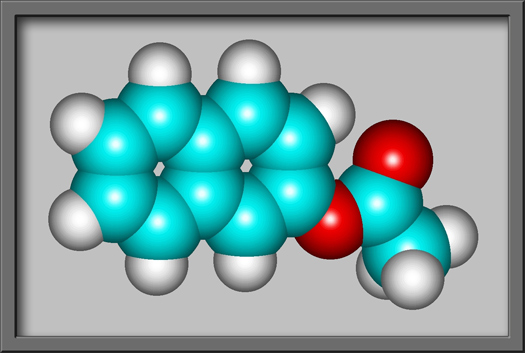
As can be seen in the structural
formula and molecular shape shown below, the structure is based upon
two attached benzene rings. (Both illustrations were produced by HyperChem Pro.)
The very low melting point of the
white powder, about 69 degrees Celsius, allows the easy production of a
melt specimen by heating a small sample between microscope slide and
cover-glass. Little research seems to have been done on the
dangers involved in the use of beta-naphthyl acetate. The MSDS
safety document contains the following.
Caution! The toxicological properties of this
material have not been fully investigated. May cause eye and skin
irritation. May cause respiratory and digestive tract irritation.
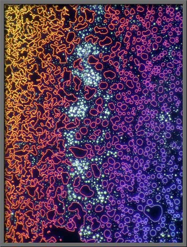
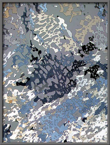
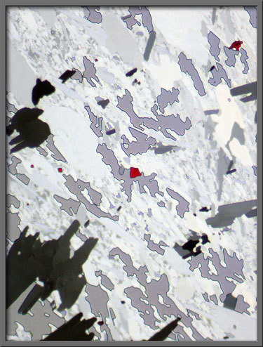
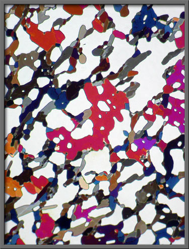
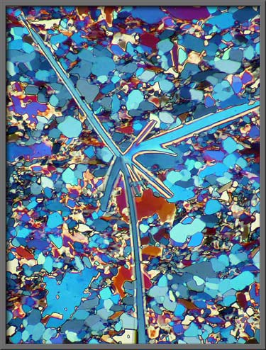
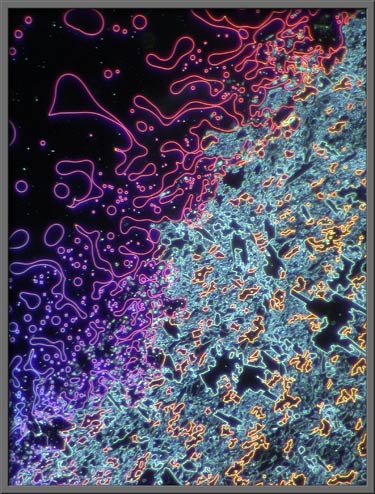
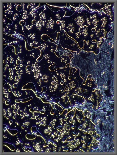
Most photomicrographs of melt
specimens of the compound show evidence of a multitude of tiny gaps
between the crystals. These gaps appear light gray in the
polarized light images below. Two lambda/4 compensators were used
to produce the gray background, rather than the normal black.
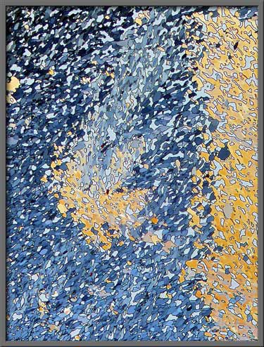
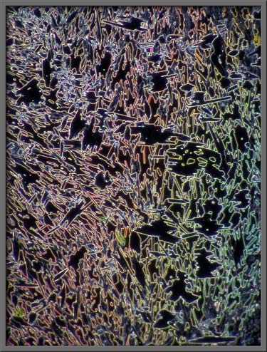
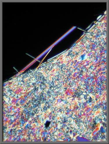
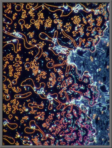
The edges of individual crystals
can be more easily distinguished when dark-ground illumination is
used. In the case of the two images that follow, a normal
objective was paired with one of the annular rings of a phase-contrast
condenser in order to produce the dark-ground effect. (A normal
dark-ground condenser would not produce the subtle colouration seen
below.)
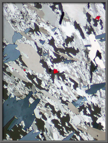
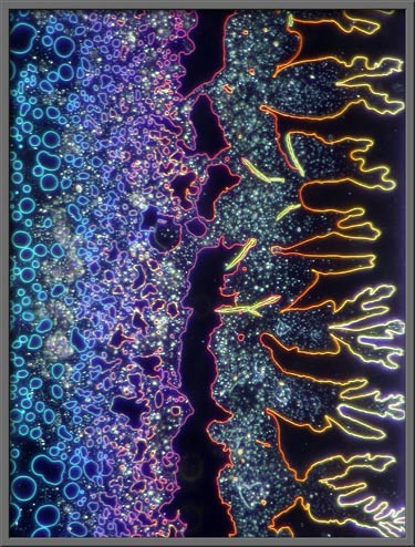
Here
again, polarized light, with two lambda/4 compensators helped produce
the colouration. Can you see that both photomicrographs are of
exactly the same field? The circular lambda/4 compensator was
rotated slightly to produce the dramatic difference in the appearance
of the two images.
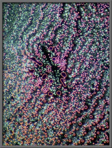
Three more images using the same
illumination technique follow. The higher magnification shows the
gaps between crystals more clearly.
Larger structures sometimes
form. The left image uses the same illumination as the images
immediately above. The right image uses crossed polars with no
compensators.
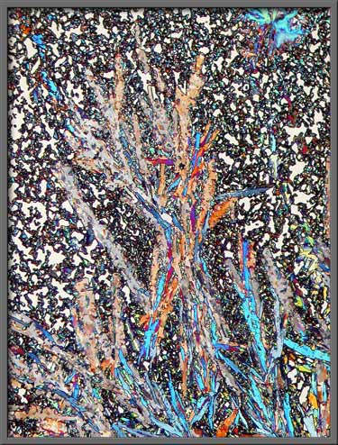
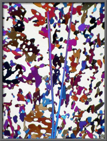
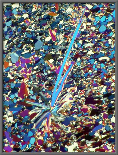
Needle-like structures sometimes
occur.
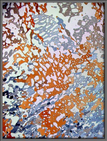
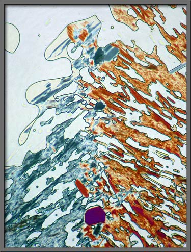
In the image that follows, one of
the two lambda/4 compensators was replaced by a lambda
compensator. In this case, the gaps between crystals are orange
in colour.
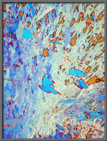
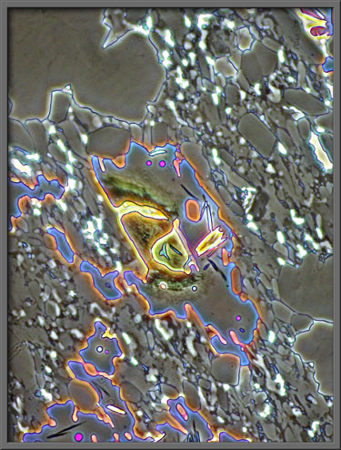
A much higher magnification reveals
the detail at the edge of a gap (black).
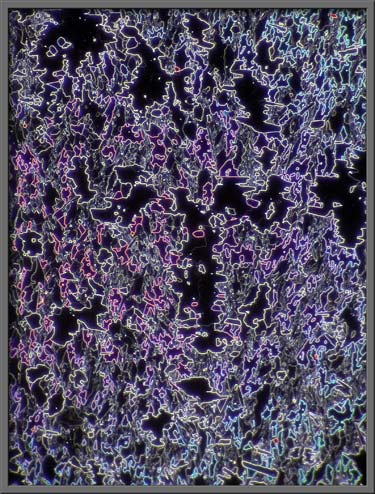
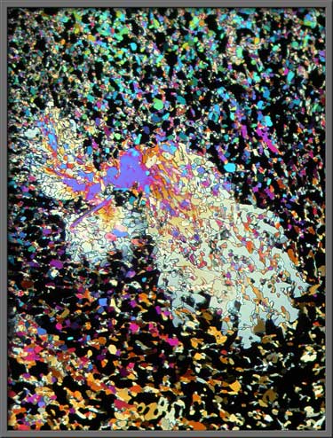
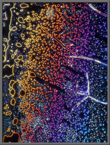
In the next group of five images,
the polarizing condenser has been replaced by a phase-contrast
one. A normal, (non-phase) objective was used to form the
image. By experimentation, a particular annular ring of the
condenser was chosen to produce the desired effect.
For comparison, here is a similar
field using a normal dark-ground condenser.
Again for comparison, a similar
field at higher magnification is imaged by using a normal
phase-contrast configuration (with phase objective).
Since compounds like beta-naphthyl
acetate have been removed from high school chemistry labs for safety
reasons, it has become almost impossible to obtain tiny samples for
microscopic examination.
Photomicrographic
Equipment
The images in the article were
photographed using a Nikon Coolpix 4500 camera attached to a Leitz
SM-Pol polarizing microscope. Images were produced using several
illumination techniques: dark-ground, phase contrast and polarized
light. Crossed polars were used in all polarized light
images. Compensators, ( lambda and lambda/4 plates ), were
utilized to alter the appearance in some cases. A 2.5x, 6.3x, 16x
or 25x flat-field objective formed the original image and a 10x
Periplan eyepiece projected the image to the camera lens.
Published in the March
2007 edition of Micscape.
Please report any Web problems or
offer general comments to the Micscape
Editor.
Micscape is the on-line monthly magazine
of the Microscopy UK web
site at Microscopy-UK
© Onview.net Ltd, Microscopy-UK, and all contributors 1995 onwards. All rights reserved. Main site is at www.microscopy-uk.org.uk with full mirror at www.microscopy-uk.net .