The Impressive Bausch & Lomb
Research Microscope DDE
Manuel del Cerro, Pittsford, NY, USA
Eight years ago, Dan Kile and I wrote an article on the Bausch and Lomb microscope DDE (Kile and del Cerro, 1999); that year there were two additional publications on the same model (Bishop, 1999; Solliday, 1999). This was a clear indication of the interest
on that particular instrument among collectors and historians of the microscope. That interest was well justified then and it remains so today. This is why I would like to present here a review on it that, while shorter than our original article it does incorporate
pertinent information collected during the intervening years.
THE ORIGINS
By the late 1920s, the Bausch & Lomb Optical Company of Rochester, NY (B&L), had become a world leader as a microscope maker. The volume of production was enormous, possible larger than that of any of the other first-class makers, such as Leitz, Spencer, or Zeiss. Competition was fierce, but the unchallenged notion that microscope optics had reached an unsurpassable limit succeeded in stagnating the creativity of optical designers. Thus, the efforts to improve the microscope were directed towards the mechanical components of the instrument. That was the frame of mind that brought about the creation of the Model DDE (figure 1). It should be noted that the design of the DDE was not, at least initially, a product of the B&L Engineering Department. Just as all the other major makers, B&L was open to suggestions from distinguished users of the instrument. As we will see below, they were also quite willing to give credit to their consultants.
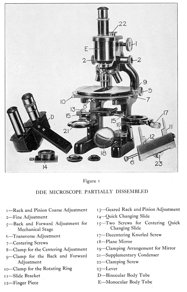
Figure 1. The anatomy of the DDE.
MAKERS DESCRIPTION
In 1929, B&L introduced the Model DDE and described it using the following (abridged) text:
“ DDE – A New Research and Photographic Microscope. The word “new” is so often used to describe a revamped apparatus that one hesitates to use it but in the case of this Research Microscope, the word “new” can certainly be used in the fullest sense of the word.
This instrument is a radical departure from any existing type of microscope.
When we took under consideration the design of a new Research Microscope, we adopted the suggestion of Dr. Lester W. Sharp of Cornell University and of his associate Dr. L. F. Randolph of the U. S. Department of Agriculture and Cornell University of placing the arm at the front of the instrument, so as to give free access to the object, stage, objectives, sub-stage and mirror and thus offer greater convenience and comfort to the user. With the further aim of providing extreme rigidity we have eliminated the usual inclination joint for arm and pillars, which are made in one piece. The stage is thus always horizontal except when the instrument is used photomicrographically, when the entire instrument is placed in horizontal position resting upon three bosses provided on the two arms and base, thus leaving each part intact and in the vertical stage.” (figure 2) [Italics in the original]
Lets note that of course, the DDE could be used in the more conventional, upright position (figure 3). In either case a powerful lamp was recommended as a nearly indispensable accessory (at extra cost, naturally).
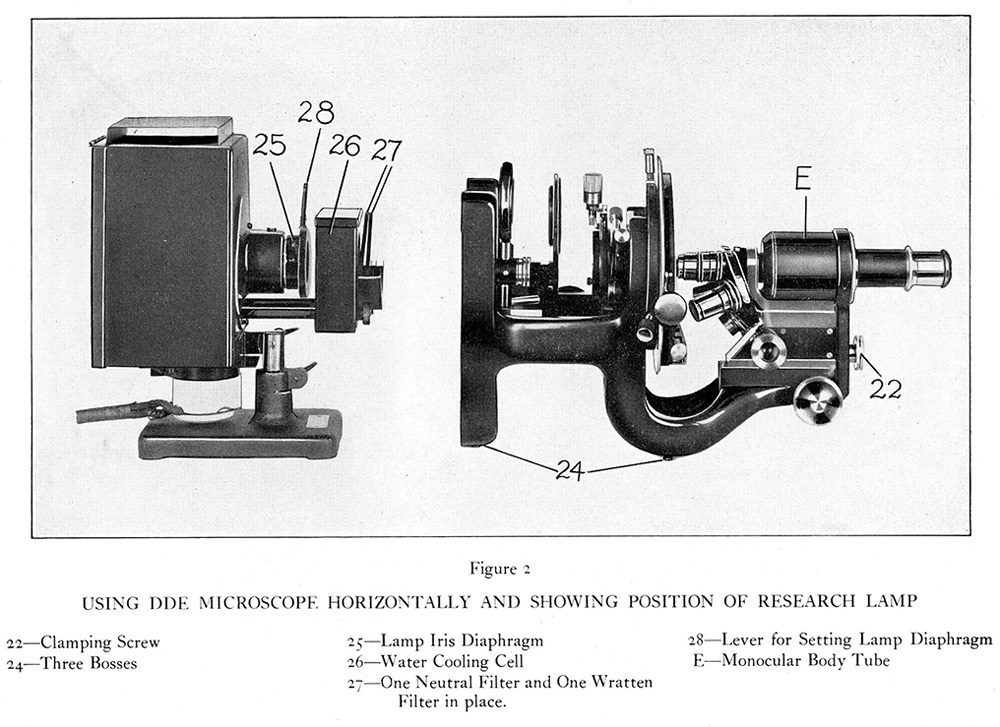
Figures 2. The DDE positioned horizontally for photomicrographic use with its mirror removed from the optical path. Notice the special illuminator.
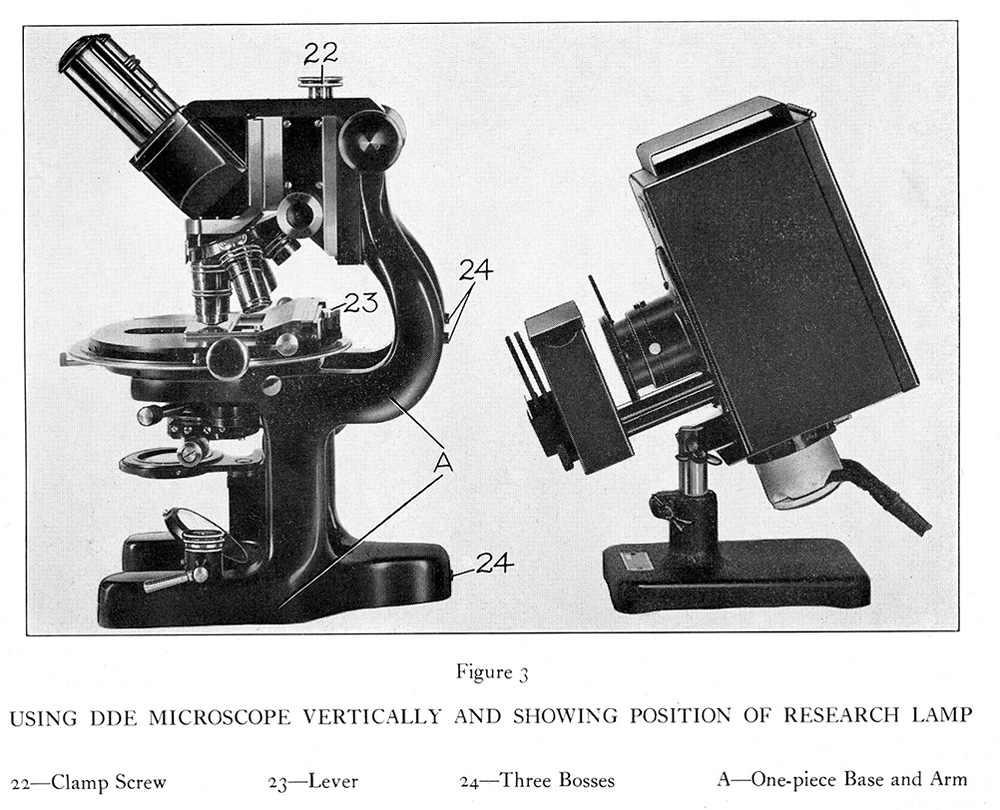
Figures 3. The DDE positioned for visual work with its binocular head in place. The illuminator sends light through the opening between the double pillars.
FAME, GLAMOUR, AND SERIOUS RESEARCH
During the last decade of the 19th century and the first halve of the 20th, the Bausch & Lomb microscope operation was ahead of its times in developing aggressive marketing strategies and advertising campaigns. They were not timid in promoting DDE that was for three decades their flagship instrument. B&L advertising department was quite successful in their efforts and the DDE reached iconic status. This fact was not lost on Hollywood. “Topaze,” a satire by Marcel Pagnol with John Barrymore and Myrna Loy in the leading roles, portraits a chemist (Barrymore) who makes some sensational discovery assisted by his microscope. The microscope was a DDE. Later, Warner Brothers had a DDE assisting the criminal investigations carried on by the elegant detective who is the protagonist of their film “From the Headquarters.” (Corrington, 1939).
Informally speaking one could say that the DDE was not just about fun and games, quite the contrary, it was used for some very serious research, even at the time when it was going out of production. The work of Zeller (1951) provides a particular good illustration of this point. This researcher took advantage of the easy accessibility of the DDE stage and of the microscope tremendous stability, to use micropipettes to seed single live bacterial cells on agar, and then to study the genetics of these clones as the cells divided – all under direct observation using the 44x objective (figure 4).
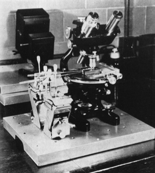
Figure 4. Dr. Zelle’s setup using the DDE for the study of bacterial genetics.
LATE VERSIONS OF THE DDE
The DDE model evolved little from the beginning to the end of its production time. The first novelty was the introduction of the single-objective, centering nosepiece that was offered as an option in 1947 (fig. 5) and was of assistance particularly in chromosomal research (Grun, 1959). Another change was the absence of the so-called “operating head” (B&L nomenclature) in the binocular head of late-production models (compare figs 6 and 7). That operating head was a chrome-finished knob located on the right side of the binocular body; when turned 90º it diverted all light to right-side ocular. It was not particularly useful and it was quietly removed in instruments manufactured after WW-II, although some old, unsold stocks with it were still in the market in the late 1940s (personal communication from ex-B&L employees).
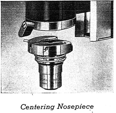
Figure 5. The single-objective centering nosepiece for the DDE. This format was preferred for studies that required repeated observation
of some minute details in the specimen, such as in chromosome work.
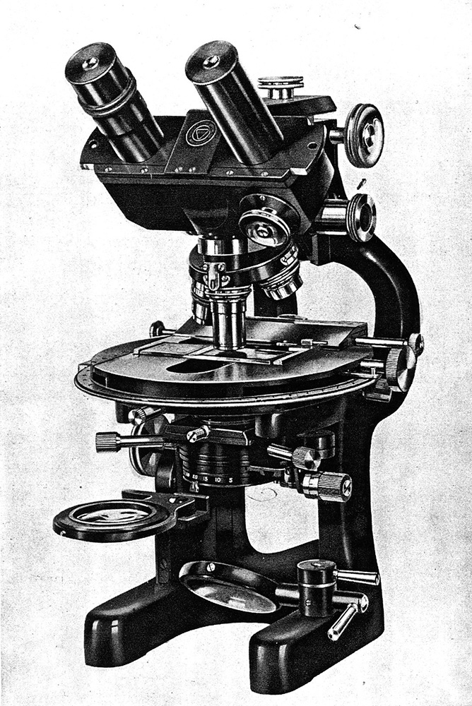
Figure 6. A DDE with the original binocular head. Notice the light-diverting knob located on the right lower corner of the head; contrast it with the late 1940s version shown in figure 7.
I have examined closely a 1946 DDE, signed Bausch & Lomb Opt Co U S A, serial number MT36, MdC Collection #405, (figure 7). This is a mono-binocular microscope with original wooden case and accessories. With the draw tube extended to the required 16 cm mark, and the 10x objective focused on the specimen, the microscope is 33.2 cm tall and it weights 13 kilograms. The horseshoe base is 12.1 cm wide and 19.0 cm long. The mirror has only a plano surface, a typical feature of the DDE stand. It can be moved vertically, held in place, or be entirely removed from the optical path by means of a clamping device located on the right extension of the horseshoe base. Removing of the mirror while the microscope is in horizontal position allows the light from the illuminator to reach the condenser directly. This arrangement that called for using the monocular tube as well, was considered optimal for high quality photomicrography (fig. 8). The base, the double pillars, and the double arms are part of a single metal piece. The curvature of the arms is away from the observer. The understage has an aplanatic condenser with a NA of 1.30. It can be centered by the action of two side knobs, and can be focused by an exquisitely precise movement controlled by a large knob located on the left side (item 13 in figure 1). A rack and pinion mechanism allows de-centering the condenser and its attached iris diaphragm, so as to provide oblique illumination to the specimen (17 in figure 1). Finally, an auxiliary lens located below the condenser (21 in figure 1) provides full-field illumination when using a 4x objective, then it can be swung aside when switching to the higher power objectives. In 1932, B&L was issued a USA patent 31,860,430 to protect the design of this condenser. The circular stage rotates and it has graduations in degrees and a vernier. There are also verniers to measure the x and y movements; separate, horizontally arranged knobs control this movements. Both the rotation and the x and y movements can be clamped in order to keep the stage at a fix position. The arms converge on a robust, rectangular piece that houses the mechanisms for fine and coarse focus. The respective side knobs are located high above the table surface (items 1 and 2 in figure 1). The oculars in the binocular head are 15x compensators; the one in the monocular is a 10x Hyperplane, recommended for photography. All the four objectives are brass-finished apochromats. They are a 10x 0.30 N.A., a 20x 0.65 N.A., a 45x 0.95 N.A. with correction collar, and a 90x oil immersion 1.30 N.A. OPTICAL PERFORMANCE: The test was run by examining five paraffin sections of normal human tissues stained with hematoxylin-eosin, and mounted with a synthetic resin, and four Golgi slides of monkey cerebellum. The images given by all the dry objectives are very good centrally, 4/5 in a subjective scale (Oldfield, 1994, p. 59). The 10x and the 20x objectives suffer from moderate field curvature. Field curvature is negligible in the case of the 45x. The field diameters are 1,110 µm, 550 µm, and 210 µm respectively. Although this is a sixty-year old instrument, the fine focus control retains a silky quality of movement. The fine control of the condenser focus is superior to that of any microscope I have used, modern or antique. COMMENTS: Few DDE microscopes are known to exist at this date; with the exception of the one in the Billings Collection, Washington, DC (Purtle, 1974) all that are known to me are in private collections. Additionally, I am not aware of any surviving example of the late binocular body than the one illustrated in figure 7.
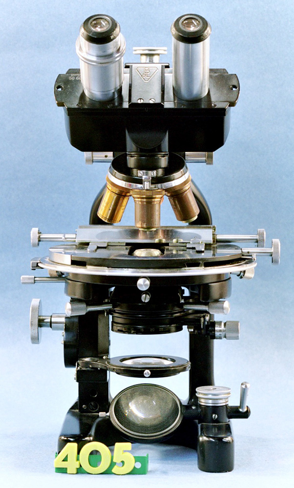
Figure 7. The 1946 DDE number MT 36, from the author’s collection. The light-diverting knob in the binocular head has been removed in this late-production example;
contrast this with figure 6.
THE DEMISE OF THE DDE
Why did the DDE become obsolete in the 1950s? The creation of the DDE in the late 1920s represented the materialization of some very innovative concepts. It was designed to be the ultimate research microscope, and in a way that is what it was, at least in the American scene. That was, at the same time, its strength and its weakness. It was an expensive microscope. Equipped with the quadruple nosepiece, apochromatic objectives, the optional (but very necessary) monocular tube, and using the lamp specially designed for it, the total cost in 1929 was around $800. That was a very significant expense at the times and it was not a happy coincidence that 1929 was the year of the DDE introduction and the year the Great Depression started.
Designed specifically for advanced research and photomicrography; the DDE is a grand microscope with poor ergonomic features. It is tiring to hold one’s hands 22 cm above the desk surface for periods of time while fine adjusting focus as required when the higher power objectives are in use. This was not a microscope for the busy clinician or for the pathologist. This is not the microscope one will choose for evaluating 100 slides in one afternoon! Setting it up for photography (figure 8) must have required considerable time and effort, not to mention dedicated laboratory space.
Cost and extreme specialization conspired against the sales. The DDE sold in modest numbers, a fact that accounts for the scarcity of examples known to have survived to our days.
As the decade of the 1950s started it was clear that the DDE had become hopelessly outdated. Its brass barrel objectives had all the appearance of a 1920s item, which was what they were. More serious that appearance was the lack of anti-reflective coatings on the optical surfaces, a feature that was becoming standard in all research instruments. As we all know now, metallic coating of the surfaces of lenses reduces internal glare and makes images crisper, a most desirable feature for a photographic microscope. The capability of the DDE to be set horizontally for photographic work became irrelevant as the large-format, long-bellows cameras (figure 8) were replaced by the small (“miniature”) 35 mm cameras. There were other limitations, such as the lack of a built-in, or attached to, illuminator, the lack of a trinocular configuration, etc. Clearly, the DDE could not compete with the more practical and modern research microscopes that were becoming dominant in the market such as the Leitz Ortholux, to mention just one.
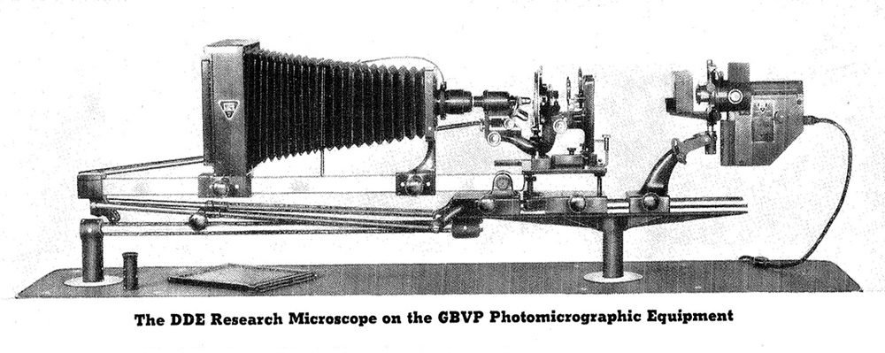
Figure 8. The impressive, optical-bench style, photographic set up was considered optimal to record images with the DDE. The camera back and the illuminator display the characteristic triangular B&L factory logo. A plate holder and the focusing loupe rest on the table at the left.
LEGACY
Of the two most innovative features of the DDE stand, the dual arms and the open stage design, one never become too popular, while the other has met with overwhelming success.
Only two other makers adopted the dual-arm configuration for research microscopes. One was Spencer for their failed Model No. 8, of the 1930s (Purtle, 1974; Kile and del Cerro, 1999). The other was Steindorff for their Microbe Hunter (sic) Model which had a trinocular configuration, was more reasonably priced, and survived both the DDE and the No. 8 (Mach and del Cerro, 2004). A few other makers, such as Enuro, used the dual arm configuration in their hobby, or student microscopes but it was for aesthetic rather than functional reasons.
The second distinctive feature of the DDE was the placement of the stage directly in front of the user, with the arms away from him or her. That was a revolutionary innovation and the one that constitutes the lasting legacy of the DDE. Today, every laboratory, clinical, or research microscope known to me, has its stage directly in front of the user. That is to say, every time we use a microscope we are benefiting from the innovation that Drs. Sharp and Randolph brought into microscope design and that B&L implemented in their DDE Model.
ACKNOWLEDGEMENTS
I thank Dan Kile, Colorado, USA, for the many fruitful discussions we had on the DDE. I learned much of what I know on the subject of dual-arm microscopes from Martin Mach, Munich, Germany. I thank several B&L retirees who shared with me the memories of their microscope making years.
Comments to the author are welcomed.
BIBLIOGRAPHIC REFERENCES
• Bishop, Allen (1999) The Journal of the Microscopical Society of Southern California, vol. 12, December issue.
• Corrington, Julian D. (1939) The Microscope a Pictorial History. Science Magazine, February Issue. (Reprinted in The Journal of the Microscopical Historical Society (1999) 7:29-38.
• Grun, Paul (1959) Variability of Accessory Chromosomes in Native Populations of Allium cernuum. American Journal of Botany, 46: 218-224. doi:10.2307/2439280.
• Kile, Daniel E. and Manuel del Cerro (1999) The Microscope Model DDE, Bausch & Lomb’s Flagship for the 20th Century. 7: 50-63.
• Mach, Martin and Manuel del Cerro (2004). Between Glamour and Glory: The Steindorff “Microbe Hunter.” The Journal of the Microscopical Historical Society 12: 30-46.
• Oldfield, Ron (1994) Light Microscopy. An Illustrated guide. Wolfe, Mosby-Year Book, London, Oldfield, 1994, p. 59.
• Purtle, Helen R., (Ed., 1974) The Billings Microscope Collection. Armed Forces Institute of Pathology, Washington, DC, p. 148, fig. 278.
• Solliday, James D. (1999) The Journal of the Microscopical Society of Southern California, vol. 12, December issue.
• M. R. Zelle. (1951) A Simple Single-Cell Technique for Genetic Studies of Bacteria. J Bacteriol., 61: 345–349.
___________________________________________________
Published in the March 2007 edition of Micscape.
Please report any Web problems or offer general comments to the Micscape Editor .
Micscape is the on-line monthly magazine of the Microscopy UK web site at Microscopy-UK
© Onview.net Ltd, Microscopy-UK, and all contributors 1995
onwards. All rights reserved.
Main site is
at www.microscopy-uk.org.uk
with full mirror
at www.microscopy-uk.net
.