|
Review of a Celestron LCD microscope |
By
Jean-Marie Cavanihac |
|
|
|
|
Celestron announced a new microscope with LCD screen for March 2008. In France the same microscope was commercialized by Bresser around the 2007 Christmas holidays. This scope is not a toy, nor a research microscope, of course! But, probably, a first step in a new form of modern microscopy. It's more pleasant to use than a single monocular microscope, indeed, with screen, many users can see real time pictures simultaneously without the need of a micro computer! |
|
Some advantages: built in 3,5 '' LCD screen (78 x 53 mm) , a 2 mega pixel camera AND.... 128 Mbyte of picture storage. An SD card (= 1 gigabyte) can to be used in conjunction for extended memory and a USB link allows transfer of pictures to a computer. (The main memory and SD card are recognized as 2 independent external drives); It's not possible to see real time pictures on the computer, only after downloading from memory. The LCD screen is larger than a screen encountered on a digital camera and is comfortable to use. |
|
Some features from Celestron: Compound (Biological) Microscope: Objective Lens - 4x, 10x, and 40: x 40 to 400 Power - up to 1600 Power with Digital Zoom, Built-in Digital Camera - 2 Mega Pixels, 3.5" (88mm) LCD Screen with 4x Digital Zoom, Top and Bottom LED Illumination, 128MB Internal Storage Memory + SD Card Slot, Mechanical Stage - 3.5" x 3.5" (88mm x 88mm). - Six Position Color Filter Wheel , USB Cable for Transferring Images to a PC, 5 volts AC Adaptor to Power the Microscope. - Carrying Case Included ,- Weight - 51oz (1446g) , Two Year Limited Warranty, -Five Prepared Slides + 10 empty slides, |
|
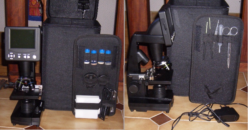
First impressions: The LCD Microscope is sold in a rigid plastic case with removable strap, easy for transportation. Inside, a separate panel contains some sampling accessories, a well written small manual with examples of preparation, a kit for Artemia culture and a gum medium to prepare slides, plus a box with prepared slides: onion cell, wood stem, fly leg and 10 empty slides. |
|
|
|
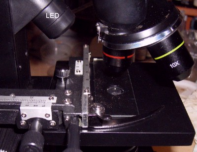
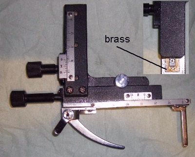
Almost all the scope is made of black anodized aluminum: only the screen housing, focusing knob and illuminator are plastic material . In the right hand picture below you can to see the X-Y stage with a simple but smooth motion: the X-Y assembly is easily removable if you want to put a Petri dish, for example, on the stage: | |
|
|
|
|
In the base, a white led with dimmer is covered by a removable lens or ground glass in front of the illuminator A 4 position switch is used to power episcopic led or diascopic led or both. |
|
Objectives are well made and carefully centered on a brass revolver: x 40 has a retractable frontal lens! Of course they are not apo chromatic and some chromatic aberrations are present on edge of field of view . Note: Objectives do NOT have a standard RMS thread. |
|
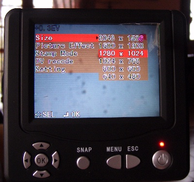
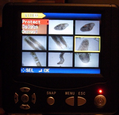
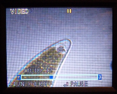
Below are some shots of screen menus: 1 - main menu with sub menu : - image size selection, 6 sizes (in fact it's best to use 1600 x 1200, the full camera resolution, and you can save more than 500 pictures in internal memory). - Picture effect: image processing (normal, sepia, black and white, inverse). - Stamp mode (insert date or time, or both in picture or none..) - DV record : video recording (320 pixels) in ASF format. - Setting : time setting (real time not saved) and an interesting feature: possibility to take pictures with time lapse (minimum 5 seconds). 2 - Slide show menu: you can select picture (or video) in thumbnail format, protect, or delete them. (Note : if you insert an SD card from your digital camera, you can see pictures of your past holidays on the microscope screen!!) 3 - Video menu with elapsed time and fast forward/pause commands. | |
|
|
|
|
| |
|
Others features: zoom function between 1 and 4 times (in fact 2 times is sufficient to obtain intermediary magnifications of 4 to 8 ... 10 to 20 and 40 to 80 ) using right and left arrows on navigation knob. Up and down arrows adjusts screen lighting (and picture lighting too). A filter wheel with five colored filters and one neutral, which would increase contrast, is located under stage (usable mainly on colored specimens). But I think you are impatient to see pictures!! The pictures are more contrasty on the LCD screen than on computer screen, probably automatic exposure is optimized for the little screen, but lighting of pictures can to be adjusted by up and down arrows on navigation knob. | |
|
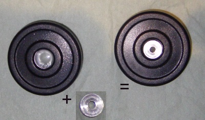
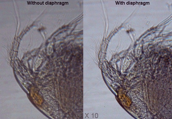
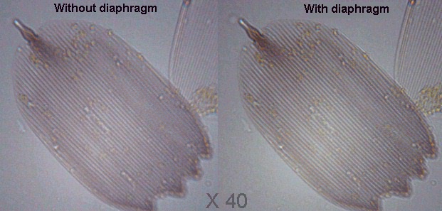
A little regret: the illuminator does not have a diaphragm: A removable diaphragm (made by me: in fact a simple washer with 3 mm hole!) located on the ground glass on the illuminator, improves greatly picture contrast and depth of field, mainly with the x4 and X10 objectives, a little less with x40. | |
|
| |
|
See improvements in pictures below for 2 magnifications (copepod and butterfly wing scale). Note: all pictures taken in 1600 x 1200 format are reduced 4 times. | |
|
| |
|
| |
|
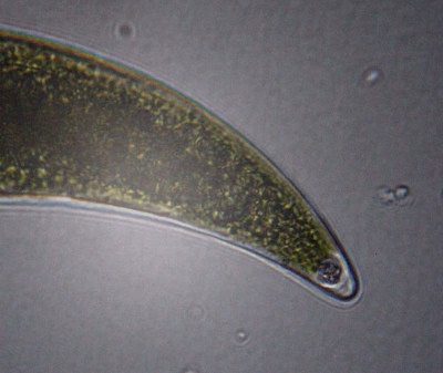
Other trials: actinarian (eating a diatom! )(x 10) , - Detail of end of desmid Closterium showing small crystals (x 40). | |
|
|
|
|
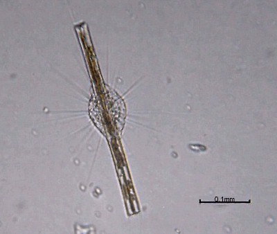
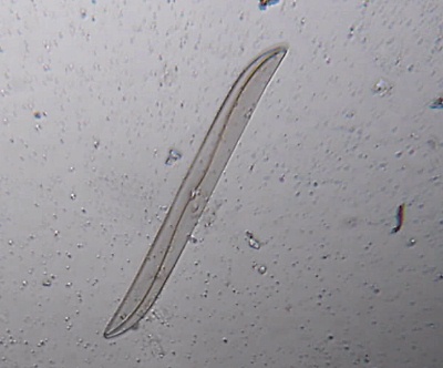
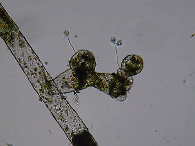
Diatom Gyrosigma (x 10), - Vaucheria (x 10) , | |
|
|
|
|
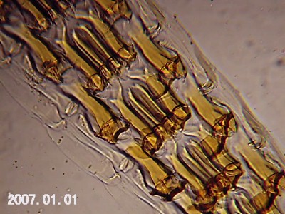
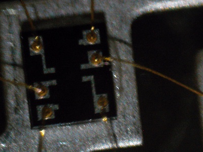
Snail radula (x 10) (note date stamp inside) - episcopic image: detail of photo detector device on CD reader (x 4 ) | |
|
|
|
|
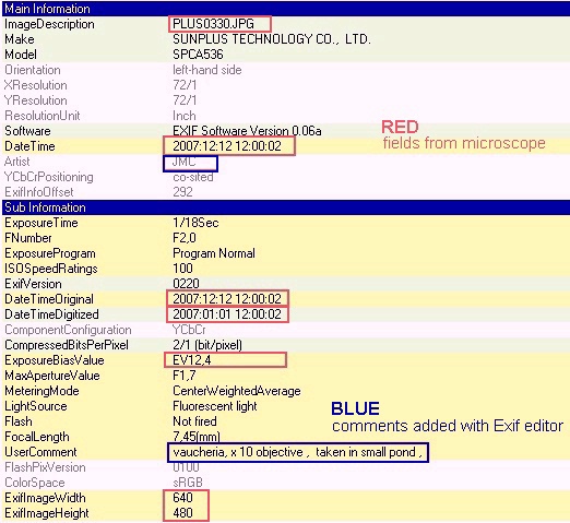
Suggestion for the manufacturer: to improve contrast and depth of field, replace 3 filters on the filter wheel by holes with 1, 2 and 3 mm diameter (a better solution than mine with the washer on illuminator!! ). Note: An EXIF file is embedded in the pictures; in red are values which are changing according to picture size, exposure, date etc... With an EXIF editor you can modify items (in blue) "user comments", "author" etc into the file to annotate your pictures. |
|
|
|
The zoom is very handy, mainly to adjust fine focus on small detail. I prefer not use more than 2 times of magnification, to conserve image quality. Real size of field covered by objectives are: 1,6 mm for x 4 objective, 0,65 mm for x 10 and ... 0,16 mm for x 40. The x 40 objective gives astonishingly good pictures: it's often the "weak link" on this kind of microscope. A slight "hot spot" of light, is seen in picture center with this objective (may be an internal reflection?). |
|
A small video clip (ASF format, 680 kbytes) of a protozoan. (Editor's note: In Explorer 6.0 (not 5.0) or Firefox the clip is automatically played in Windows Media player.) |
|
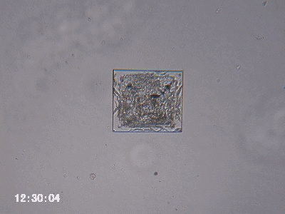
An animated sequence: salt crystal growing, obtained with time lapse function: 20 seconds between shots and 20 pictures assembled in a animated gif file (with PSP software ). |
|
|
|
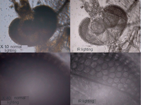
Some simple improvements you can make: The 5 volts external power supply can easily be replaced by a 5 volts power supply made with integrated regulator (7805) from cigar lighter on a car (or a boat!): (and probably you can power directly the scope with a single 4,5 volts battery): it becomes a fully autonomous scope for studies in the field!! Other trials: just replace the white led by an external infrared led (led in TV remote control) and you can see through opaque insects, for example this mosquito head (detail of eye). |
|
|
|
In conclusion: a very pleasant microscope including picture taking and usable without need of computer! But don't forget: like for other microscopes, if you want to make good observations (and pictures ) some little preparation of specimens are necessary! You can see a French translation at Microscopies.com |
|
Llink: Celestron site: http://www.celestron.com/c2/product.php?CatID=31&ProdID=516 Editor's note: Typical street price $299. |
Comments to the author Jean-Marie Cavanihac are welcomed.
All drawings and photographs © Jean-Marie Cavanihac 2006
Published in the August 2006 edition of Micscape Magazine.
Please report any Web problems or offer
general comments to the
Micscape
Editor,
via the contact on current Micscape Index.
Micscape is the on-line monthly magazine of the Microscopy UK web site at http://www.microscopy-uk.org.uk