|
Nineteenth Century British Microscopy and Natural History: Part 4 by Richard L. Howey, Wyoming, USA |
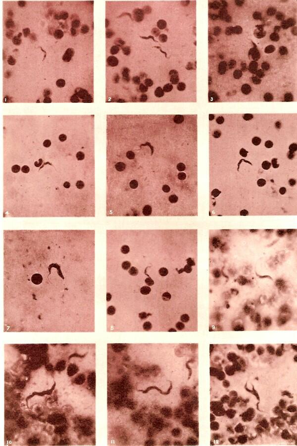
In this part, we’ll take a look at the December, 1886 issue of the Journal of the Royal Microscopical Society and this time we find two lead articles. The first of which is by Edward M. Crookshank and is titled “Flagellated Protozoa in the Blood of Diseased and apparently Healthy Animals.” The second by T.B. Rosseter is “On Trichodina as an Endoparasite.” Each is accompanied by a plate and I will include them at the appropriate spots. The plate for the first article is a photographic isochromatic plate, instead of the more usual plate with drawings, and the organisms have been tinted with magenta.
For those of you who have any familiarity with protozoan parasites, you will immediately recognize the organism as a trypanosome. In figure 7 especially, one can see the flagellum which is really an extension of its undulating membrane. Crookshank refers to a report on this organism which underscores its military and economic significance.
“In the year 1880, Dr. Evans presented a report to the Indian Government on a fatal disease know by the natives as Surra, occurring in horses, mules, and camels. The importance of this disease may be realized from the fact that the 3rd Punjab Cavalry alone lost no less than 300 horses from it.”
Trypanosomes were, at this time, not widely known and early investigators thought that they were a spirillum type of bacteria. Trypanosomes are intriguing, but nasty organisms which have been found in the blood not only of horses, camels, and mules, but monkeys, dogs, bison, caribou, elk, moose, certain fish, rats and humans among others. The most famous trypanosomes that infect human beings are undoubtedly those that cause African Sleeping sickness. These odd little creatures have an enormous impact in the world. Evans was, naturally, interested in finding the mode of transmission and Crookshank reports the following.
“The disease was not observed to be contagious or infectious in the ordinary sense, but the possibility of its conveyance by means of large brown flies was suggested. These flies attack the horses so vehemently that the blood frequently streams from the bites; and the opinion that they propagated the disease was prevalent among the natives. At the same time, it was particularly noted that at outposts where the disease originated the water was very impure.”
In Africa, the tsetse fly is the transmitter of sleeping sickness. Biting insects are vectors for a wide range of diseases and flies and mosquitos, in particular, are primary villains. Yellow fever, dengue, and malaria are transmitted by mosquitos. The second “War to End All Wars” created a false optimism that there would never again be a necessity to send troops into impoverished, disease-ridden tropical countries; there was a naive belief that either there would be no more such wars or that they would be conducted by largely mechanical means relying on machines and high-tech weapons. As a consequence many institutes and hospitals which had specialized in tropical medicine were phased out. Unfortunately the optimism was not justified. Furthermore, annually, millions of people still die of malaria and we have discovered new biological horrors, such as HIV and the hemorrhagic fevers like Ebola and in the age of rapid air travel such diseases can be spread from remote areas to cosmopolitan centers in a matter of hours. Sell your cell phones and donate the money to tropical disease research.
The organism in the second article, Trichodina, doesn’t have the kind of economic and medical impact of the trypanosomes but is, nonetheless, an intriguing little beastie with some peculiar characteristics. I’ll include the plate here, so that you can get an idea of what it looks like.
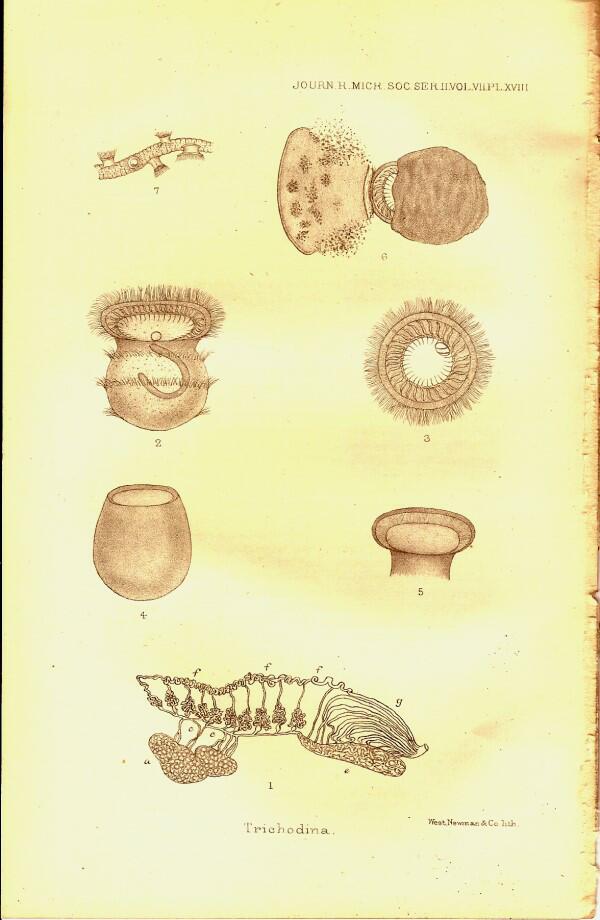
Mr. Rosseter is convinced that he discovered a new species of Trichodina and indeed he makes a very good case. The famous species of this organism is Trichodina pediculus which is an ectoparasite on Hydra.
It probably is not a parasite at all, but a commensal; that is, it attaches itself looking for a free ride, a free lunch, and protection. Ehrenberg, who originally named it, seemed to think that it was emphatically a parasite since the Latin pediculus means “louse”. It can and does detach itself and has periods as a free-swimming ciliate. Hydra are not particularly fastidious feeders and after devouring a Daphnia or small cladoceran, they eject the remains and there is an abundance of micro-food particles which Trichodina can feed on by creating ciliary currents. Trichodina can attach to the tentacles of Hydra and gain advantage from the protection of the nematocysts or “stinging cells” that are lodged in the tentacles like a battery of weapons. Why doesn’t Trichodina trigger the nematocysts? It may be that the size and weight of these organisms is insufficient to provide a stimulus to trigger the mechanisms or perhaps they have learned how to “tickle” the tentacles in a manner pleasing to the Hydra. I’m not being altogether facetious here. There are many strange, complex, and only partially understood relationships between organisms of radically different character. Consider the various fish that take up refuge among the tentacles of anemones. Some of them apparently nibble on the tentacles and acquire an immunity to the toxins in them–rather like mini-marine versions of Rasputin. Others seem to secrete a mucous coating over their skin which protects them. Still others swim through the tentacles as though they were caressing and being caressed.
Mr. Rosseter, interestingly, was studying newts when he discovered this new endoparasitic species of Trichodina and found them to exclusively inhabit the kidneys and seminal glands. By means of a set of well-designed experiments, he was able to demonstrate that this new species was not ectoparasitic on Hydra and would not attach to them. Clearly, if this species had adapted to the very different internal environment of the newt–the salt balance, pH, temperature, and oxygen levels–it would be living in a significantly different environment than that of Hydra, which in itself would suggest that it is a distinct species. It is, however, still possible that this organism is an opportunistic endocommensal rather than a parasite. In other words, it may live and thrive within the newt and benefit from the environment in terms of food and protection, but without doing any harm to the newt. Mr. Rosseter’s experiments are an excellent example of careful, clear investigation and lucid, convincing deduction. Since Mr. Rosseter’s day, species of Trichodina have been found as endoparasites or endocommensals in 2 genera of frogs, a surgeonfish, and a marine gastropod.
Now, I’m going to turn to the February, 1886 issue of the journal. Most amateur microscopists love old books on microscopy and natural history and there are certain classics by authors such as Carpenter, Hogg, and Beale that many of us have or covet. Occasionally, I wonder who these people were and what they looked like. In this issue there is a photograph of Lionel Beale who wrote How To Work With The Microscope, so we can satisfy a bit of our curiosity since I will scan it and reproduce it here.
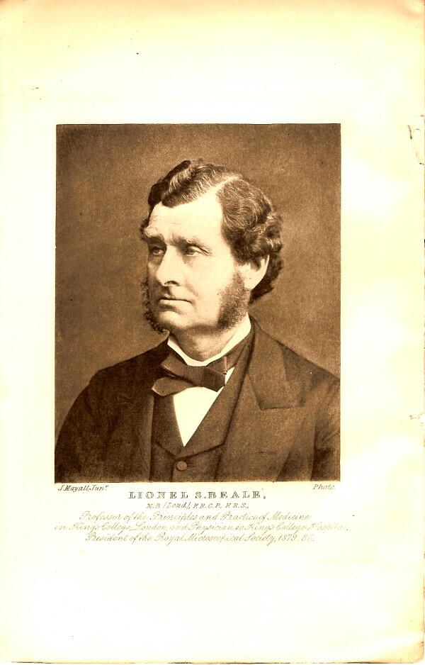
Definitely a serious-looking sort, but it’s difficult to say whether he looks severe, a bit sleepy, or just rather bored with the process of having his photograph taken.
The first of six initial papers is by Alfred W. Bennett titled “Fresh-water Algae (including Chlorophyllaceous Protophyta) of the English Lake District; with description of twelve new species.” Mr. Bennet, F.R.M.S., F.L.S., was also a Lecturer on Botany at St. Thomas’s Hospital; I’ll bet you there aren’t any lecturers on botany in hospitals today.
The article is accompanied by 2 splendid plates with drawings of desmids, with some Nostoc and filamentous algae thrown in.
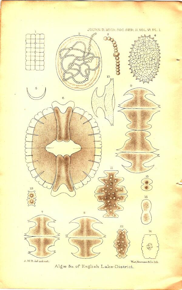
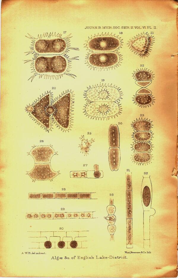
In the text, Bennett refers to those who specialize in desmids as desmidiologists. Imagine how pleasing it would be when filling out bureaucratic forms, to write in the blank for profession: Desmidiologist. Desmids are some of the loveliest organisms which the aquatic microscopist encounters. Closterium is a common and wonderful example.
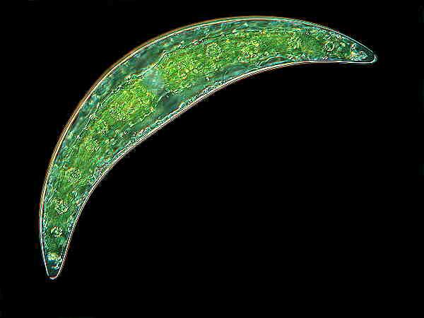
Wim van Egmond has produced a series of beautiful images of desmids. 'Galleries of desmids'.
I shall not discuss the next 2 articles since they are only of middling interest (at least to me).
The next article by our old friend Edgar Crookshank is of special interest for the 3 color plates, the first one containing 2 images which look like abstract art.

Figure one is reminiscent of an abstract painting and shows a series of patterned curves which Crookshank describes as typical of what he calls Bacillus figurans. This a cover glass impression made from an agar plate and stained with fuchsin. Figure 2 is a magnified view of a portion of a curved section showing how it is composed of rod-like bacteria.
Plates II and III show an infection of the fungus Actinomyces in a tumor in a cow. They have been stained using two different techniques. The intriguing thing in all 3 of these plates is the distinctive patterns which the bacteria and fungi make. Ordinarily, we don’t think of bacteria and fungi as having a distinctive colonial form. One is perhaps much more inclined to think of randomness, that such colonies are amorphous like clouds. In fact, however, colony structure is sometimes an important aid in identifying certain types of micro-organisms. Many of us have seen, either directly or in photographs, colonies of the mold Penicillium notatum with its distinctive green center and white circular fringe.
Over the years, when prowling around in protozoan cultures with my stereo-dissecting microscope, I have repeatedly come across different types of bacterial colonies with definite but, by no means, identical “structure”, even within the same species. Here we run up against the boundaries of language in trying to describe what is transpiring. Off the top of my head, I am inclined to introduce the term “semi-morphous”.
Next, let’s turn to the December 1881 issue, wherein we find a brief article by the Reverend G. Mills, LL.D. F.R.M.S., &c., titled Diatoms from Peruvian Guano. Clearly 19th Century English clerics had a lot of time on their hands to be mucking about with diatoms in guano. Let’s look at the first two paragraphs.
“For many years past it has been my custom every winter, to make a few large preparations of Peruvian guano, for the sake of obtaining diatoms usually to be found in that deposit. I procured the supplies of the material from extensive warehouses, and from time to time, as samples of several importations.
I have observed within the last fifteen years, certain changes have taken place as respects the diatoms to be found in this guano, and my object in writing this short paper is to point out a few of these changes, and to direct attention to a few remarkable forms that have come to my notice when mounting the slides from my many preparations.”
The plate shows three specimens, two of one species.
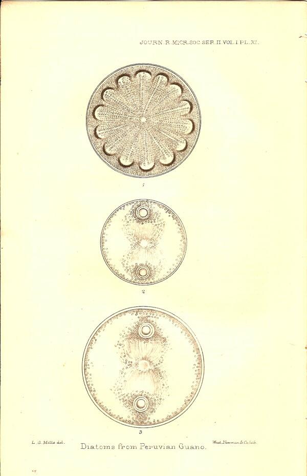
EXPLANATION OF PLATE XI
Fig. 1–Aulacodiscus kittoni var. x320
Fig. 2–Auliscus contellatus n. sp. x320
Fig. 3– " " large specimen x320
A splendid plate! Clearly the Rev. Mills knew how to draw.
It’s fairly unlikely that diatoms would show up in bat guano, so this was almost certainly guano collected from sea birds which was shipped in great quantities to Europe and America as fertilizer–yep, it’s bird poop.
I can’t resist imagining the Rev. Mills wandering around the warehouses in the harbor district, wearing his clerical collar, and negotiating for a few pounds of bird droppings.
‘Ah, no father. There’s no charge fer you. Just put a good word fer me with the Lord.”
Did the good Reverend arrive with a bucket? A wheelbarrow? Did he hire a horse-drawn cab and threaten the driver with excommunication if he didn’t agree to fecal transport?
And what of his parishioners? How did they feel about him spending time preparing avian dung to look at diatoms?
The second lead article is 4 1/3 pages written by B. Will Richardson and the article has the magnificent title: “Multiple Staining of Animal Tissues with Picro-carmine, Iodine, and Malachite-green Dyes and of Vegetable Tissues with Atlas-scarlet, Soluble Blue, Iodine, and Malachite-green Dyes.” A true Victorian title for an article which strongly expresses a Victorian sensibility. Of animal tissues he writes: “Beautiful treble stainings may be obtained with the picro-carmine and iodine-green after the method described by Stirling.” He goes on to mention modifications that can produce quadruple and even quintuple stainings.
Regarding vegetable tissues, Richardson remarks: “Strikingly beautiful, as sections of some animal structures undoubtedly are,...sections of many vegetable tissues are also susceptible of very fine double staining, and occasionally of fine treble staining with atlas-scarlet, soluble blue, iodine and malachite-green dyes.” Aesthetics were a significant consideration for Victorian microscopists, in large part, because the structures of nature they were investigating, they regarded as the creations of an omnipotent and perfect God, so why shouldn’t they be beautiful? To produce slides that demonstrated this beauty was a way of showing laymen that the glories of God were manifest in even the smallest details. Perhaps that’s why in Britain there were so many clerics who were naturalists.
In the summaries of researches, there are, as always, fascinating tidbits. For example, you can discover that the cochlea of the monotreme Ornithorhynchus platypus (in other words, an ear bone of the duck-billed platypus) is “a somewhat curved tube, about 1/4 inch in length and about 1/ 20 inch in diameter” Or you can find out the composition of the ink of cephalopods, in this case Sepia officinalis–a squid.
“Of 100 parts ink, the constituents are:—
Water 60 parts
Mineral substances 8.614 parts
Insoluble organic substances 30.546 parts
Extractive matters 0.851 parts”
“The mineral substances, as determined after calcining, include calcium, magnesium, potassium, iron and the acids, carbonic sulphuric, and hydrochloric acid, the absence of phosphoric acid being noteworthy.” Fairly sophisticated analytic chemistry for 1881. However, I must admit, I’m always a tad suspicious when presented with results to 3 decimal places.
Some early investigators were willing to take substantial risks in their researches. E. Yung is reported to have examined the influence of poisons on hearts of certain lamellibranch mollusks trying electricity, fresh water, increases in temperature, curare, strychnine, digitalis, potassium sulphocyanide, and upas anitar. (A very toxic latex from a type of evergreen found in southeast Asia sometimes used as an arrow poison–and yes, I had to look it up.)
Ordinarily, we don’t think of fresh water as a poison, since it is beneficial to us. However, for a large number of saltwater organisms, it is rapidly fatal, just as oxygen is a poison to anaerobic organisms and as saltwater is to us if we drink it. What this reminds us of is the very important fact that there is no single set of standards for “normal” conditions for life. It is a deeply ingrained tendency for us to anthropomorphize such issues as what are “good” conditions for living things. That’s a major reason why the recent attention which extremophiles have been receiving is very intellectually healthy, because we are reminded of the extraordinary environments to which life forms have succeeded in adapting–pink algae that live on snow banks, bacteria that survive in thermal mudpots at over 212 degrees Fahrenheit, giant red tubeworms that metabolize sulfur compounds in the depths of the sea, blind salamanders in caves, lichens that grow on rocks, and the list goes on and on.
There is an interesting, brief discussion of an article on the development of Doliolum. Doliolum is a fascinating creature. It’s like a miniature glass sculpture. Imagine a tiny, transparent barrel with a thin coil running through it from one end to the other. Now, add to that the fact that this lovely little being moves through the water by jet propulsion! It has an incurrent siphon at one end and an excurrent siphon at the other. The coil is a band of muscles which the organism can use to contract, expelling water from the excurrent siphon, thus propelling it forward. Clever little Doliolum! It is a pelagic (free-swimming) tunicate related to salps and appendicularians and more distantly to ascidians. If you don’t know about these various groups of organisms, don’t worry about it; they won’t help you corner the stock market. However, if in only 2 or 3 people, I’ve sparked enough curiosity to look up some information regarding these strange and wonderful creatures–try Google–then, I’ll be content.
There is also a full page description of Darwin’s book The Formation of Vegetable Mould through the Action of Worms, with Observations on their Habits. Darwin was an extraordinary naturalist with exceptionally broad interests and he wrote extensive accounts on subjects that resulted in books which relatively few biology students today have even heard of.
There are two summaries of investigations on rotifers and a very brief one on Agassiz major study (over 300 plates) on the echinoids from the Challenger expedition. Regarding specimens obtained from the Challenger expedition, there is an important, but highly technical, summary titled : “Soft Parts of Euplectella aspergillum.” This is the elegant glass sponge, the Venus Flower Basket.
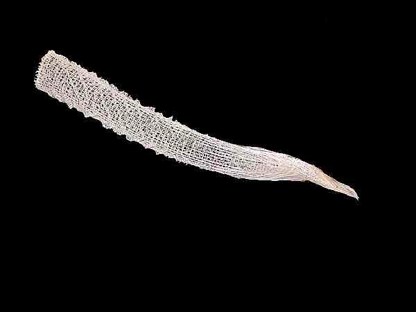
The beautiful skeleton of this organism had been widely studied, but the nature of the tissue was virtually unknown. Professor F.E. Schulze obtained specimens which had been preserved in absolute alcohol and was able to do careful histological examinations. The summary is written in dense, almost impenetrable, Biologese. This is a subject still of considerable interest to biologists and perhaps the original paper by Schulze is somewhat more accessible. If you want to look it up, it can be found in the Transactions of the Royal Society of Edinburgh, xxix. (1880) pp. 661-73.
As usual, toward the back of the issue, there is a section devoted to microscopes and accessories and I want to call attention to a quite ingenious instrument–the Ross Tank Microscope.
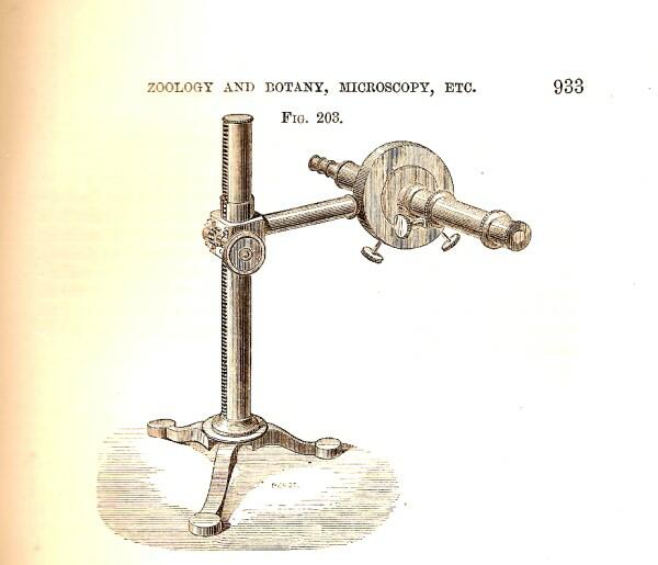
This was primarily designed for examining organisms on the walls of aquaria. However, it could be rotated to a vertical position or inclined at an angle to observe large specimens that wouldn’t fit onto the state of a traditional compound microscope. It’s a pity that some microscope manufacturer doesn’t now produce an aquarium microscope at a modest cost for the amateur.
In Part 5 next month, we’ll look at some mites and desmids.
All comments to the author Richard Howey are welcomed.
Editor's note: Visit Richard Howey's new website at http://rhowey.googlepages.com/home where he plans to share aspects of his wide interests.
Microscopy UK Front
Page
Micscape
Magazine
Article
Library
Please report any Web problems or offer general comments to the Micscape Editor .
Micscape is the on-line monthly magazine of the Microscopy UK website at Microscopy-UK .