Robert Pavlis, Girard, Kansas USA
It is possible today to purchase digital microscope cameras. Most of these seem to be either extremely expensive or of extremely low quality. Some, unfortunately are both!! The reality is that one can, with some ingenuity and care, obtain images from standard digital cameras that are as good or better than the ones obtained with the best dedicated ones.
There are many extraordinary advantages of digital photography over the old silver halide photographic processes. Unfortunately one often cannot simply take old photographic equipment and substitute new digital cameras. The photosensitive area used to obtain the image is typically much smaller in digital cameras--this fact has substantial consequences when systems intended for photographic film cameras are modified to use digital cameras.
It is essential that one be able to focus the image before making the exposure. Virtually all digital cameras are either single lens reflex or else they permit viewing the image before exposure on a LCD screen. With film cameras one was generally forced to use either a single lens reflex camera, or a special camera adaptor with some sort of reflex device built into it.
Whether one use digital or silver halide photography there really are three optical techniques to connect the microscope to the camera. All three of these techniques require some sort of device to hold the camera in position above the microscope.
It generally does not make sense to attempt to make digital images with extreme pixel counts because the microscope optical system's resolution is limited by the wavelength of light. It is in the general order of perhaps 1500 to 2000 pixels across the image where typically higher resolution becomes useless. However, the exact point depends on how the optical system is set up. The most usable pixels are obtained using low power (like 2X) projection oculars.
With a fixed lens camera the image that is formed on the sensor may be circular. Depending on camera resolution and magnification, the resulting images can often be cropped and still produce excellent images.
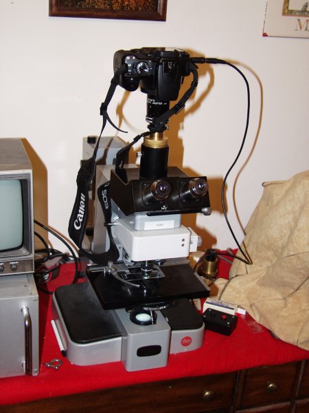
There are plethoras of microscope adaptors available for cameras. Unfortunately there are also plethoras of camera systems. For interchangeable lens cameras there are several adaptors that connect either M42 thread cameras or T-ad apter equipped cameras to the microscope and hold a projection ocular inside. These are probably the least expensive microscope adaptors in existence! Because there are both M42 an T adaptors available for virtually all interchangeable lens cameras, this is a simple solution that usually is not only simple but also the best! With digital cameras it may be difficult to find that low power projection ocular, however. One CAN use normal low power oculars, but more aberrations will be introduced generally degrading the image. Moreover, unless the ocular be very low power, the projected image quality will be degraded by the finite wavelength of light.
The image to the right of this text shows an Orthoplan microscope with one of these adaptors being used to connect a Canon digital single lens reflex camera.
Both M42 and T systems are 42mm in diameter. M42 has a M42x1 thread whilst T has an M42x0.75 thread. A fast way to tell whether a threaded 42mm device is T or M42 is to take a standard M6x1 screw and compare the thread pitch. If they match it is M42!
The same adaptors can be used for direct objective projection--just omit the projection ocular. (Note however that the results are apt to be not so good on most systems except on stereo microscopes.)
Caution! Most of these adaptors are designed to come apart just above the ocular to permit changing the oculars and to avoid accidentally removing the camera from the microscope and pointing the camera upward--which would result in EXTREME damage to the camera from having the ocular fall into the camera mechanism. DO NOT EVER remove an adapter of this sort from the microscope with the ocular inside!!!!!!!
Adapting cameras without removable lenses to microscopes can be a very trying experience. The easiest way to do this, most would agree, is to purchase a special system for doing this. An excellent, though somewhat expensive way to do this is to purchase something like the Scopetronics Maxview Plus system. This includes a special ocular designed for use with cameras and a series of tubes for mounting to many different types of optical instruments including both the standard microscope size and the 30mm stereo microscope size. (The kit also includes fittings for standard telescopes.) The system currently sells for $US299. One, however, also needs a threaded adaptor for one's camera's lens size. These generally sell for $US30.95 for each size. The included ocular is first rate, both on microscopes and telescopes, and results generally are excellent. The ScopeTronix web site can be located by clicking here.
It is far better and far simpler to use a digital camera that has threads for attachment of lens accessories. One very bad characteristic that many digital cameras have is having lenses that retract when the camera is turned off. A camera of this sort cannot be attached to the microscope by its threads because the retraction mechanism would have to move the camera when it was turned on. Sleeve adaptors are available for such cameras, however, that fit over the entire camera lens assembly.
The images below this text show the ScopeTronics system:
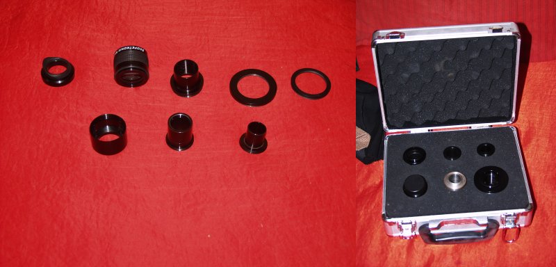
Notice all the tubes that attach to the ocular to fit into various optical systems. There are also two camera adaptors to the right side of the left image.
The other solution to this problem is to try several oculars with one's camera until one finds one that has optical characteristics that are compatible. When this is accomplished one needs to obtain an adepter. This either requires access to machine shop equipment, or locating a machinist to make one. Adaptors of this sort can be turned from machinable grades of aluminium or brass. The adaptor will need to have threads cut with proper pitch, and the spacing between the ocular and the camera will need to be optimised. (When machining parts like this it is generally best to cut the threads first, because it is very easy to err whilst cutting threads. It is best to err at the start of a machining project so not to have to start over anew on a nearly finished part!)
There are two alternative materials that can be used to make these adaptors. 360 Brass is extremely easy to machine, but is dense and somewhat expensive. Aluminium is the other alternative. It is much lighter, less expensive, but many aluminium alloys are difficult to machine because they stick to the lathe tools. A trick to making short thread sections is to turn the lathe chuck by hand with the lathe power turned off, and to make multiple passes with the cutting tool taking great care not to keep the piece in place between passes.
There are some really wonderful things about digital photography and microscopes. For example when making images of metallurgical samples with through the lens illumination there is always some problem with scattered light. Because the scattered light is generally uniform across the image, the digital values of each pixel can be reduced to compensate completely by image editing software (or by writing computer programs--a lost skill in recent times it seems!).
Some digital cameras have a "raw" mode that permits recording unprocessed images. Because light conditions used for microscopy tend to be unusual there are occasional situations where this capability can dramatically improve images. (LED light source images provide much better colour balance and less of these problems.) Camera manufacturers generally provide software to edit and convert these images. There is a free POSIX program written in C called "dcraw" which will convert the raw images from many cameras into a file format called ppm. Image editing software capable of handling 16 bit ppm images can be used to edit these files and ultimately convert them into a more standard format.
A very large portion of digital images obtained using microscope setups need to be edited. They may require cropping, and they may require gamma, contrast, and intensity modifications for each of the primary colours. Images taken using monochrome radiation generally are also best converted to monochrome. There are a variety of programs available to accomplish these tasks. The many free image editing tools available for POSIX (Linux, BSD, etc.) are often at least a match for the best and most expensive commercial programs. The gimp program is extraordinarily good, and it now has good documentation. It is an unusually user friendly program. Unfortunately it does NOT handle 16 bit colour images. The ImageMagick program package has certain capabilities lacking by the gimp (including 16 bit colour) but is also a bit user hostile. The netpbm package has still other capabilities but can be savagely user hostile.
Also for POSIX operating systems, there is also an interesting X window program called digiKam. This program manages collections of images, and has quite good built in image editing capabilities. The wonderful thing about this program is it not only allows viewing of the images in large collections of them, but it can also edit the images. POSIX operating system users might also consider using ksquirrel--a very clean image viewing program.
One should note that commercial image editing software is expensive, costing at this point substantially more than a very good computer. The free programs generally are designed to run on POSIX operating systems. Those who do not want to use POSIX operating systems for their general computing needs are probably best advised to get a second computer and install a decent POSIX operating system on it for image handling rather than purchase dramatically overpriced software. They can also partition their existing computer's hard drive and place a POSIX compatible operating system on part of it and use "dual boot."
Perhaps the most user friendly POSIX operating system, both from an installation point of view, and from a users point of view is Ubuntu. It is free and available in several flavours. It includes a powerful system for downloading and installation of an enormous library of available free software including the packages discussed above.
All comments to the authors via Robert Pavlis are welcomed.
Microscopy UK Front Page
Micscape Magazine
Article Library
Please report any Web problems or offer general comments to the Micscape Editor.
Micscape is the on-line monthly magazine of the Microscopy UK website at Microscopy-UK