Review of a Watson & Sons brass student
microscope of the late 19th century
Introduction
I bought this stand about
five years ago, mostly an impulsive eBay
buy. I don't exactly remember the price, but it should have been
something around 200 euros a rather expensive price based on the
poor state the microscope was left in. The lacquer was literally
peeling off, the fine focus didn't work and one lens was almost totally
opaque due to a thick dust cover! Fortunately, except for the lacquer,
the microscope could be brought to a better state.
Some historical background
I'm not at all an expert in
microscopes history. Here are some relevant
information I found on the arsmachina.com website :
"In 1854 the London Society
of Arts awarded a prize to G. Fields of
Birmingham for an inexpensive microscope for medical students. The
design requirements outlined a microscope that had two lenses, and
could be disassembled and stored in a compact box. The instrument had
to sell for a specified low price and the prize winner had to agree to
keep the microscope always available for purchase. The popularity of
the design was such that most manufacturers produced their own models
with slight variations.
In 1858 the Royal Microscopical
Society standardized the thread size for
screwing the objective into the microscope. The thread pitch is
measured at 36 to an inch with a diameter of .8 inches.
Modifications have been made to the standard over time, but it is
essentially the same today."
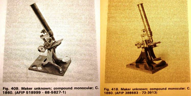
The Watson microscope is a
very typical example of a student microscope
from the second half of the 19th century, several other makers had quite
similar stands. For instance, here are two examples of such stands I
found in the Billings microscope collection:
Since the Watson microscope
reviewed here has both the London Society
of Arts pattern and RMS thread, it is typically dated after 1860. See
further below for some discussion about its possible age.
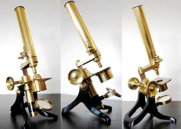
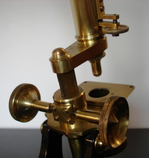
The stand
This stand is simple, yet
sufficiently stable and robust. It's quite
straightforward to understand the role and the operation of each part
(except maybe the fine focus). It's a clever design for students
starting the use of a microscope, and it is not a toy !
The total height is 31cm
(12.4 inches), the base width is 12 cm (5
inches), base has the typical British horseshoe design, which also
appealed to me while buying the scope.
I have to point out that
the lacquer in the pictures you see here
is NOT original. Because the scope was in
very bad condition when I bought it, I had to carefully
remove the remaining lacquer and add a transparent lacquer to protect the
brass. I hate to do this, but on this microscope it was really
necessary to restore the look and protect from corrosion. Believe me:
I could clearly see a finger print on the microscope tube. I'm pretty
sure it was where the last owner put his thumb before storing the
microscope for a few decades. The acidity of the grease that
naturally covers our fingers was enough to locally attack the original
lacquer, I'm now deeply aware of this problem, and always clean up my
hands before touching any of my microscopes !
The coarse focus is by
triangular rack and pinion, as shown on the picture below. The four
screws can be tightened so as to adjust the focusing resistance. This
is a very nice feature, as I like the focusing to move firmly but
smoothly.
The fine focus acts only on
the microscope objective, it is incorporated directly into the tube.
Mechanically, it is a simple design with balance and spring. Though thefine focus does not provide the feeling of more modern scopes, I found it sufficient for its use. Actually, as the objectives have low
NAs and large depth of field, I found the coarse focus to be largely
enough for normal use. I almost never touch the fine focus. Another
feature I particularly like is that you can always see where the fine
focus is set, and ensure you never go too far while moving it.
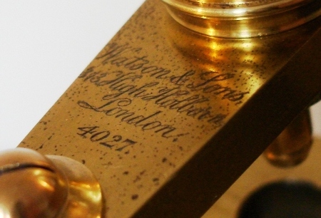
The microscope is signed on
the limb with "Watson &
Sons, 313 High Holborn, London" and the serial number 4027. On the
picture below, you can also see the damage on the brass and has all sorts
of black spots (I didn't want to over-polish this zone to keep the
markings as close to original as possible).
The optics
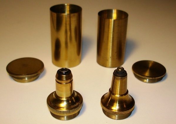
Two microscope objectives
(with brass canisters) came with it. They have
no labels, but I assume these were the original ones fitted to the
microscope. I estimate the following characteristics :
- for the one on the left:
10x, 0.15 NA
- for the one on the right:
25x, 0.25 NA
The 10x shows some minor
signs of delamination on the front lens, but due
to low magnification and NA, this doesn't affect much the final image.
The 25x came completely
blurred with some brown grease on the back of the lens. This grease was
resistant to normal cleaning with isopropanol or acetone, the objective
top element had to go into an ultrasound sonicating bath to clean all
traces of grease (I don't recommend this cleaning method at all!). At
that point, I was very surprised to notice the 25x objective was
actually a SINGLE lens element, with a shape close to a half-ball. I
felt even more surprised when I noticed it provide fairly decent
imaging capabilities, see sample images below.
The eyepiece is fixed
(screwed into the microscope tube). It is still a
compound eyepiece, with both field and eye lens. The magnification is
about 8x. A niggle here: because the eyepiece field lens is located
deep into the tube, it is very difficult to clean up, yet it tends to
be quite prone to dust while the scope is stored into its box
with separate tube and stand. As the field lens is close to the image
plane, any dust on it will appear sharp on the final image, so regular
cleaning has to be undertaken.
There is no illumination condenser, but the below the stage it s
designed to hold a 39mm
condenser. Actually, I find no use of such a condenser with the two low
NAs objectives provided, the concave mirror is largely sufficient.
The mirror is concave, single
face. Its support can be tilted along two axis, and the black mirror
support can also be turned and raised to adjust critical illumination.
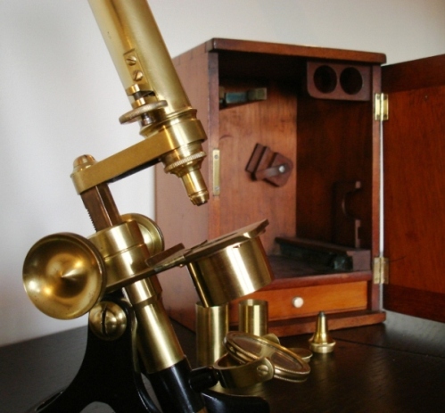
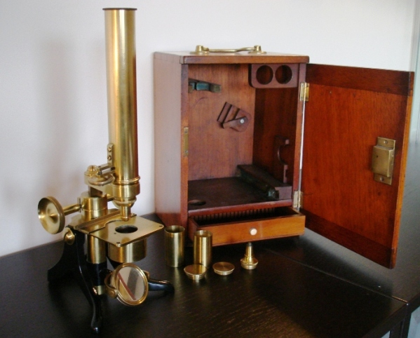
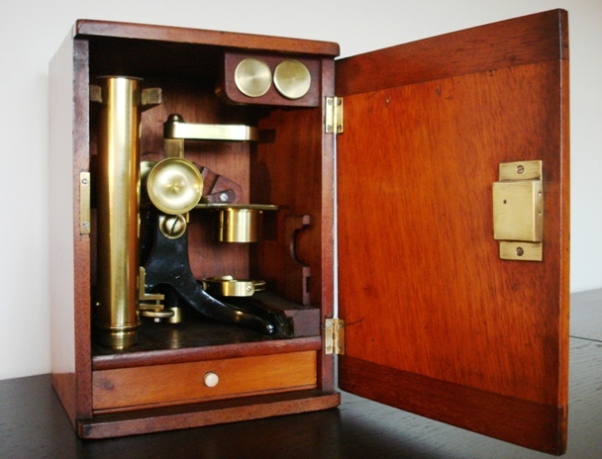
The microscope came with a
matched wood box with brass holder and key. The
box has a drawer to hold about 20 microscope slides of standard size
3x1 inches. All microscope parts store simply into this box, I really
like this design which rapidly turns the microscope into a field
microscope.
Stop discussing antiques - What
images can you expect from it ?
The two images below were
taken with the 25x objective and 8x eyepiece, using my DSC-W100 Sony
digital camera with focus set to infinity. I manually hold the
camera above the eyepiece, which worked quite well with brightfield
illumination (here I just used my desk lamp and the concave mirror).
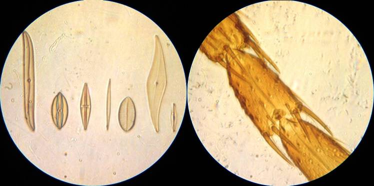
The left image is Klaus Kemp's well known eight form test plate, no
fine details could be resolved with the 0.25NA. The image on the right
is a common housefly leg, from a no-name microscope kit I bought in a
Lidl store.
The two images below are more
curious. Although they look awful, I really appreciate them a lot for their
physical content. Illumination here is provided by a Brunel Köhler
lamp with the collimated light coming from the top of the sample at
grazing
incidence. The reflected light is not accepted by the objective, only the
light scattered (or diffracted) by the object is used to form the final
image. So you may call it a variation of epi-dark field illumination
at high incidence.
The image on the left is
Morpho butterfly scales taken with the 10x objective (male Morpho
butterflies are well known for their bright blue color). The image on
the right are again Klaus Kemp's diatoms with the 25x objective (from
right to left : Frustulia rhomboides, Pleurosigma angulatum, Surirella
gemma, Nitzschia sigma). Let me emphasize that no color modification
was madeto the images, I only slightly adjusted the contrast. These are
natural colors !
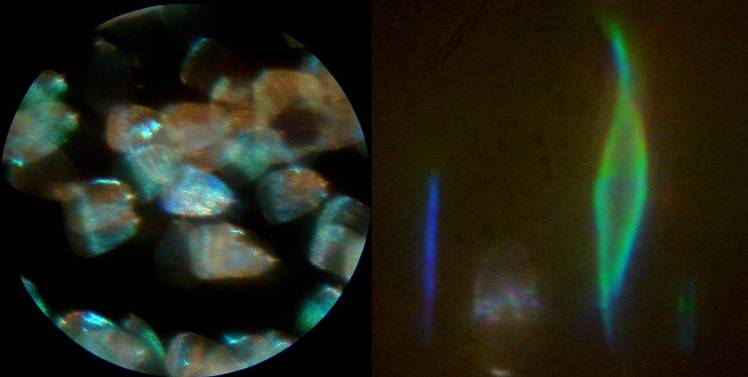
The nice feature is that the
colors stem from diffraction of light (as
from the top of a CD-ROM). The tiny 'dots' on the diatoms have a
common interaction with light to diffract a certain wavelength (colour)
into the objective NA. I like this because it provides a direct
observation of grating diffraction by a natural sample.
Let's discuss
again... Some intriguing features about this particular stand
My first estimate about the
age of this stand was around 1870, based on the simple design of the
objectives and the fixed eyepiece. However, David Walker* tells me that
there may be some intriguing features: (*Editor's note: from an admittedly limited knowledge and access to published information on old Watson stands.)
* The serial number: Bracegirdle's book 'Notes on modern microscope
manufacturers' gives a serial list 1894 - 3527 and 1909 - 11791, which
would presumably date the microscope reviewed here to about the end of
the 1890s.
* The name / address format name. The same book indicates 1867 - 1882 'W Watson and
Son' and 1882 - 1908 'W Watson and Sons'. The microscope has 'Watson and
Sons' written, it is thus missing the leading 'W'. This may seem minor
and a confirmation of the late 19th century period for the microscope,
yet the brand name has italic script, suggesting an early one before Watson adopted a block capital
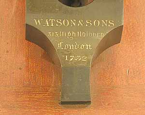
type script. Looking on the Internet, I found an early example with serial no. 1752 http://www.asiuk.net/w17322.jpg,
which also missed out this first 'W' but did use block capitals suggesting Watson and Son(s) have not always been rigorous in
labelling, and confirming the estimate of the date of the present stand from the serial number.
* The objective canisters. Another strange thing is that the canisters
to hold the objectives are not marked with Watson brand, which I
thought was the case for all of its objectives.
* The condenser holder. All students scopes of that design I saw didn't
have the condenser holder below the stage, but a ring with apertures
instead. I also wonder that with a concave mirror, setting a condenser
below the stage requires the mirror to be lifted up to enable critical
focusing, which I believe isn't practical if one wishes to take the
condenser or not.
Any comments about these open questions are welcome.
Conclusions
Altogether, this microscope
is very well designed for its purpose: teaching the basics of
microscopy to medical students. Its imaging capabilities are not as
good as microscopes from the end of the 19th century, but one should
remember it was intended to be sold to a large number of students for a
reasonable cost. About 150 years later, it is still in good working
order and pleasant to use. Moreover, it has now become an historical
piece with appealing esthetics.
Collecting different accessories for my Wild M20 has turned into an
expensive business. Therefore, the Watson
left a few weeks ago on a eBay auction, and sold for about 300 euros.
As this is a happy end story, the microscope is now part of the
collection of a Belgium academic, and will serve to teach the basis of
microscopy to undergrad students. After a long life moving from places
to places, it has now returned back to its original teaching purpose.
Thanks for reading. Comments to the author, Jerome Wenger are welcomed !
The author acknowledges stimulating discussions with the Micscape Editor, David Walker.
Microscopy UK FrontPage
Micscape
Magazine
Article
Library
Published in the March 2010 edition of Micscape Magazine.
Please report any Web problems or offer general comments to the Micscape Editor .
Micscape is the on-line monthly magazine of the Microscopy UK website at Microscopy-UK .
© Onview.net Ltd, Microscopy-UK, and all contributors 1995 onwards. All rights reserved. Main site is at www.microscopy-uk.org.uk .