|
Mysteries Surrounding Dried Stuff: Part 3 A Battered Chirodota Sea Cucumber From the Philippines by Richard L. Howey, Wyoming, USA |
Yes, I’m still fussing about the mangled, old sea cucumber from the Philippines and it’s all the fault of Mr. F.M. Hamlin and his article “The Wheel-Like And Other Spicula Of The Chirodota of Bermuda” published in the Proceedings of the American Society of Microscopists in 1882. [I want to note here that there are some who use the genus name Chirodota and other Chiridota. The latter is more frequently employed.] Mr. Hamlin was an interesting figure. His health was not good and he was compulsive and over-demanding of himself. He succeeded in getting a medical degree from Albany Medical College. He specialized in nervous and mental disorders and took a position on the staff of the Government Hospital for the Insane of Washington, D.C. (now known as Congress). However, in 1878, his health deteriorated and he had a prolonged convalescence and during the next 3 years spent the winters in Bermuda. He was a naturalist, a microscopist, and a keen observer. In his last years, he was director of a school for mental patients in Willard, N.Y. and wrote a paper titled “Schools for the Insane”.
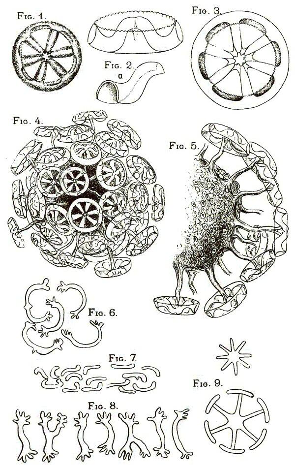
In one respect, the most intriguing part of Hamlin’s article is the very last page which is a plate with 9 figures and the drawings are sufficiently provocative to send the reader back to the text of the article for a careful re-examination. I will include the plate here.
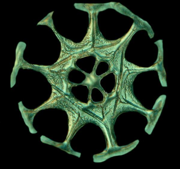
Figure 1 is straightforward enough and shows a wheel spicule typical of several species of Chiridota. Hamlin doesn’t provide a species name for his specimens, but this type of spicule is one which I have found in specimens from Maine (Chiridota laevis) and California where a species name was not available.
Figures 2, 2a, and 3 are of great interest because Hamlin is arguing that the term “wheel” is a misdescription and he is perplexed that other observers have not noticed certain characteristics. Specifically he cites the reverend Dr. William Benjamin Carpenter whose very large work The Microscope and Its Revelations has become a highly sought-after volume. Here is Hamlin’s remark:
Carpenter adds, “‘These ‘wheels’ are objects of singular beauty and delicacy, being especially remarkable for the very minute notching which is traceable round the inner margins of the ‘tires’.” (Hamlin p. 139)
Hamlin goes on to specify his perplexity:
“That they should receive the name ‘wheels’ when viewed as commonly mounted in balsam is not surprising, for then their resemblance to a wheel is most striking; but when one comes to see them [as] opaque objects and understand what their structure really is, the comparison to a wheel seems unsatisfactory and far-fetched. The only kind of a wheel to which they can properly be compared is that technically known as a ‘crown wheel’–that is a kind of wheel with a broad tire or rim, with cogs upon one side of the tire. To me it seems more like a flattened crown. In attempting to make out the structure accurately, many difficulties present themselves from the very transparent and glass-like material of which they are composed. (p. 140).
[As clarification, I just want to add that although the spicules are indeed transparent and glass-like in appearance, they are calcareous and not siliceous.]
Hamlin then proceeds to give a more detailed account and analysis of his conception of the structure of these spicules. Here it is worth noting that Hamlin in 1882 speaks of “tires” and cites Carpenter’s reference to “tires” which had to have been even earlier. Now, at this period, neither of them could have had a very extensive acquaintance with tires. Nonetheless, these figures along with Hamlin’s comments are of considerable interest because they direct our attention to the complexity of these structures and to the fact that, microscopically speaking, these wheels, tires, or flattened crowns (pick your own favorite description) are quite thick which leads us to Figure 9 and an intriguing puzzle.
Hamlin contends (and he could very well be right) that these 2 spicules, one having 8 spokes and the other having 6 are developing forms which he discovered on very young Chirodota “about the size of a very small pin-head”. He adds that the fully developed animals were over an inch long and had only the 6-spoked wheels. However, there may be another aspect to this morphological oddity.
A month or so ago, I sent some images of spicules from my mangled Philippine specimen to Dr. Phillip Lambert, Curator Emeritus of the Royal British Columbia Museum of Natural History and who, among other books, has produced a splendid volume on the sea cucumbers of British Columbia and Puget Sound. In a reply, he commented that some of the spicules looked as though they might have suffered some erosion damage from a preservative fluid. Even though this specimen was dry when I received it, it is fairly common practice for collectors and dealers to preserve these specimens in formaldehyde and then dry them for shipping. Formaldehyde, unless it is buffered, can readily turn acidic and etch or dissolve parts of calcareous structures. Over a long enough period of time, it can also simply dissolve these structures completely and lead one to conclude that a particular species has no spicules when, in fact, it did at one time.
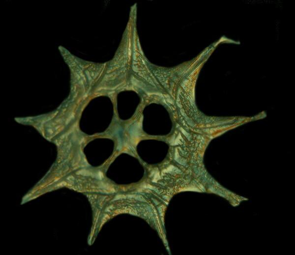
The specimen I have been dealing with is adult and yet I have found spicules that are very similar to the large drawing in Figure 9.

There is also an adult spicule which I found with no “T-bars” and which resembles the smaller spicule in Figure 9 with 2 significant differences: 1) my spicule has 9 spokes, whereas Hamlin’s has 8 and 2) mine has 6 fenestrations on the central disk and there are none on Hamlin’s.
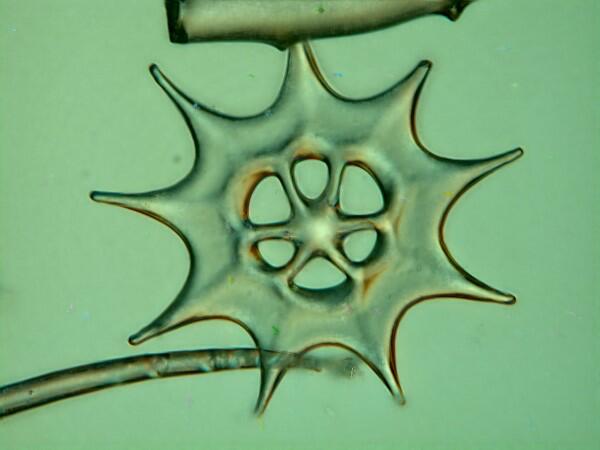
In this particular slide sample, I wasn’t finding any “T-bar” forms nor the spoke forms like those in the image immediately above. I observed the sample over a long enough period that the water under the cover glass began to evaporate. I mentioned before, and Hamlin also remarked on the fact, that these spicules are microscopically rather thick and, I am beginning to suspect, layered. I didn’t add any additional water, because I noticed an intriguing phenomenon. As the water evaporated and the cover glass put more pressure on the spicules, they began to break apart in a manner that suggested that they were doing so along lines of fracture or stress points which indicated something about their construction.
Now, let’s go back to Figures 2 and 3 and remind ourselves that this is indeed a complex, layered structure and as the cover glass exerts pressure on the projecting structure rising from the center, the resulting breakage reveals spokes with T-bars. So this could mean that as these spicules develop, calcareous material is deposited in stages which creates stresses or fracture points. This might also explain why, in an adult specimen, I found incomplete wheels, but with the symmetry of spokes or T-bars preserved.
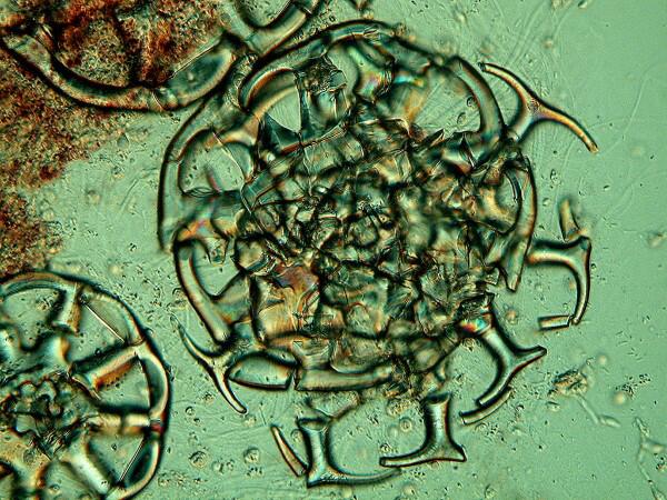
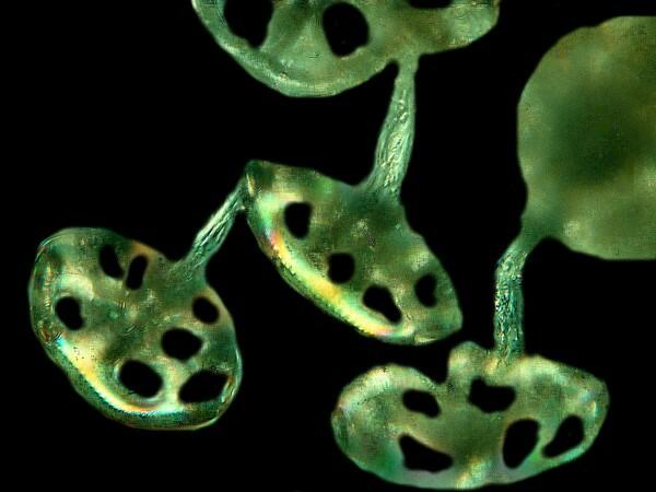
Next, I want to turn to Figures 4 and 5. Hamlin observes and reports that the spicules on the Bermuda Chirodota occur in clusters just under the surface and are covered by a delicate membrane. With the specimens I have from Maine and California, my experience has been similar. In the Philippine specimen, however, the spicules are widely scattered and don’t appear to occur in clumps, although again that could be a consequence of the damage to the specimen. I am going to try to obtain another specimen or two of this species from the dealer to see if I can sort out this issue.
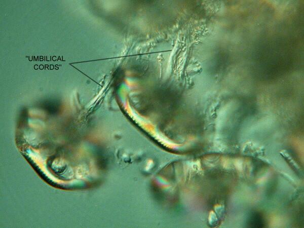
I took a spicule cluster from a California specimen and using micro-dissecting needles, removed the clump from its encasing membrane. It had never occurred to me that there might be what Hamlin calls “umbilical cords” connecting the spicules to the central mass and I don’t recall any other investigators giving such an account. At this point, I had a minor cerebral event–an idea-- although some of my cynical friends may say that I suffered a Transient Ischemic Attack. The standard protocol for preparing these spicules for observation is to use a caustic to dissolve the surrounding tissue–Sodium hypochlorite (or household bleach), Potassium hydroxide, or a powerful oxidizing agent such as Hydrogen peroxide. The result: any trace of the “umbilical cords” would disappear. So, I took a clump and broke it apart mechanically with the needles and took some images. They’re not as good as I would like but, they do show the connecting tissue. I’ll show you 2 images. The first is essentially unprocessed and in the second, I used the graphics software to eliminate the distracting material and focus on showing the connections. Not an ideal image by any means, but it does make the point.
It was a delight to see this and realize just how good an observer Hamlin was over a century ago without all of the technical refinements that we have in microscopes today. Here are these little creatures creating these marvels of engineering and to what purpose? Wrong question!!! No teleology allowed! Nature is full of countless more or less random experiments; some of them work and lots of them don’t. No more dinosaurs, Wooly Mammoths, trilobites, or graptolites and the list goes on and on. Sometimes structures long outlast their usefulness, but the genetic machinery that produces them continues to grind on.
Today, while searching the Internet, I found 2 SEM images of Chiridota spicules which you can see here. Click on images #7 and #8.
http://www.eol.org/pages/72997
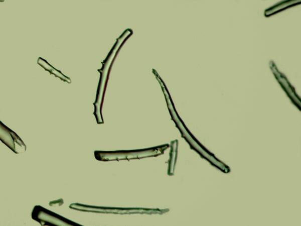
As for the spicules in Figures 6,7, and 8, the ones I have found so far are rather different but, I have not yet completed my investigations. The ones I have uncovered are curved rods with minute, thorny projections at one end.
As I continue to examine this fascinating specimen, I may yet come upon some other structures that will once more prompt me to take pen in hand and scribble away. As an endnote, I will add–Yes, I am truly that old-fashioned. I use the no-longer-produced Sheaffer Targa fountain pen to pleasurably write all my essays prior to undergoing the agony of typing them into a computer.
All comments to the author Richard Howey are welcomed.
Editor's note: Visit Richard Howey's new website at http://rhowey.googlepages.com/home where he plans to share aspects of his wide interests.
Microscopy UK Front
Page
Micscape
Magazine
Article
Library
Please report any Web problems or offer general comments to the Micscape Editor .
Micscape is the on-line monthly magazine of the Microscopy UK website at Microscopy-UK .