A close-up view of Forest Frost Hens and Chicks


A Close-up View of

Forest Frost Hens and Chicks


Sempervivum arachnoideum 'Forest Frost'


by Brian Johnston   (Canada)


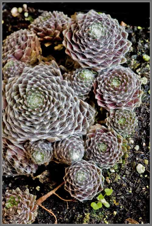
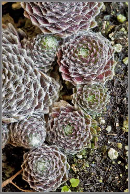
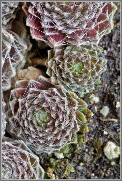
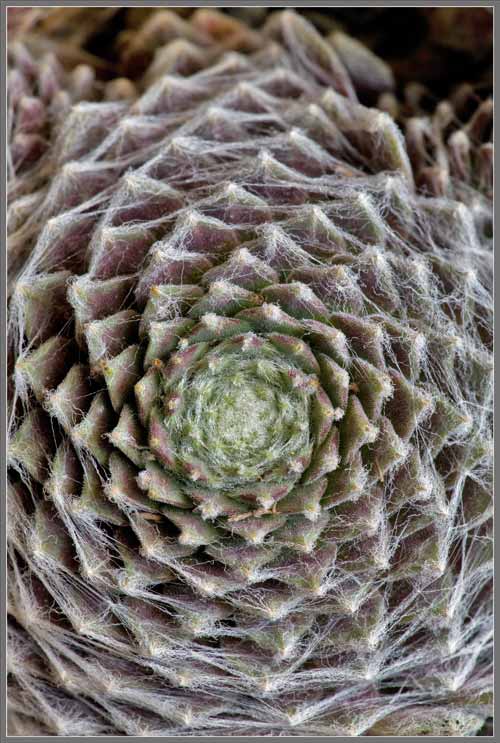
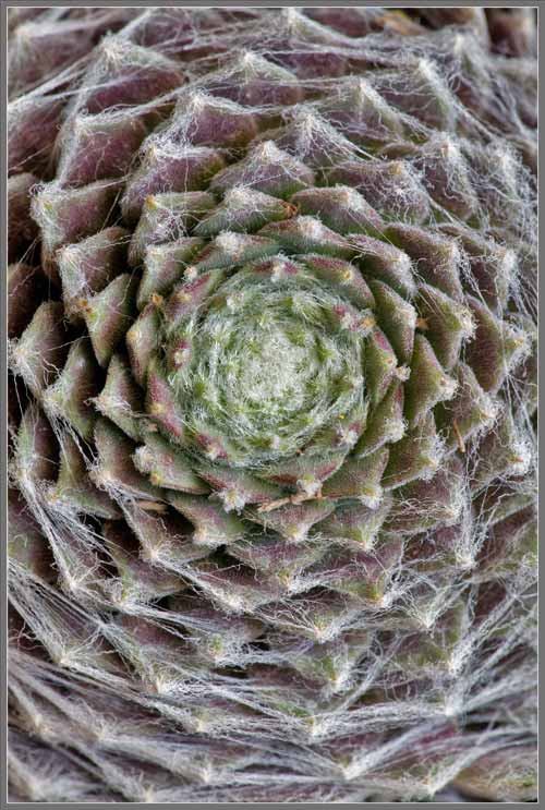
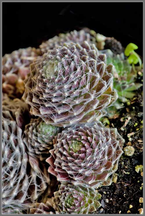
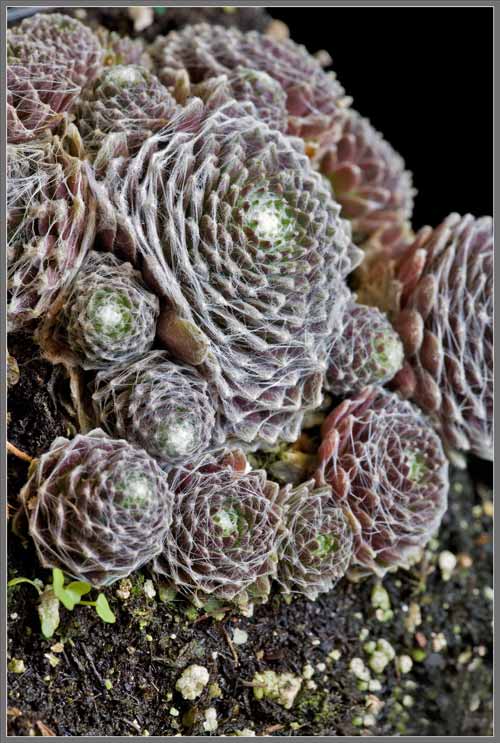
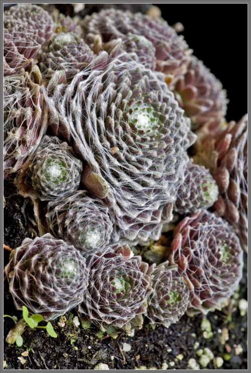
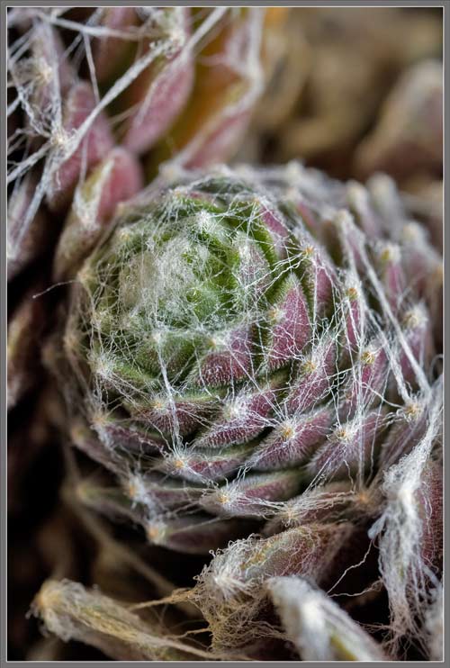
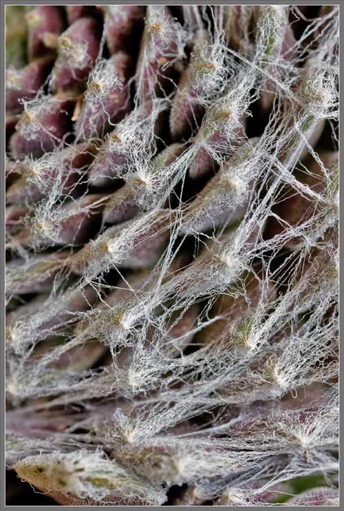
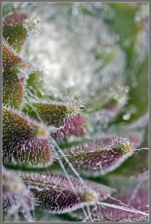
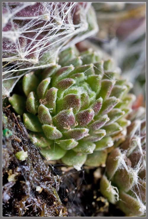
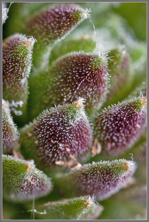
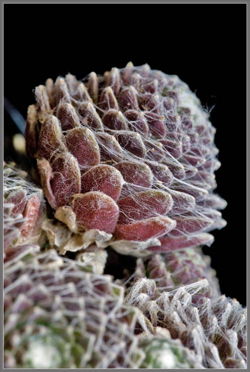
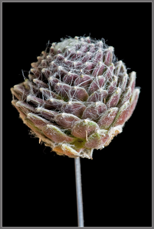
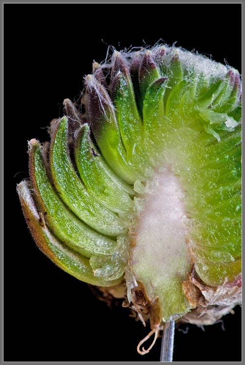
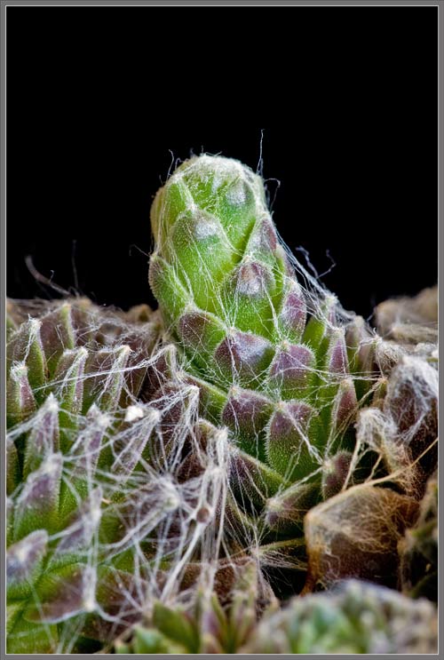
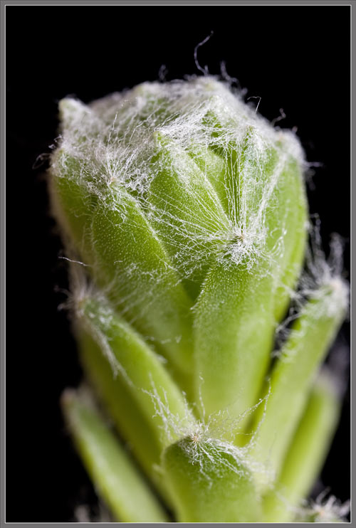
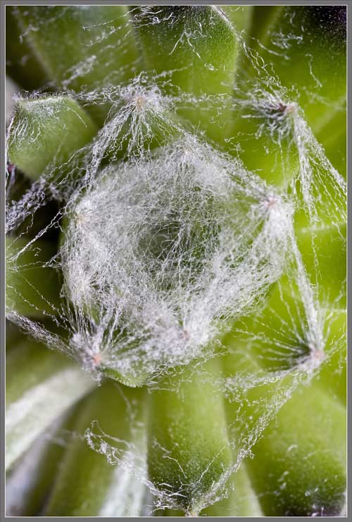
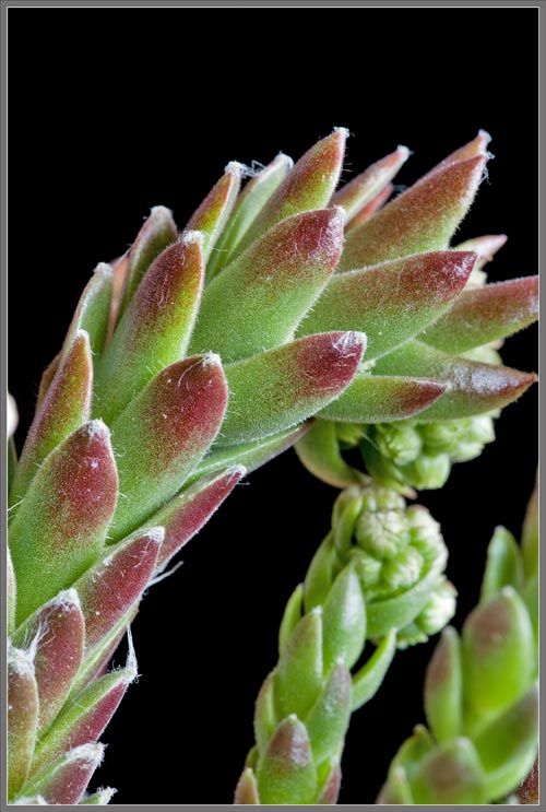
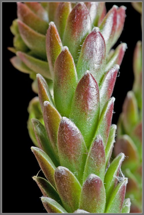
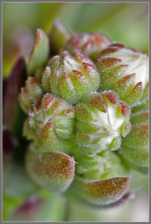
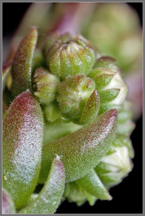
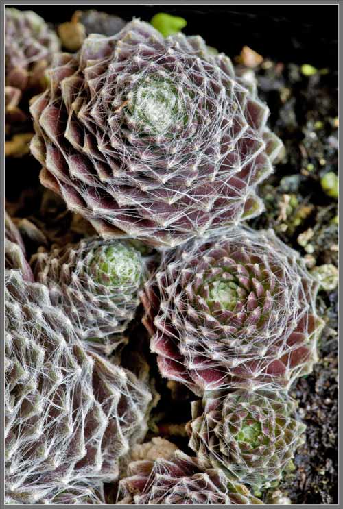
“Of all the Houseleeks neatest far

The jolly Cobweb Houseleeks are.”

Walter Ingwersen 1943
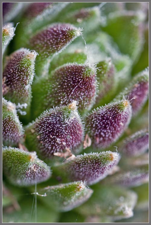
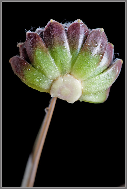
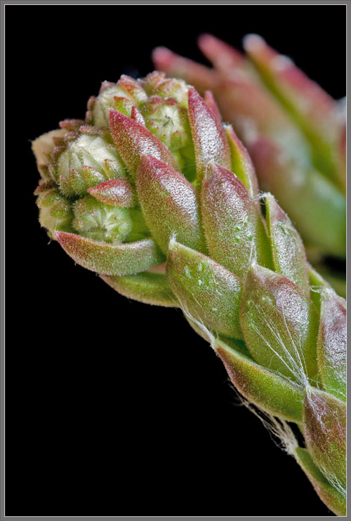
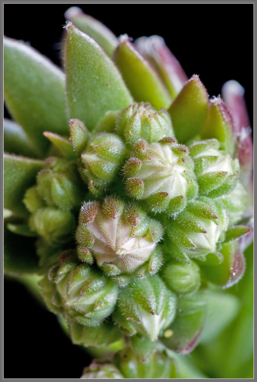
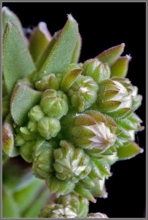
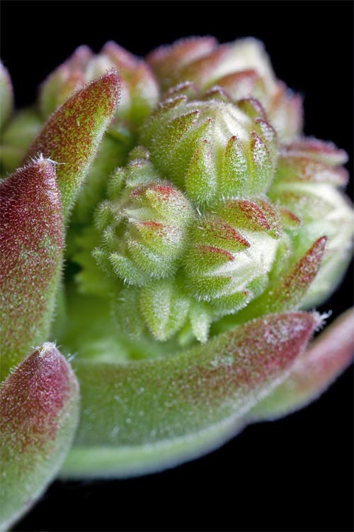
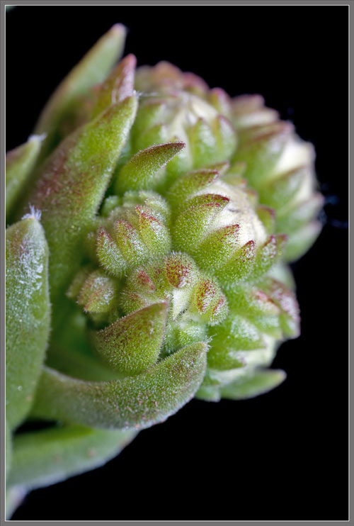
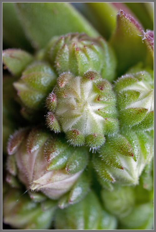
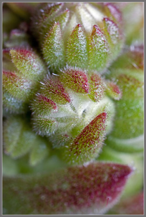
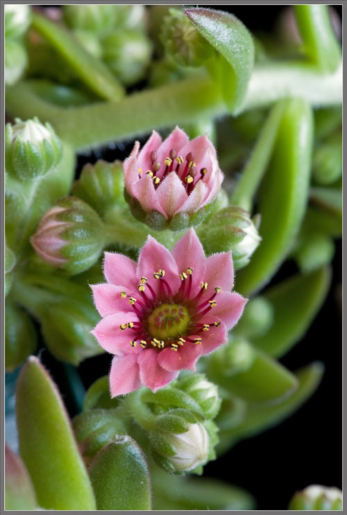
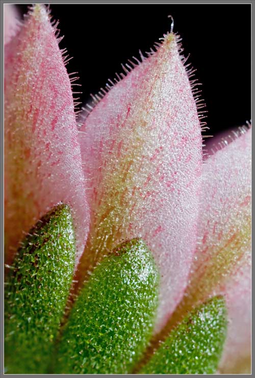
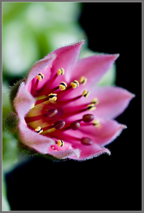
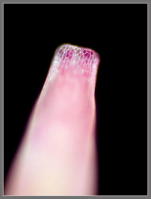

Sempervivums are extremely hardy, mat-forming succulents of the family Crassulaceae, commonly known as ‘Hen and Chicks’ or ‘Houseleeks’.  Native to the mountains of Europe, the Middle East, and Central Asia, they grow close to the ground, with fleshy leaves grouped around one another in a very tightly packed rosette.  The ‘hen’ is the main plant, and the ‘chicks’ are the offspring, which start as tiny buds on the main plant, and soon sprout their own roots, taking up residence close to the ‘hen’.

Hens and chicks have a very long history of cultivation.  It is said that the Frankish King Charlemagne (742-814 AD) suggested to his subjects that they grow the plant on their thatched roofs in order to guard against fires caused by thunderbolts, storms and sorcery.  Since succulents do retain water in their leaves, they may help in the first two cases, but as to sorcery – perhaps not!

The genus name Sempervivum is Latin for ‘always alive’ (evergreen).  Arachnoideum, the species name, relates to ‘spiders’.  In this species the tips of the leaflets are connected by thin hairs that closely resemble cobwebs.  Sempervivum arachnoideum has many cultivars, of which ‘Forest Frost’ is one.  Some arachnoideum hybrids have very dense cobwebbing, while others have little.  ‘Forest Frost’ seems to be positioned somewhere in the middle of the pack in terms of this characteristic.

When I first found this unusual cultivar at my local greenhouse, I had no expectation that it would bloom during the spring or summer period.  I was correct!  However, later in the summer, the same greenhouse had a couple of blooming examples, which I obtained in order to show its entire life-cycle.  This article has been produced by using images taken of the three ‘Forest Frost’ plants.

It should be kept in mind that Sempervivums are monocarpic, meaning that a particular hen flowers only once, and then it dies.  Fortunately by the time that this happens (usually several years), it has already produced many chicks to take its place.

The first image in the article, and the series shown below, show several ‘hens’ (4 - 6 cm diameter), and their smaller chicks (1 – 2 cm diameter). 


Two higher magnification images show the dense tufts of cobwebbing at the leaflets’ tips, and the strands that connect each tip to its nearest neighbours.


Although Sempervivums can reproduce by seed when the plant finally flowers, a much more common reproductive technique is vegetative reproduction.  This occurs when the hen ‘offsets’.  Here, the base of the hen produces what look like thin, root-like stems called stolons with tiny chicks at their ends.  Each offset develops roots of its own and becomes independent of the parent when the stolon withers and dies.  Some Sempervivums produce offsets on the ends of long stolons, which produces a less tightly packed family group.  Vegetative reproduction produces offspring with the same characteristics as the parent plant.  The four images below show the tight packing of a family group.  Notice the light coloured stolon visible in the middle left of the first image.


Another view can be seen in the images that follow.


The leaflets at the centre of a chick rosette are green in colour, while those further out have a deep purplish-brown coloration.


Cobweb density is very variable in different hens and chicks.


When viewed at a very close range, it is evident that the threads comprising the cobweb originate from the tip of a leaflet.  While the chick is still very tiny, the threads become tangled and caught by nearby leaflets, and as the offshoot grows, a cobweb is produced.


For some unknown reason, the occasional chick has many fewer threads.  Notice that the lack of threads allows us to see the outer surfaces of the leaflets much more clearly.  Hair-like projections cover all surfaces.


Many of the chicks are not solidly rooted to the soil in the container, and they jiggle when touched.  This one was precariously balanced between a couple of siblings.


When removed from the container, it is evident that there is no cobwebbing underneath.


A longitudinal cross-section of a chick reveals that the leaflets grow from a colourless, cone-shape core.


The four images that follow show what appears to be the formation of a new chick, but the shape isn’t right.  Instead of a ball, a stalk appears to be growing away from the base of the rosette.


A couple of weeks of patient observation resulted in the view below.  It appears as though several of the container’s hens have almost reached the end of their life-cycle, and are preparing to bloom.  The light coloured spherical structures on the tops of stalks are immature buds.


Before the buds appear, the top of the lengthening stalk has the appearance shown below.  At a later stage, the stalk’s leaflets will take on the normal purplish colour of the species.


Many immature buds push their way through the packed leaflets in the top section of the stalk.


In some cases the bud location is limited to the very tip of the stalk.  By this point the leaflets’ green colour has transitioned to purplish-green.


Notice the great variation in bud size within a particular group.  There is just a hint of the flowers’ final pink colour in a few of the larger buds.


A sequence of images taken with increasing magnification follows showing the buds at the top of the stalk shown at left.


Each of the buds is ringed by many green sepals which have pink, sharply pointed tips.  These sepals surround pink flower petals.  The whorl of sepals is referred to as the calyx.  In the last few images, taken at the highest macrophotographic magnification possible with my equipment, it is evident that the hairs on the sepal have bulbous red tips, which indicates that they are glandular in nature.


Eventually some of the buds begin to bloom.  A closer view of the flowers reveals that they are (actinomorphous), shaped like a star, with about 12  reddish-pink petals.  The most noticeable characteristic of a flower is the ring of stamens arising from the disk at the base of the petals.  Anthers are yellow in colour, and their supporting filaments are bright red.


Sempervivum arachnoideum ‘Forest Frost’ buds are strikingly beautiful both in form, and colouration!


At the 
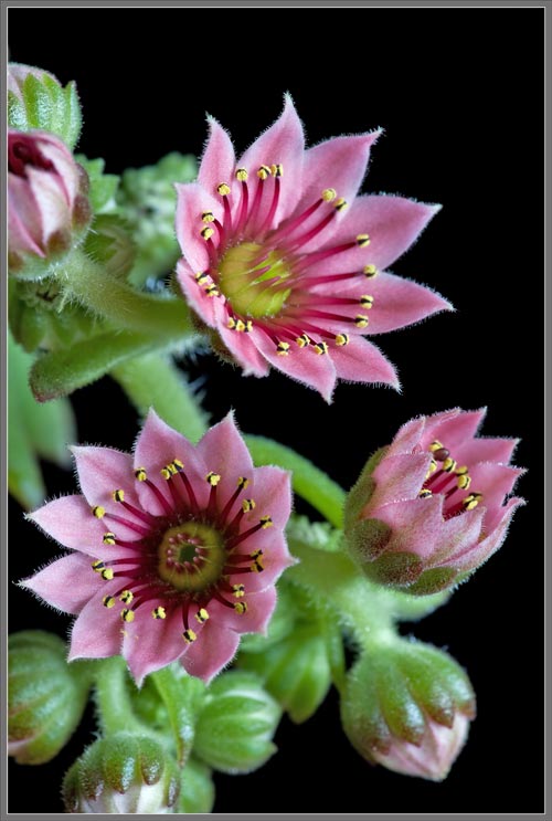
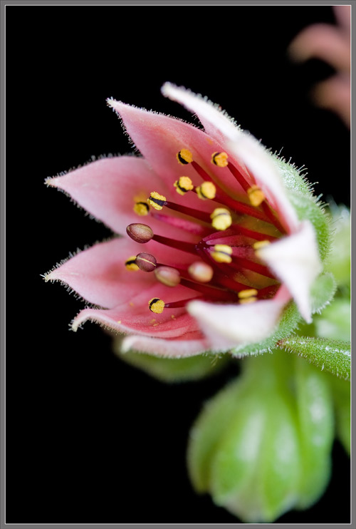
magnification shown below, it is apparent that both the sepals, and the flower’s petals are covered with fine hair-like projections.  
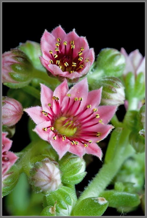
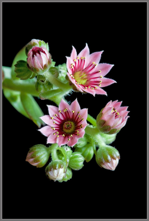
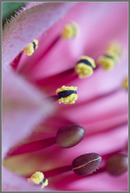
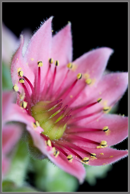
The whorl of pink petals is referred to as the flower’s corolla.


The petals of the flower grow from the edge of a yellow dome-shaped disk, and the male stamens grow from a ring where the petals meet the disk.  Emerging from the disk itself is a group of bright red female pistils.


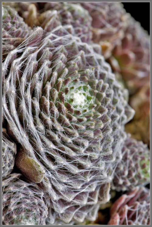
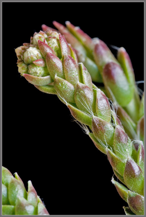
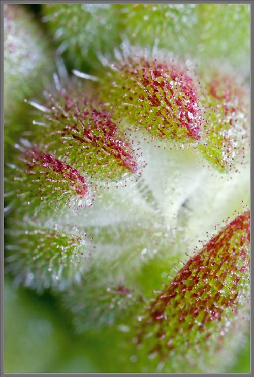
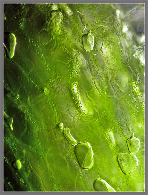
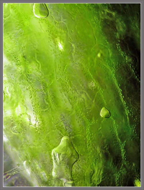
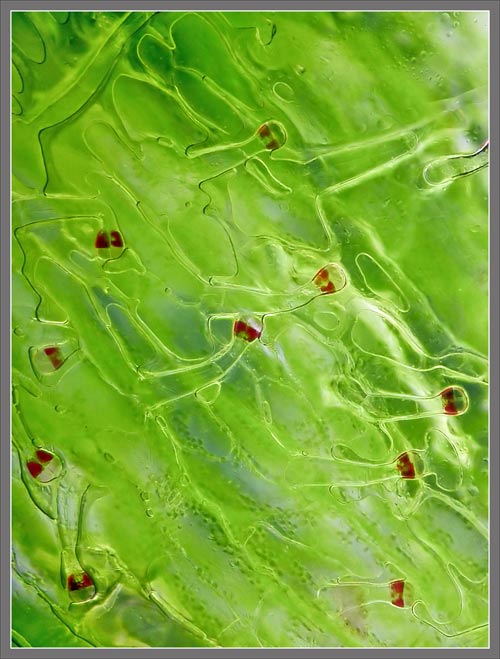
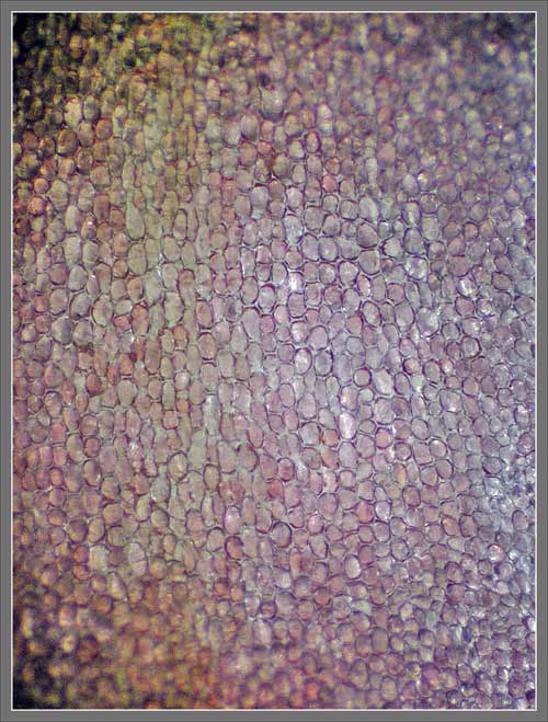
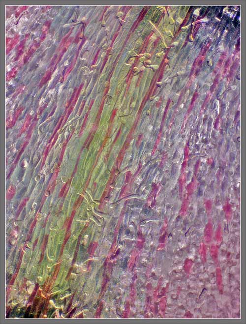
If the surface of a green sepal is examined under the microscope, the subtle green colouration is reminiscent of a pastel painting.  Note that a water mount was used, and this accounts for the occasional bubble in the field of view.


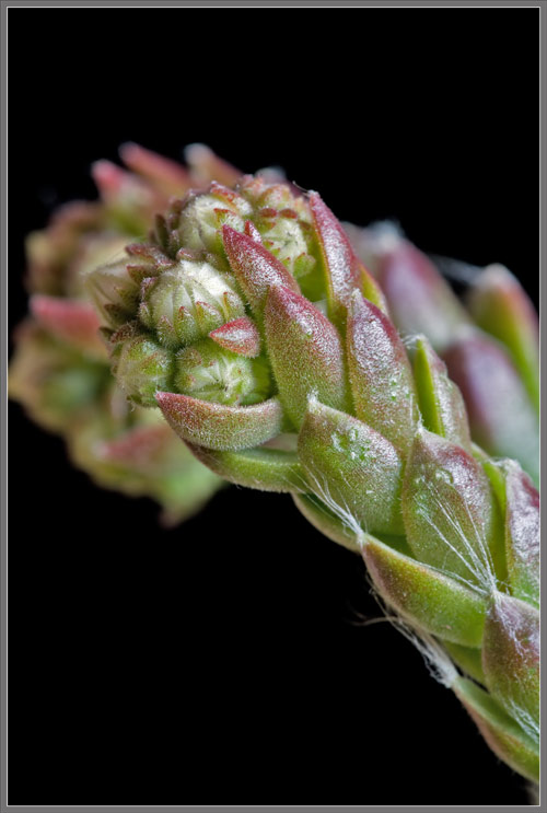
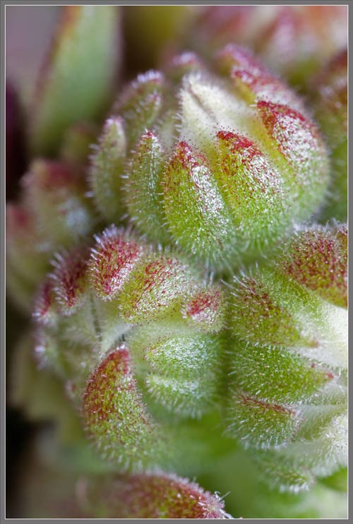
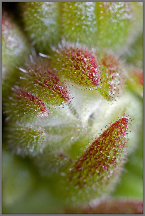
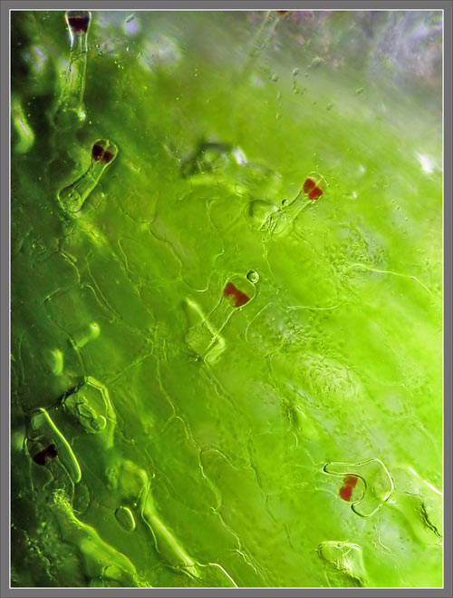
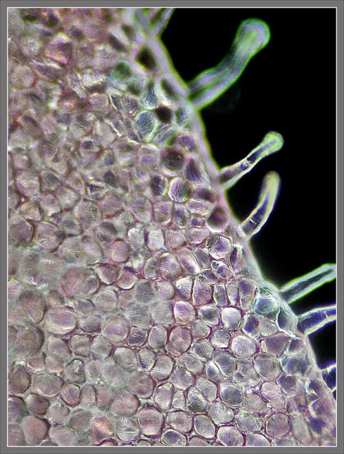
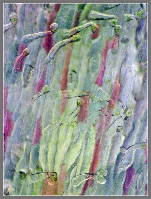
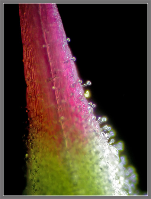
Near the edges of a sepal, some of the glandular hairs seen earlier are visible.  Notice that the red colouration in the glandular, bulbous tips is localized in two distinct areas!


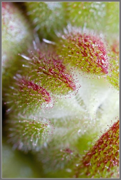
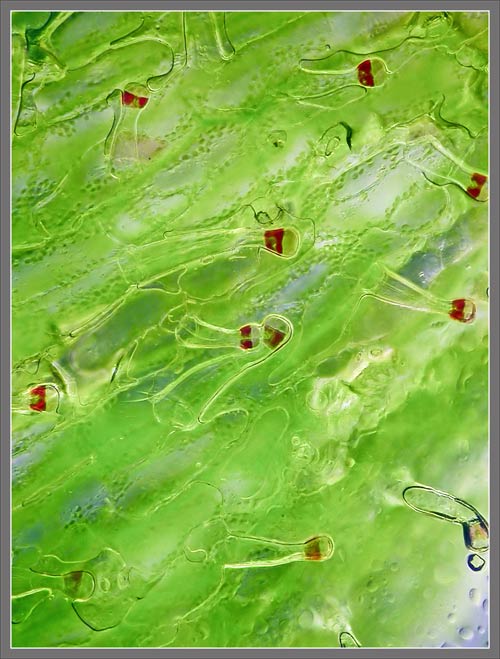
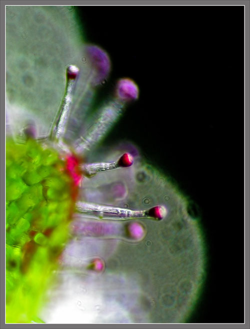
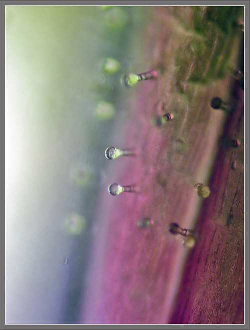
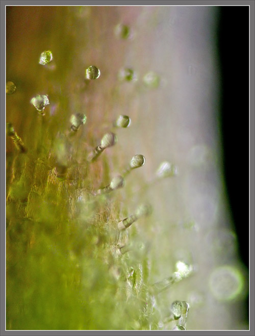
The use of a different lighting technique emphasizes the bulbous tips of the hairs, but does so at a lower resolution.


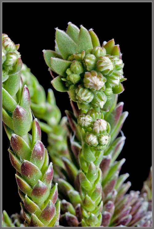
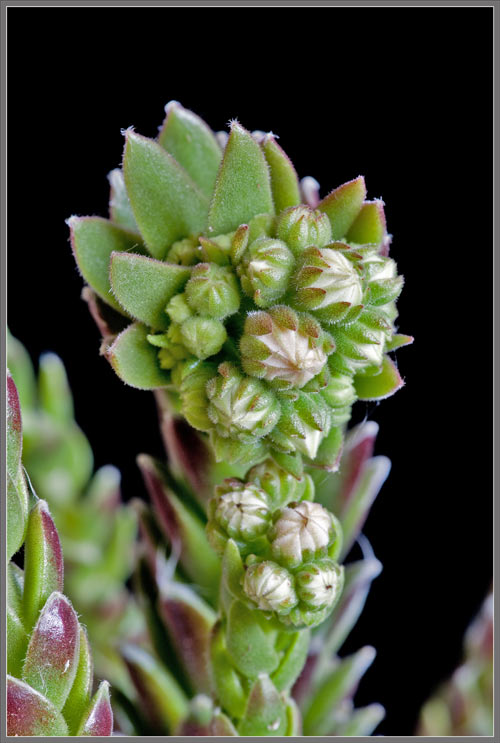
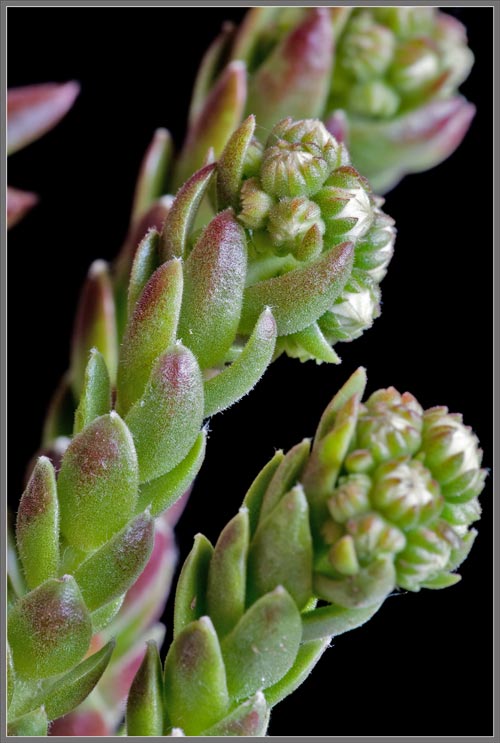
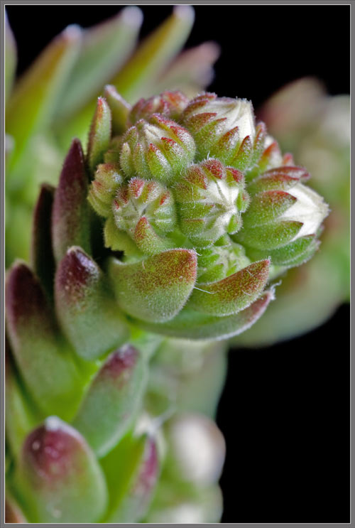
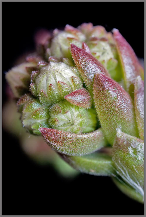
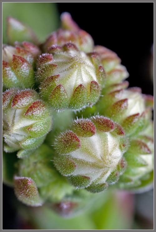
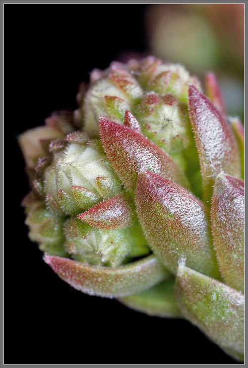
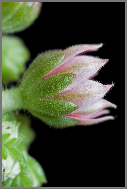
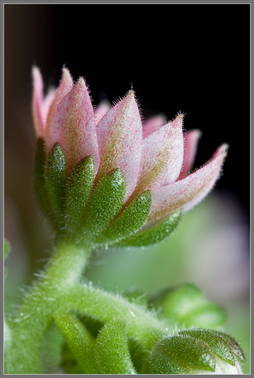
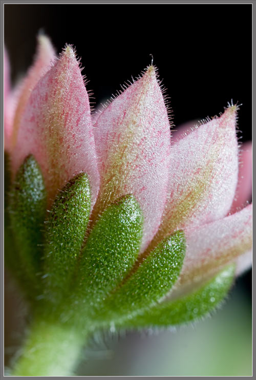
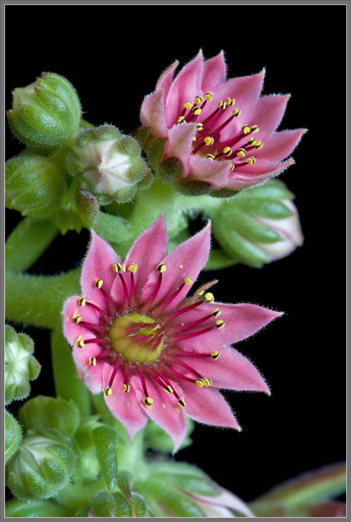
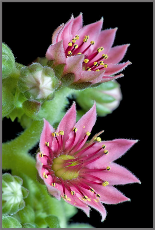
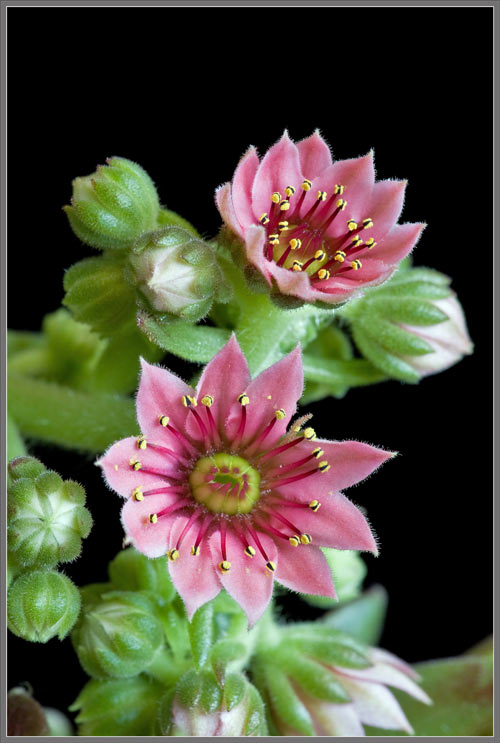
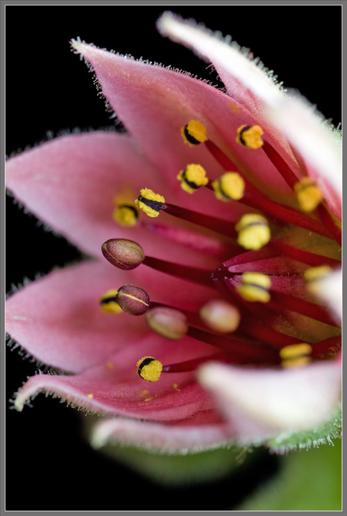
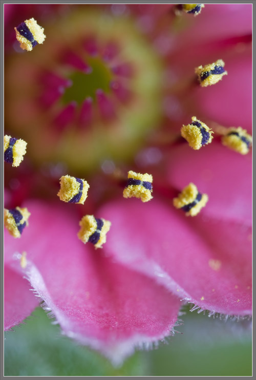
More images of flowers at different stages of development follow.  Next, we will look at one of a flower’s pink petals with the aid of the microscope.


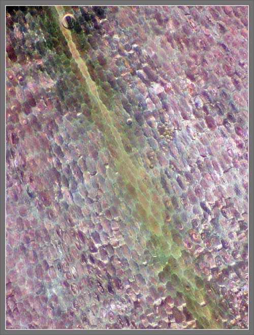
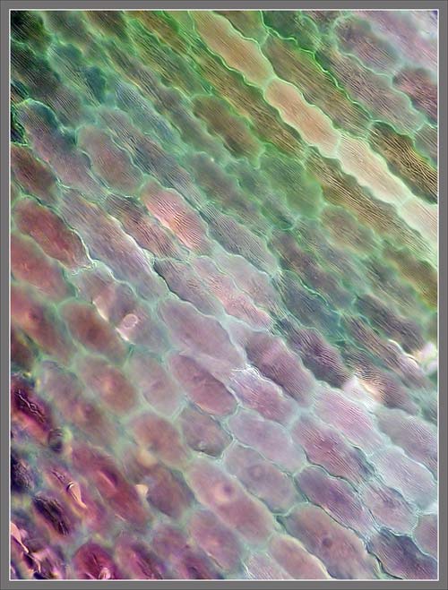
Photomicrographs of the surface and edge of a petal can be seen below.  Its cells are irregular, both in shape, and pink colouration.  Hairs growing along the petal’s edge are visible in the second image.


A petal vein can be seen in the image on the left below.  The image on the right reveals the presence of glandular hairs on the petal’s surface.


A higher magnification shows the longitudinal striations on the surface of individual cells (left image), and the bulbous tips of glandular hairs (right image).


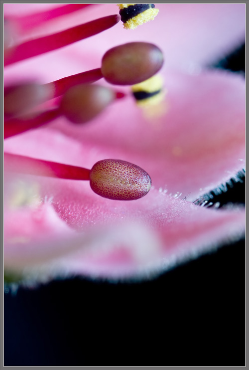
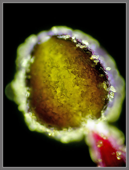
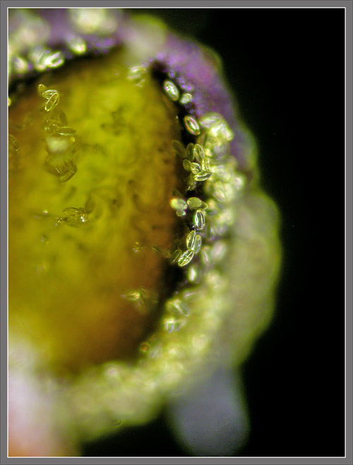
In the sequence of three images that follows, some of the anthers are covered by a thin purple membrane which hides the developing pollen grains beneath.  In more mature anthers this protective purple membrane has disintegrated.


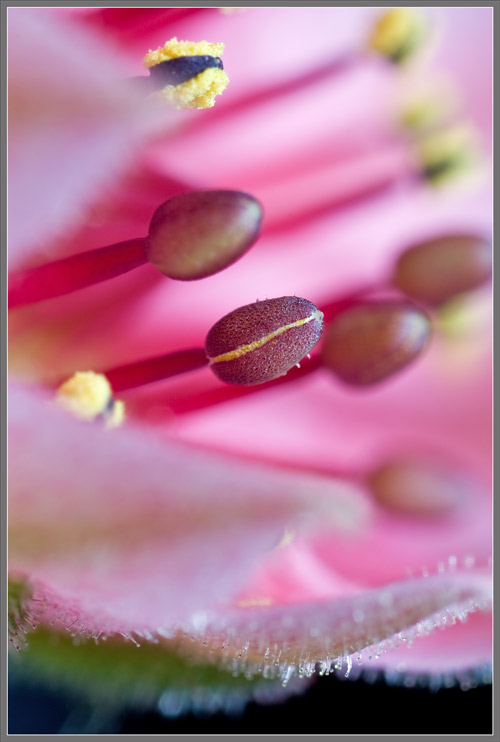
Notice the unusual surface texture of the anther at the centre of the left image.  The image at right shows a membrane that has split longitudinally to reveal the yellow of underlying pollen grains.


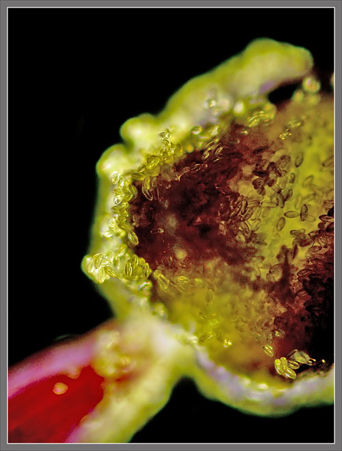
When the membrane has completely disintegrated and fallen away, the remaining anther appears considerably smaller.  Each anther has two pollen releasing pads, with a darker central section that is connected to the filament.  This forms a sort of anther ‘sandwich’.


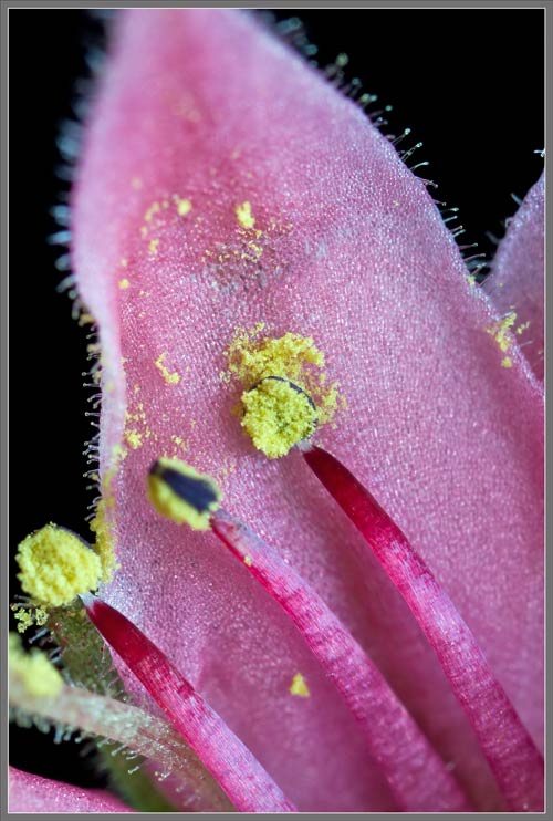
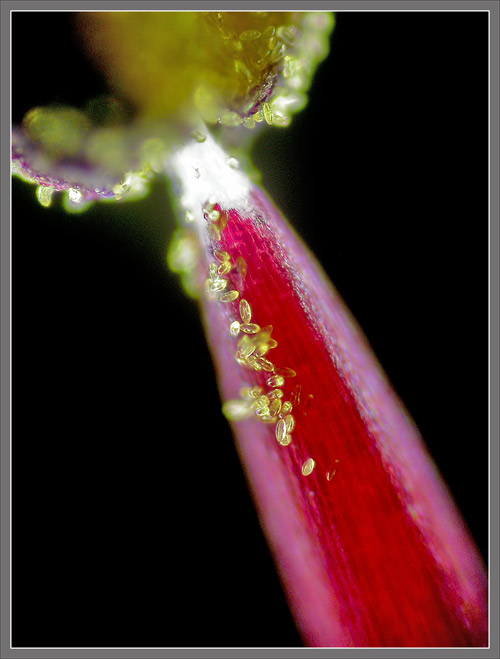
The slightest contact is able to dislodge pollen from the anther, as can be seen in an area where one has touched the surface of a petal.


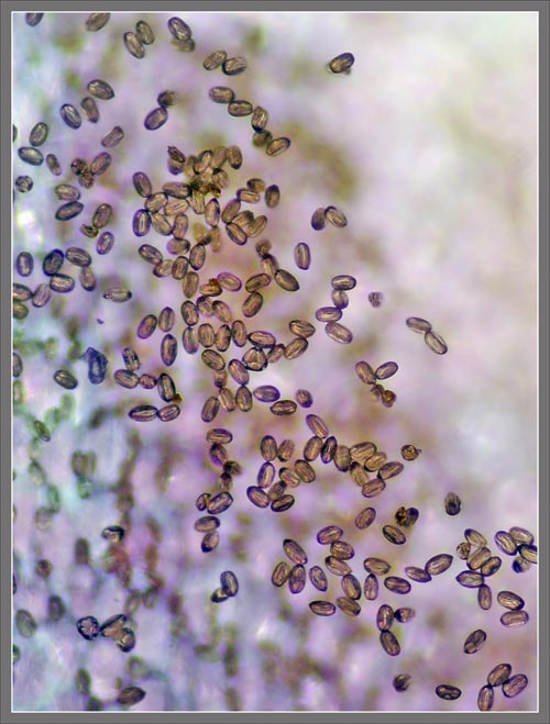
Below are photomicrographs showing the surface of a mature anther.  The pollen grains can be seen to be ellipsoidal in shape.


Images showing pollen grains adhering to the top of a filament (left), and the surface of a petal (right), can be seen below.


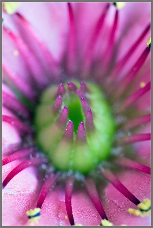
Ten bright red pistils, each connected at its base to a pale green swollen ovary are visible at the centre of the flower shown in the two images that follow.


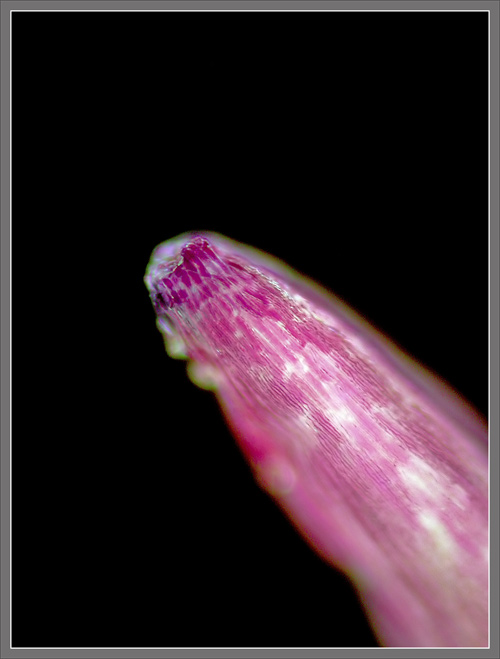
Under the microscope the cellular structure of the stigma and its supporting style can be seen below.  At the very tip of the pistil, the unusually short lobes that make up the surface of the stigma can be seen.  In my experience, these lobes are shorter than those in most flowers.


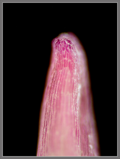
In the first image below, the base of one of a flower’s styles (red) is shown at the point of connection to its associated ovary (green).  Note, in all of the images, the large swollen heads of the glandular hairs.


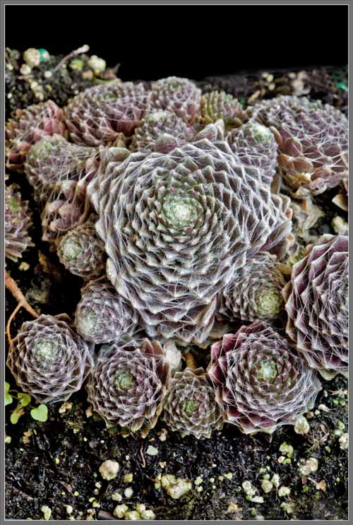
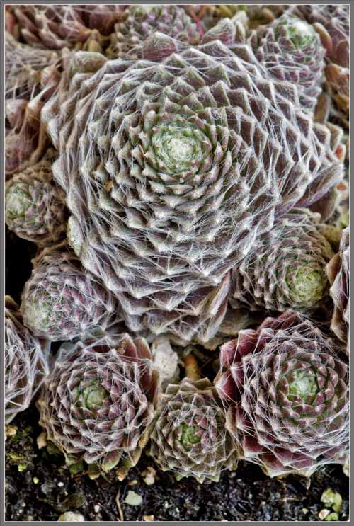
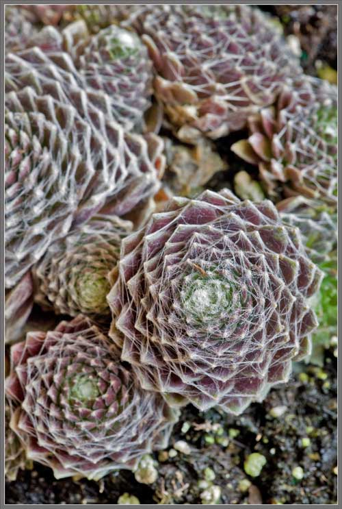
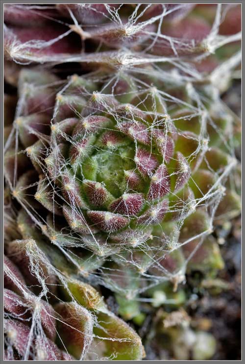
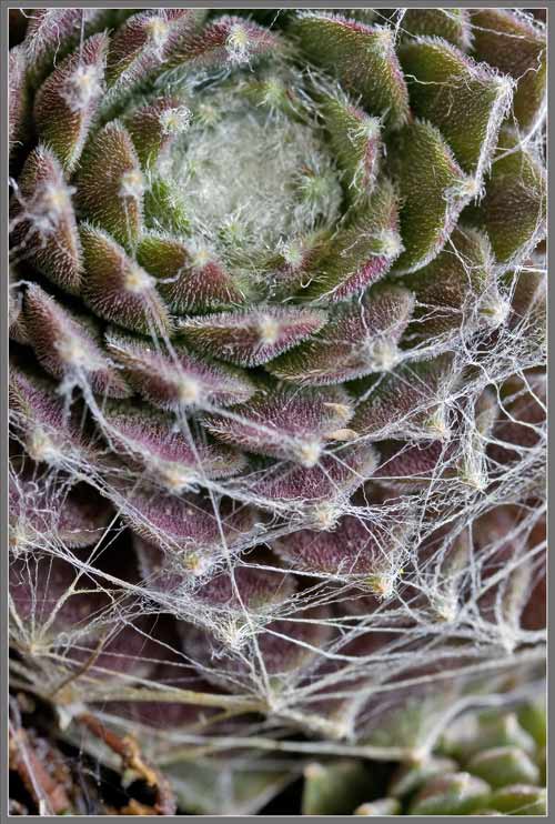
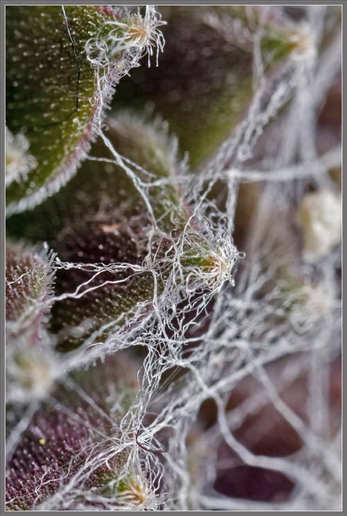
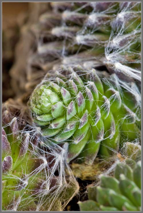
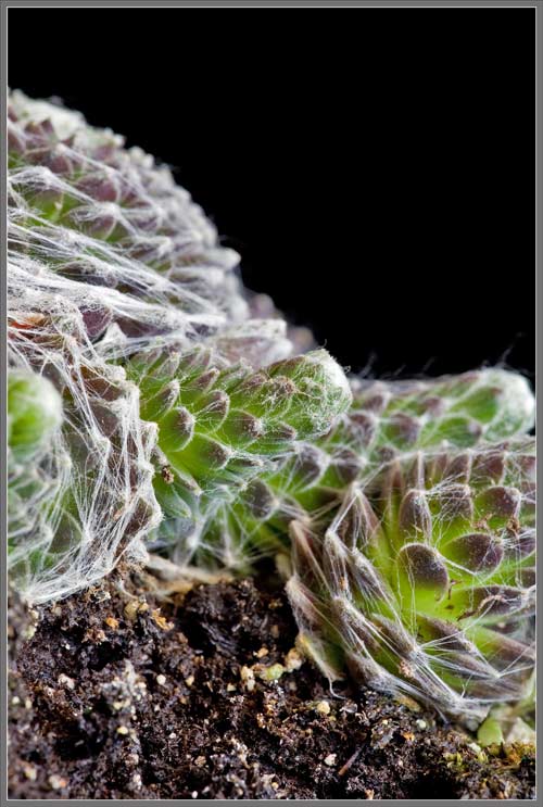
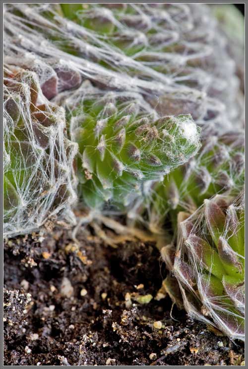
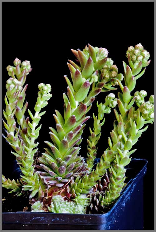
Hens and chicks are extremely popular as rock garden plants.  This popularity has resulted in an amazing number of ‘common’ names being given to the them.  A few of them are: Houseleek, Jupiter’s Eye, Jupiter’s Beard, Thor’s Beard, Bullock’s Eye, Sengreen, Ayron, Ayegreen, Aaron’s Rod, Hens and Chicks, Liveforever, and Thunder Plant!!


Photographic Equipment

The low magnification, (to 1:1), macro-photographs were taken using a 13 megapixel Canon 5D full frame DSLR, using a Canon EF 180 mm 1:3.5 L Macro lens.

A 10 megapixel Canon 40D DSLR, equipped with a specialized high magnification (1x to 5x) Canon macro lens, the MP-E 65 mm 1:2.8, was used to take the remainder of the images.

The photomicrographs were taken using a Leitz SM-Pol microscope (using a dark ground condenser), and the Coolpix 4500.


A Flower Garden of Macroscopic Delights

A complete graphical index of all of my flower articles can be found here.


The Colourful World of Chemical Crystals

A complete graphical index of all of my crystal articles can be found here.


 All comments to the author Brian Johnston are welcomed.

Microscopy UK Front Page
Micscape Magazine
Article Library


© Microscopy UK or their contributors.

Published in the March 2012 edition of Micscape.
Please report any Web problems or offer general comments to the Micscape Editor.
Micscape is the on-line monthly magazine of the Microscopy UK web
site at Microscopy-UK  


© Onview.net Ltd, Microscopy-UK, and all contributors 1995 onwards. All rights reserved. Main site is at www.microscopy-uk.org.uk with full mirror at www.microscopy-uk.net .