|
Household Microscopy: Part 5 (And the Yard) by Richard L. Howey, Wyoming, USA |
Read earlier articles in the series.
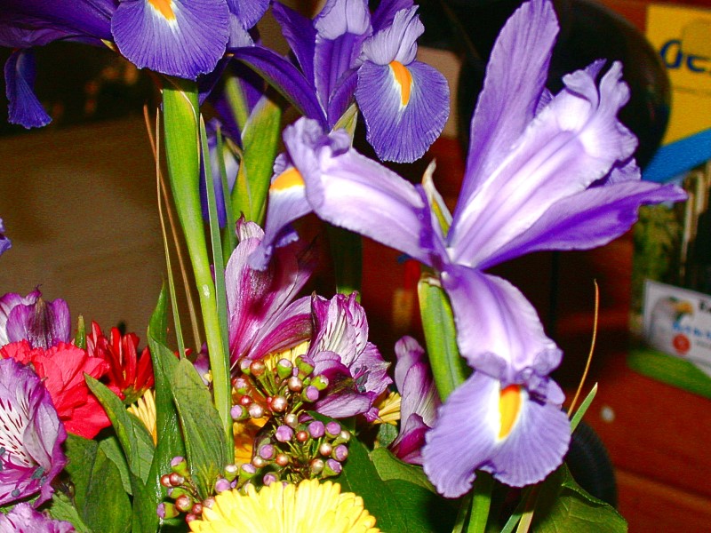
This time I wish to turn to items other than those found in a medicine cabinet. To start with, one can go to the market or florist and buy 2 bouquets of assorted blooms, one for decoration and the other to go immediately into the lab or if it’s past winter, you can go into the yard or patio and collect some flowers. Once in the lab the flowers can be examined in terms of pistils, pollen, sepals, ovaries, and sections made of leaves, stems, and anything else you encounter. One bouquet can keep you busy for weeks. However, before you start ripping it apart, take a few minutes to contemplate it as a whole and enjoy its color, fragrance, and form. Every time I do so, I think of the wonderful remark of the British philosopher and novelist, Iris Murdoch: “People from a planet without flowers would think we must be mad with joy the whole time to have such things about us.”
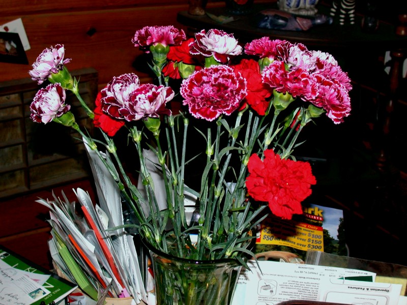
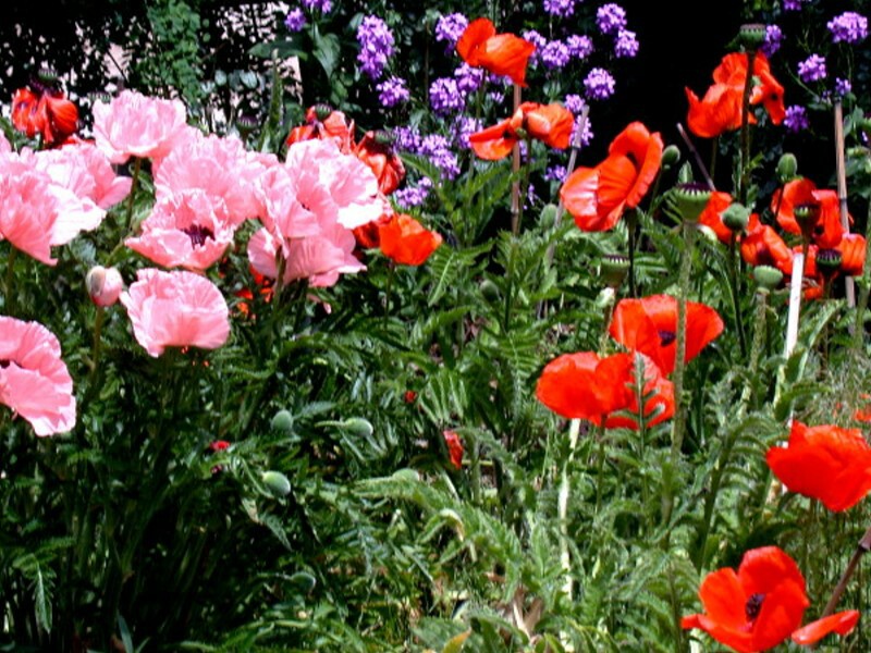
In addition to having bouquets of flowers in the house, one can select from those in the yard and patio.
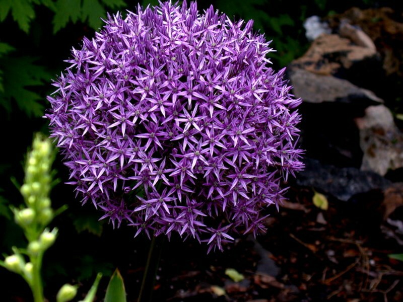
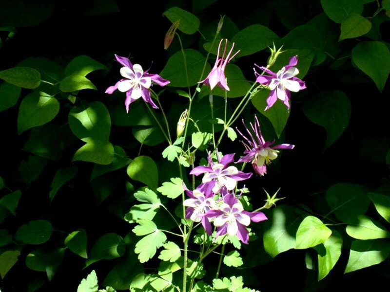
Last summer, in a bed alongside the driveway we had a giant Allium, Columbine, and Oriental Poppies. These are sources of lab material for months at a time.
Also, on the patio we had a number of potted plants, a couple of which were hanging plants and under one of the larger ones was a bucket. Because of the intense sun here at 7,200 feet they required frequent watering and with the one above the bucket, there was a gradual accumulation of water which turned a lovely green color. It turns out that inadvertently, I had created a nice pool of algae, protozoa, rotifers, and nematodes. Soil organisms are a special study in their own right and this gave me an opportunity to examine some creatures which I don’t come across very often, along with others that were old familiars.
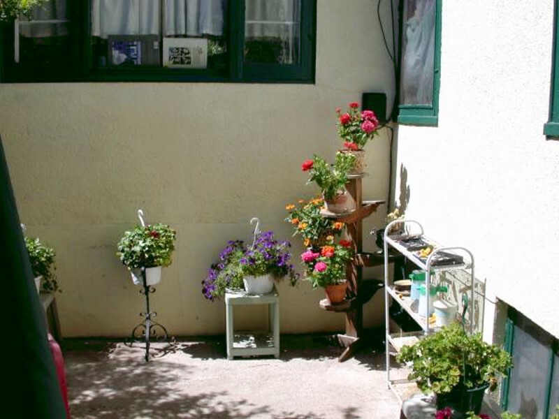
This view doesn’t show the bucket because by the time this picture was taken mosquito season was beginning to peak and I transferred the water into jars which I took inside to my lab.
The most exciting organism from the bucket was Thecamoeba which occurred in abundance. I call it the Shar-Pei of amoebae because of its surface wrinkling; it is a voracious algae eater and I want to show you a number of images of this highly interesting organism.
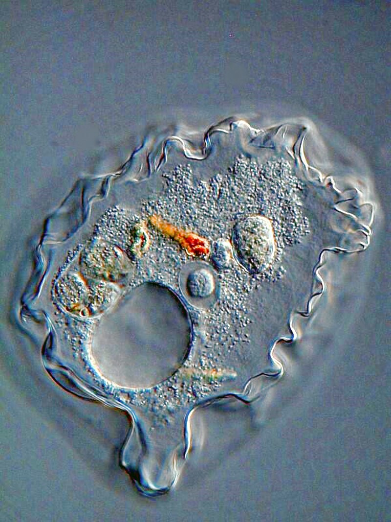
Here you can see the convolutions and layering of the pellicle and also the contractile vacuole, the macronucleus, and food vacuoles.
I removed some strands of filamentous algae and stained them with Neutral Red which in low concentrations is a vital stain and thus minimizes the possibility of toxic reaction on the part of the amoeba. Here you can see 2 different sorts of filaments in highly elongated vacuoles quite unlike the usual circular vacuoles.

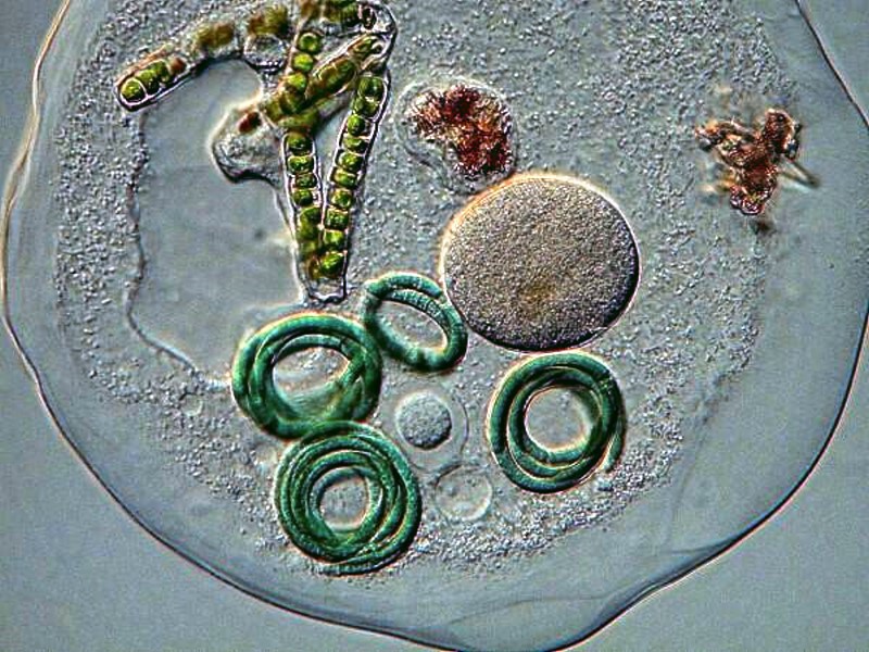
The next image is of particular interest since it shows the macronucleus, the contractile vacuole, a small circular food vacuole, a filamentous strand of algae, 2 small stained fragments, and 4 coils of yet another species of algae. [NOTE: I should mention here that some of these filaments may not be algae but rather cyanobacteria. At the time, I did not examine them to determine if that was the case.]
There were also other small soil amoeba in these samples and the first image shows a very ambitious little amoeba working to ingest a filament much longer than itself.
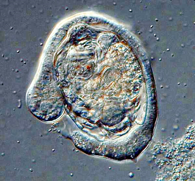
The next picture shows yet another very confident amoeba which has nearly completely surrounded a bdelloid rotifer.
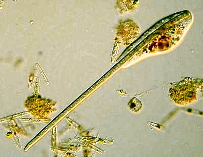
Another organism that shows up frequently in soil samples is the ciliate protozoan Colpoda which has a shape suggestive of a kidney bean.
Colpoda is distinctive in that it generally divides in a state of encystment and usually produces 4 genetically identical individuals.
In potting soil samples which I took from some house plants, I immersed the soil in water and after one day, I found some small mites, Collembola (springtails), nematodes, bdelloid rotifers, 2 species of small annelids one of which takes advantage of decaying plant stems to use as tubes, some hypotrichs and a variety of very, very small flagellates and ciliates including Halteria grandinella which is always a delight to observe since it moves gracefully through the water, stops, then suddenly springs away. It has a set of stiff cilia circling its midsection which it uses to accomplish this gymnastic feat. I suspect that there were also some amoebae but, on this occasion, I didn’t come across any.
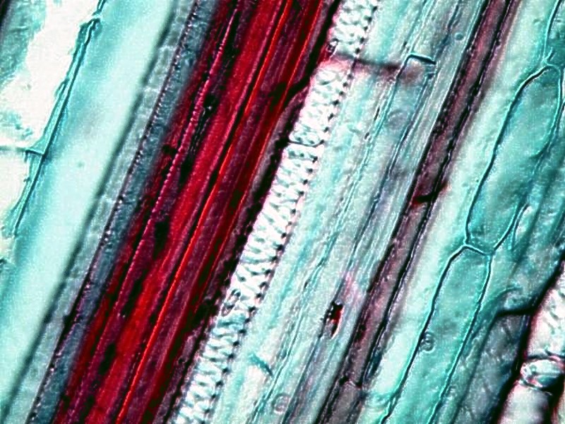
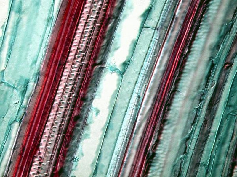
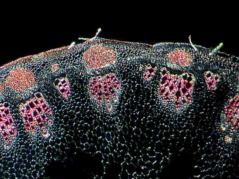
Let’s return to plants for a bit. You might not have only flowers in your garden, but you might put in some vegetables as well which will again provide you with a whole new dimension of material. Perhaps you might get adventurous and even plant some corn. The roots and small stems of Zea mays produce interesting sections and when stained can be quite attractive and show up nicely under polarized light. Here is a cross section of a root.
And 2 more of a stem.
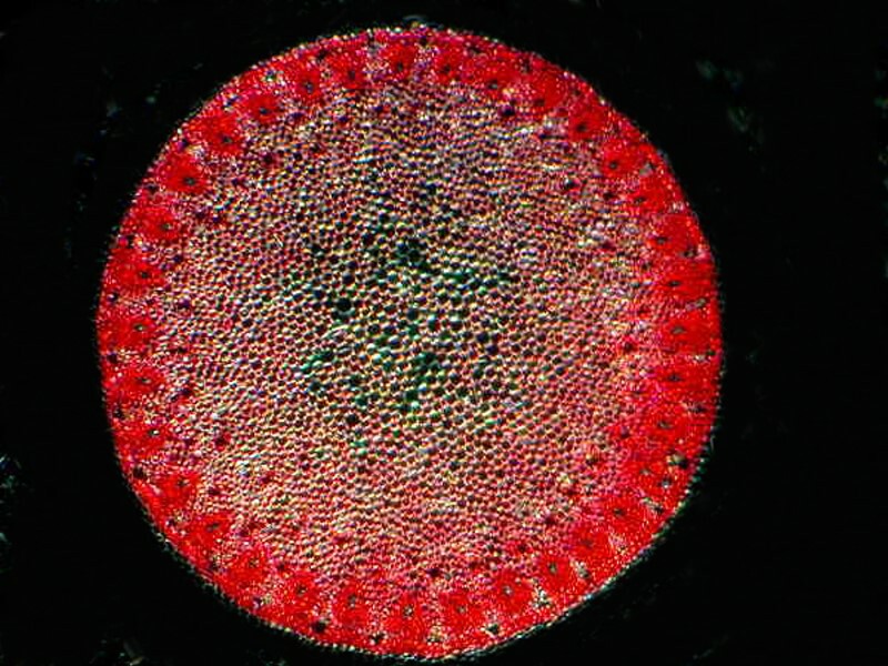
And such a garden is also a good place to have a few sunflowers which also have interesting stems. Here is a partial section.
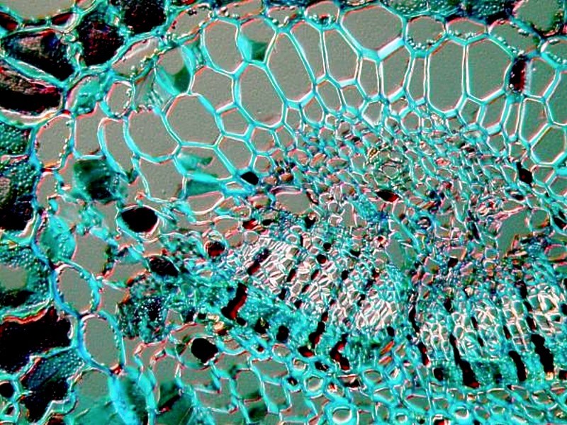
Back in the house, my wife and I have over the years kept a few orchids going. They are quite incredible plants and the ones we have had blooms for a period of about 3 months at a time. Here is a section of stem.
Pine trees also provide a variety of intriguing material. We’ll take a look at a cross section of a needle and a root in that order.
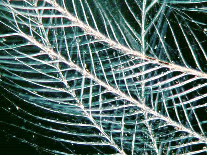
The trees and the flowers attract birds, squirrels, and insects and often when I’m out under the large pine trees, I’ll find a feather or two and usually I bring them in to examine. Here are 4 views of a single feather. Feathers are wonderfully complex and some of them are exceptionally colorful, but here what is amazing to me is the incredible intricacy.
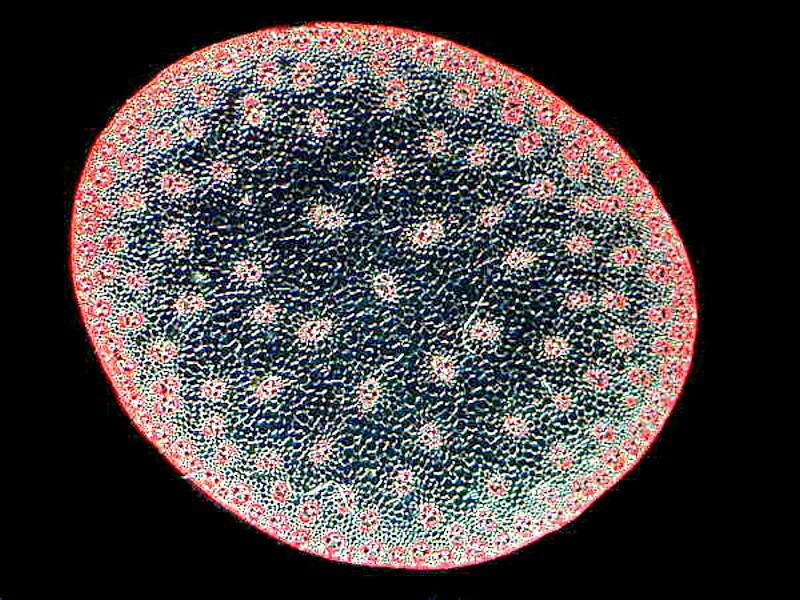
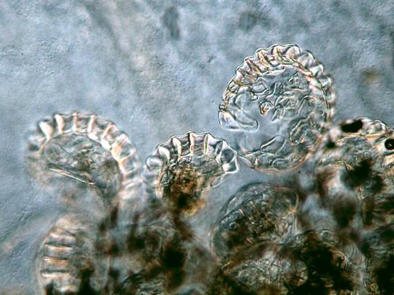
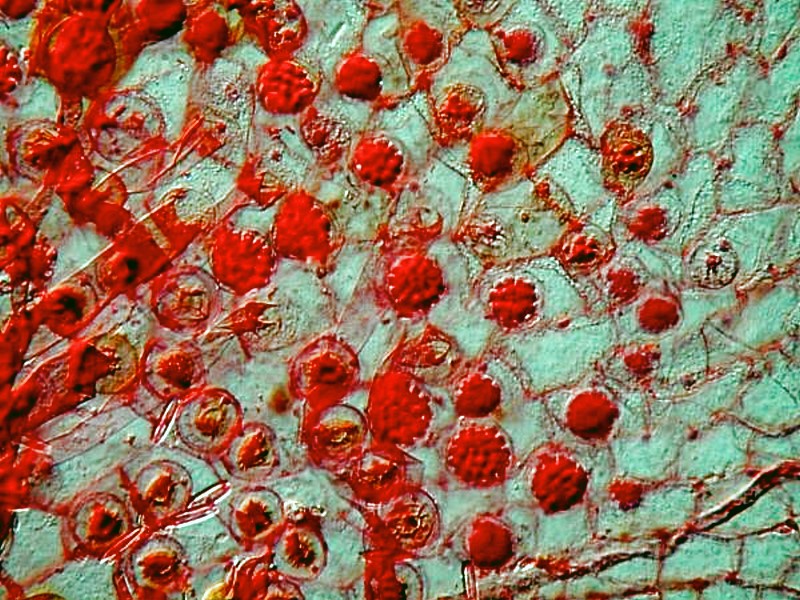
Opposite the pine trees, alongside the house, under the caragana bushes are some ferns. Ferns are splendidly decorative plants and have interesting life cycles. The first 2 images are of sori from a fern leaf. These are those little round dots on the undersides of the leaves and they contain sporangia which are packed with reproductive spores. The second image is a closeup.
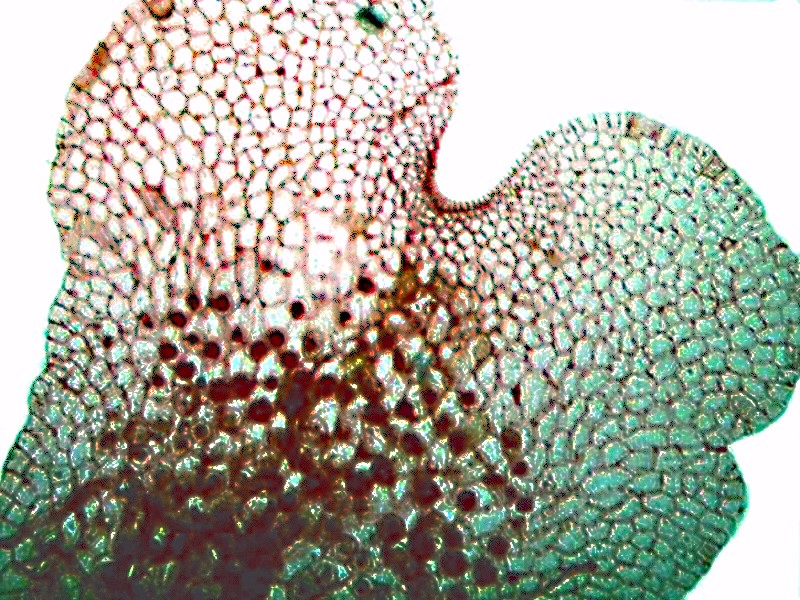
Next 2 images of the prothallium, that heart-shaped structure that produces both male and female gametes (sex cells). The second image is a closeup.
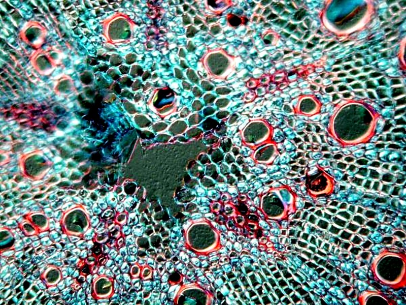
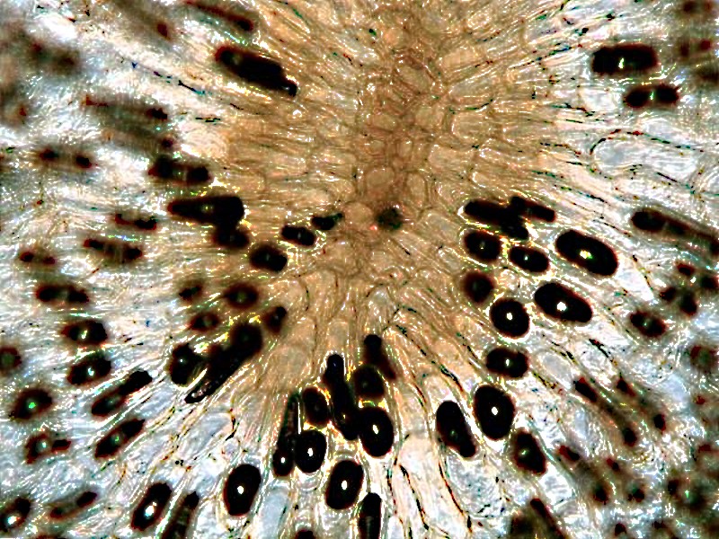
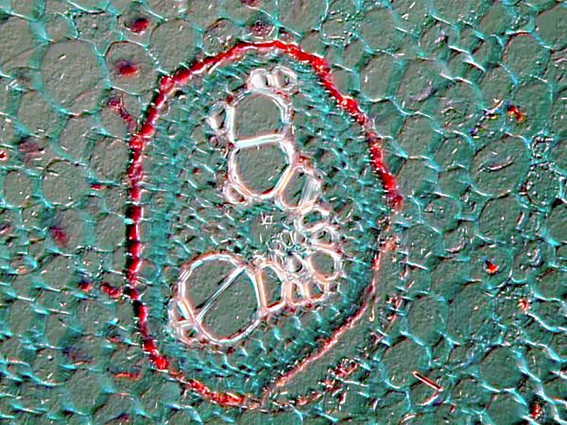
Here we have a cross section of the root of a fern.
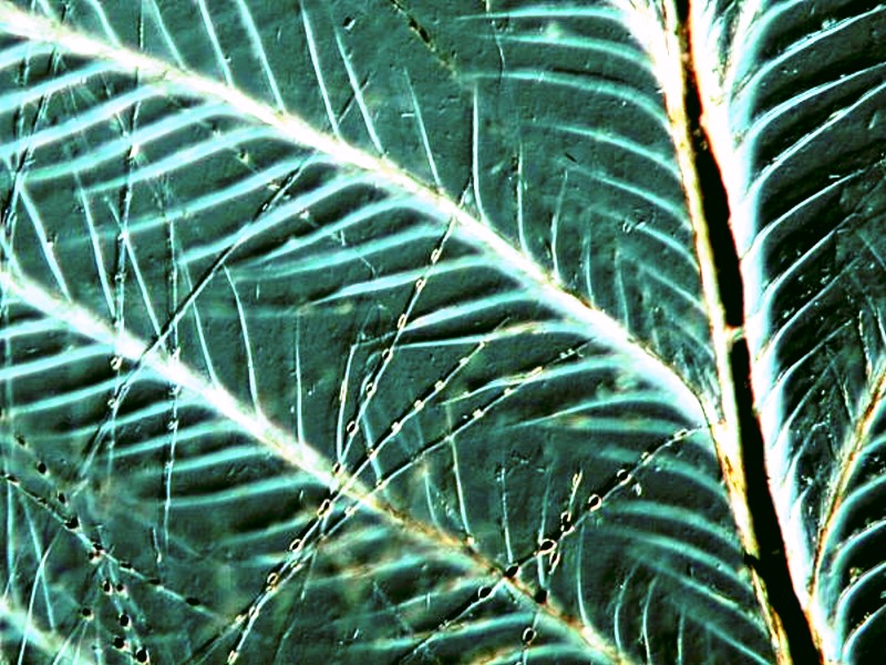
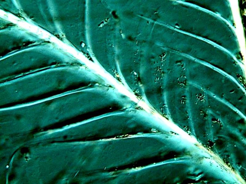
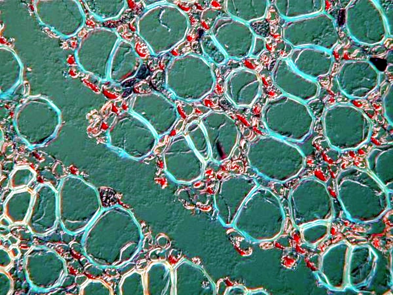
And then, a cross section of a fern leaf. Clearly, one can spend many hours just studying ferns and delighting in their intricate morphology.
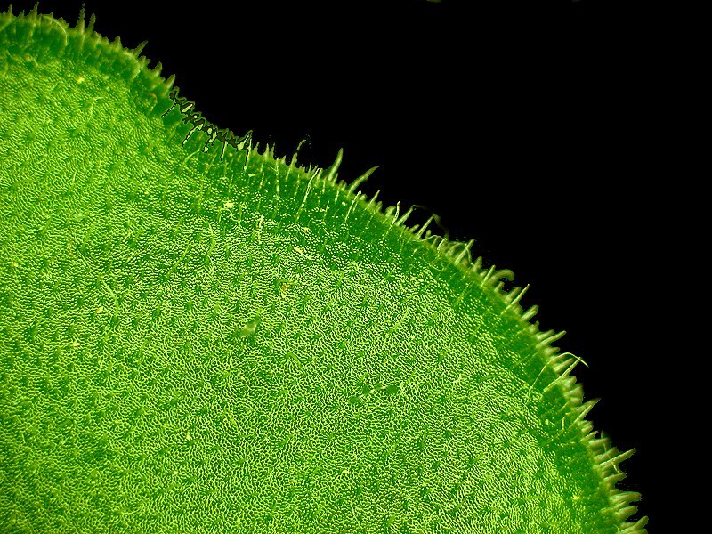
Ferns were a commonplace in substantial Victorian households and also African violets became very popular. Part of the reason for this is that both types of plants are fairly hardy and one could trust the servants or even one’s children to care for them. Violet leaves are covered with minute hairs and they are water-laden to the point of being almost spongy.
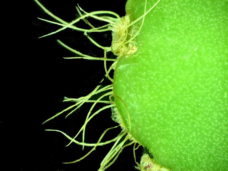
Plants which my wife is very fond of, and for reasons similar to those of the Victorians–they require little care, are the zygocacti, the so-called Easter and Christmas cacti which are never quite on track with our human holiday schedule. The branches, which extend out from the base, form stems which are composed, in most species, of flattened segments. These have numerous hairs and the terminal segment produces a quite elegant and delicate flower. Here is view of one segment with its hairs.
In the yard, the most common larger insects here are dragonflies, bees, houseflies, and mosquitoes. Now, I can hear you saying–but mosquitoes aren’t large insects. Well, you haven’t encountered Wyoming mosquitoes which are approximately the size of Blackhawk helicopters!
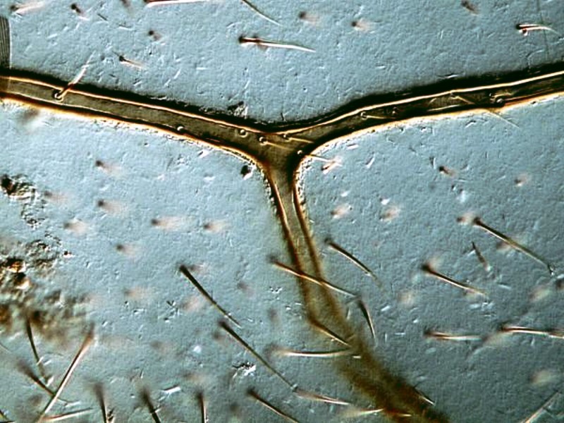
Dragonflies are aerodynamic miracles, they are beautiful to observe as they hunt and play with one another rather like fighter pilots, and they eat mosquitoes. Occasionally, I’ll find a dead one in the yard and I became mesmerized by the wings which are arranged in segments like window panes. Here are 2 views.
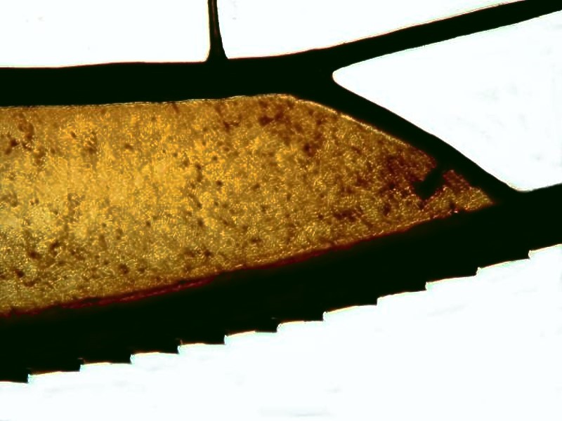
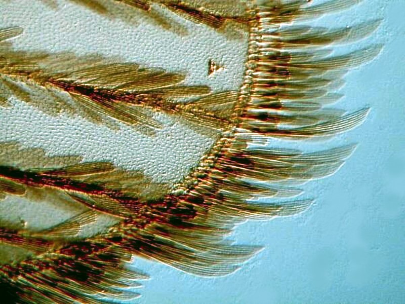
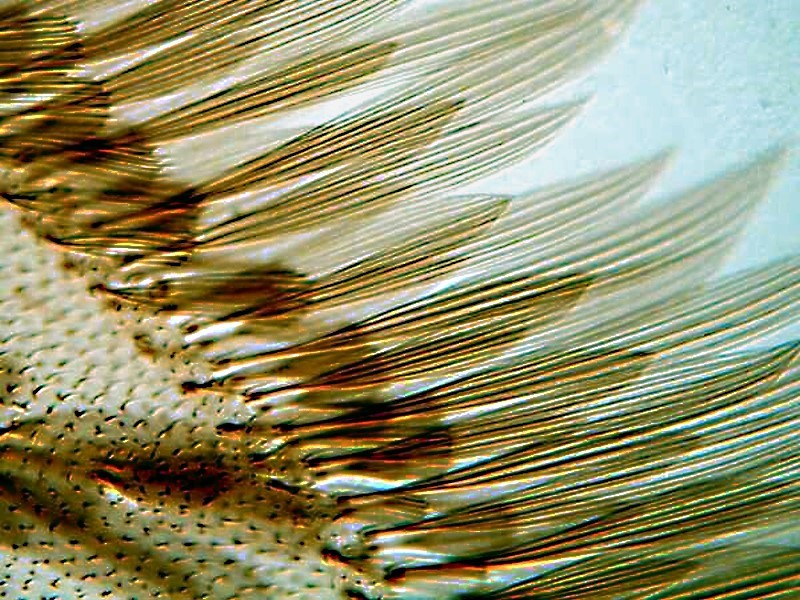
Occasionally, I will also find a dead bee or two. Their wings are quite different from those of the dragonflies. There are many tiny hairs which very likely have a sensory function.
The mouth is a complex structure which is morphologically amazing.
Houseflies (and for that matter, almost any kind of flies) can be terrible nuisances and they are not only annoying, they can transmit disease. However, from a dispassionate point of view, they are of great interest. I want to show you 3 images of the eye of a housefly, each one at a higher magnification that they previous one and then a closeup of a portion of its extraordinary mouth.
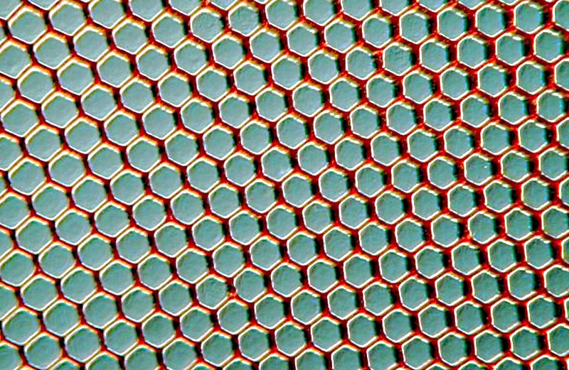
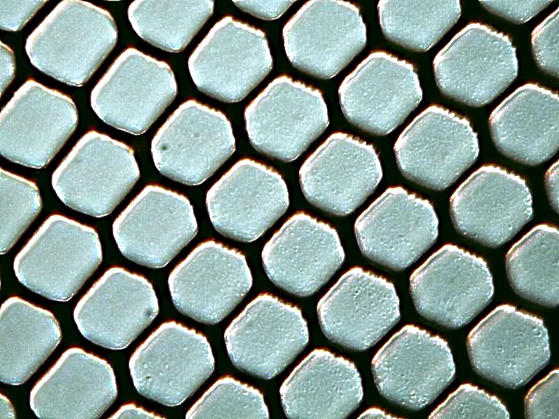
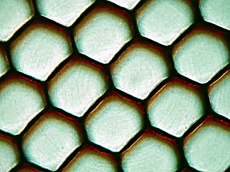
Notice the hexagonal packing which is quite typical.
Now, here is a view of a portion of the mouth apparatus.

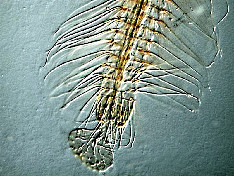
Next, a view of a portion of a mosquito wing which perhaps surprisingly is quite beautiful. The second image is a closeup which reveals even more of the detail.
Well, enough of playing rugged outdoorsman; let’s head back into the house.
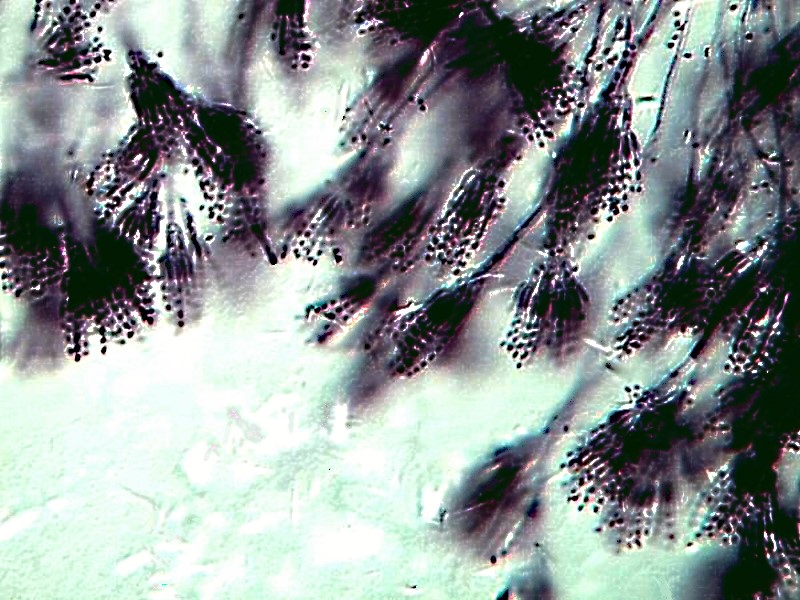
I was going to have a bite of lunch, but I discovered some mold. It turned out to be Penicillium. Ah, a friendly fungus, you say. Well, there are quite a number of species of Penicillium, such as ones used to produce Camembert, Brie, Gorgonzola, Roquefort, and Danish Blue cheeses. And then, of course, there is Penicillium chrysogenum (formerly P. notatum) from which penicillin is derived. However, there are other species that are not so friendly that can produce extremely dangerous toxins. Aflatoxins are among the deadliest biological substances known. However, I thought you might like a look at a benign species of Penicillium.
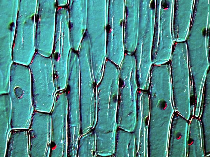
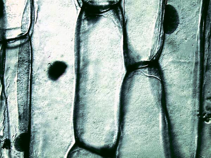
I ended up fixing myself a sandwich to which I added a slice of sweet onion. Onion is something every microscopist should investigate. It is easy to prepare and it gives one a chance to experiment with some stains. The nuclei stand out quite impressively when properly stained.
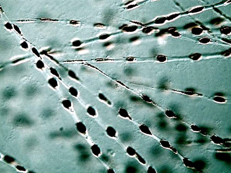
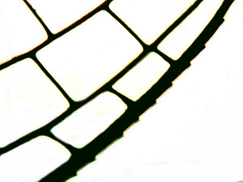
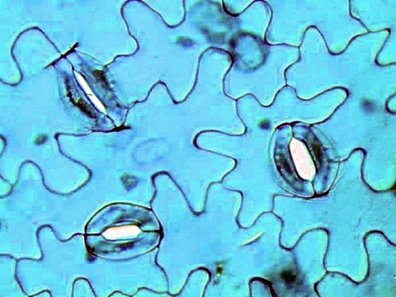
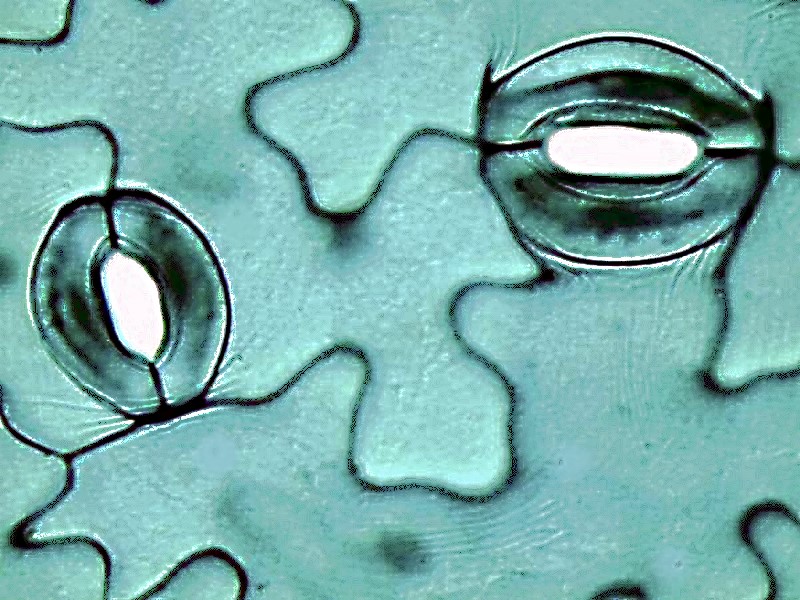
If you share Hannibal Lector’s passion–no, no, not for cannibalism, but for fava beans (Vicia faba) then you will be interested in seeing some stomata (pores) in a leaf which regulate the exchange of gases in and out of the atmosphere. This control is largely accomplished by the guard cells which surround the pore.
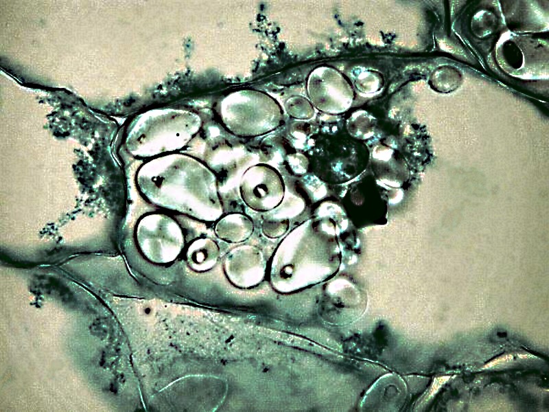
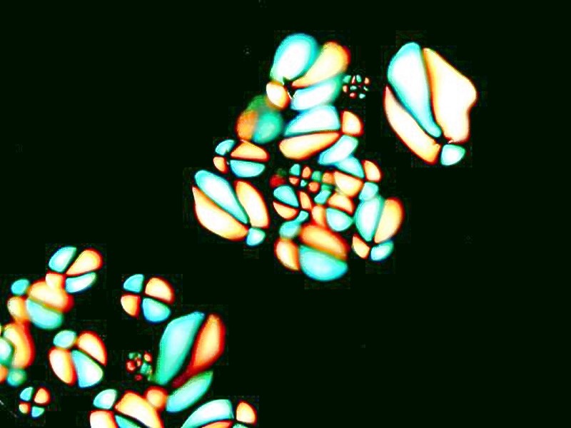
While we’re in the kitchen, let’s consider the lowly potato which medical types do their best in terms of psychological terrorism to make us avoid. How can baked potatoes with sour cream, mash potatoes swimming in butter, nicely salted potato chips be bad for us when they taste so good and produce such a satisfying gastric glow? Besides potato starch is quite impressive when viewed with polarized light. Here are 3 views; the first is with Nomarski DIC and the next 2 are with polarization.

Finally, after you have exhausted all of these possibilities, you can turn to yourself and take a look at your blood, hair, fingernails, tooth scrapings, etc. However, always make sure that you use sterile instruments when sampling yourself. A popular subject is the squamous epithelial cells from the inside surface of the cheek and when stained they reveal the nucleus quite nicely.
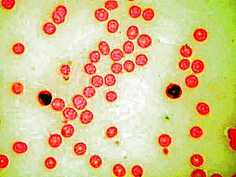
If you wish to look at blood, place a drop on the slide and then take a cover glass or another slide and smear the drop across the slide. This gives you a thin layer that allows you to examine the corpuscles.
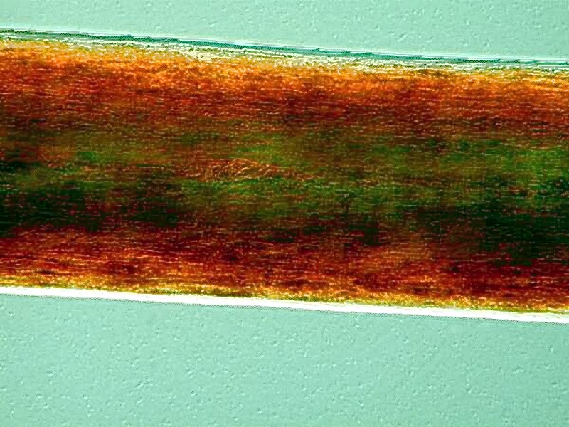
And you might be surprised to discover how colorful your hair is under polarized light.
You might even go on to pluck an eyelash or an eyebrow to look for a tiny mite, Demodex follicularium or D. brevis which is associated with the sebaceous glands of hair follicles.
They are quite common and only occasionally associated with skin rashes. Certainly, they are rather creepy looking, so you may not want to find any.
In conclusion, I would observe that we now never have cause to complain that we don’t having anything to look at under our microscopes. Just consider the kitchen which we barely mentioned; there are banana skins, artichoke leaves, orange peel, apple skin, pea pods, and while you’re at it, you can check your coffee, tea, and flour to see if it’s been adulterated. A few hints about what to look for: You Found That In My What? (Micscape, August 2001).
All comments to the author Richard Howey are welcomed.
Editor's note: Visit Richard Howey's new website at http://rhowey.googlepages.com/home where he plans to share aspects of his wide interests.
Microscopy UK Front
Page
Micscape
Magazine
Article
Library
Published in the March 2016 edition of Micscape Magazine.
Please report any Web problems or offer general comments to the Micscape Editor .
Micscape is the on-line monthly magazine of the Microscopy UK website at Microscopy-UK .
©
Onview.net Ltd, Microscopy-UK, and all contributors 1995
onwards. All rights reserved.
Main site is at
www.microscopy-uk.org.uk .