|
Some Brief Suggestions For Winter and Backyard Microscopy by Richard L. Howey, Wyoming, USA |
For those of us who live in harsh climes, winter can be a prolonged period with little or no opportunity to collect new samples for microscopic investigation. This winter, we had one night when the temperature plummeted to -40 F. and the windchill was -54 F. We also had a snowstorm which dumped a foot of snow on us. That was a month ago and the temperatures have been getting just high enough to melt the very upper few surface microns and then freeze again at night, resulting in great sheets of ice on the sidewalks and streets. Not a time for old, arthritic people with canes to be wandering about. As a result during the entire month of January, my wife and I only went out to the front porch to get the newspaper and the side porch to get the mail and February thus far is stacking up the same way.
Now, I do have a lot of preserved specimens, dried specimens, and micro-fossils, not to mention, leaves and flowers from house plants and bouquets purchased from the local grocery store, so it’s not as though I don’t have anything to look at and investigate, but sometimes I get curious about what’s hidden out in the backyard hibernating under all that snow and ice. Life, as we know, is incredibly adaptable and can be found in staggeringly extreme environments. When there are safe little paths, I venture out and sample the ice from a bucket forgotten under a faucet, skim some “dirty” snow to see what the wind has blown in and deposited on its surface, collect a few pine cones and needles from the fir trees in our yard, pull loose a few seed pods from plants that were in our garden last summer, and pull up an overlooked dried weed or two to get a bit of soil to start a culture–all of which goes up into my lab for later examination.
I also have some packets of old flower, herb, and vegetable seeds. A few of each type can be placed in a container with moist paper toweling to germinate. The roots of such plants are often very good subjects to investigate; they are easily sectioned and stained or one can make squash preparations of a bit of root tip. Some will display morphology in a very colorful fashion under polarized light. In fact, plant sections are almost always interesting, so let’s begin by looking at some examples.
This first one is Cucurbita which is from a pumpkin.
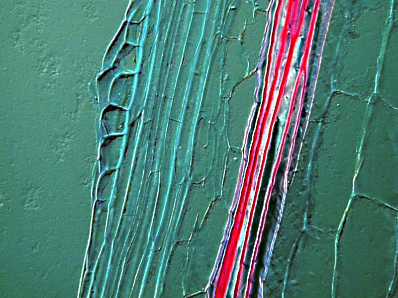
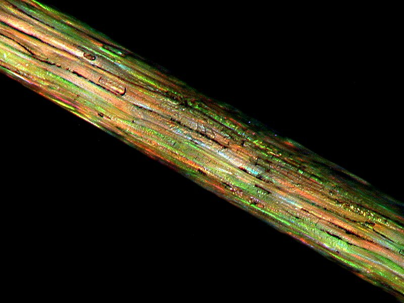
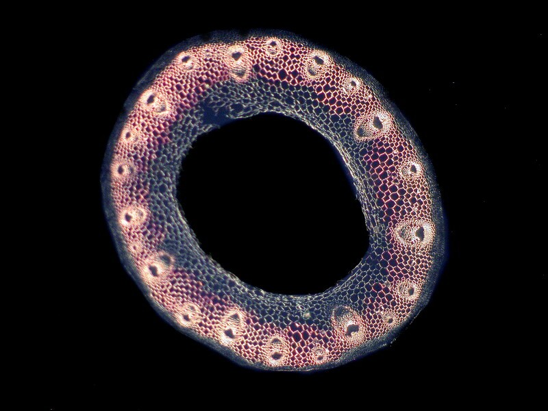
I mentioned earlier that we have some large pine trees in our yard, so it is easy enough to pick a few needles and try my hand at cutting some thin sections. Here is an example of one:
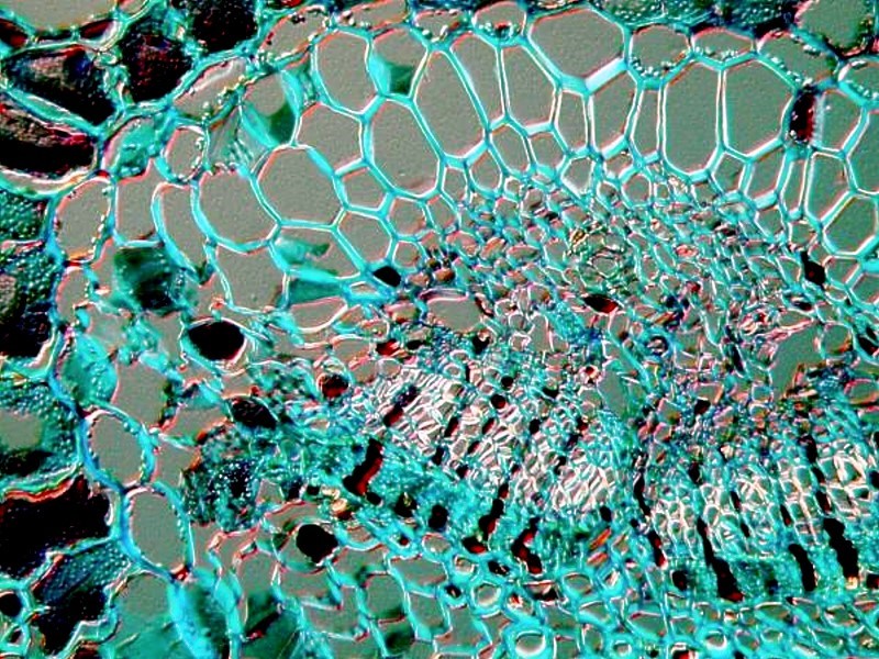
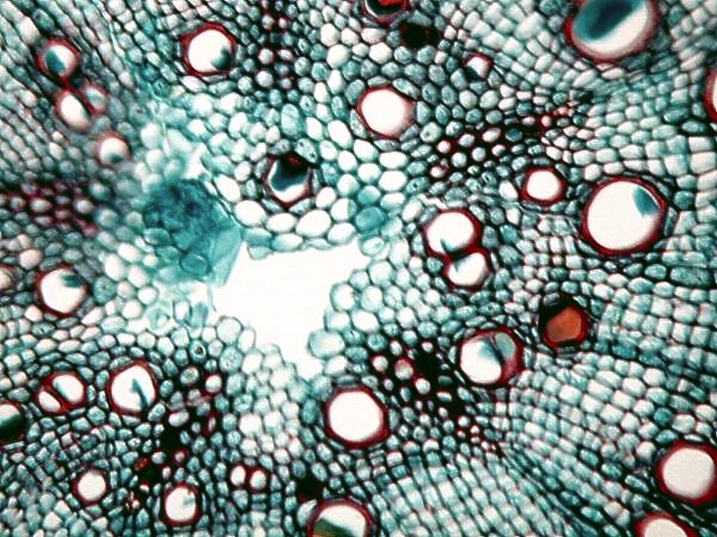
The roots of pine are also very interesting, but you can’t very readily collect a suitable piece of root from a 60 foot pine tree. You have to collect some seeds from a cone and plant them and pamper them until you get a tiny sprout with some very thin, delicate hair-like roots. These then you can section and stain. I’ll show you 2 examples. You will note that like the needle, they have lots of empty areas which are tubules for the transport of fluids and this constitutes a complicated regulatory system which provides nutrients to the very tips of the needles and also produces the sticky sap that can trap small insects and turn them into fossilized specimens embedded in amber.
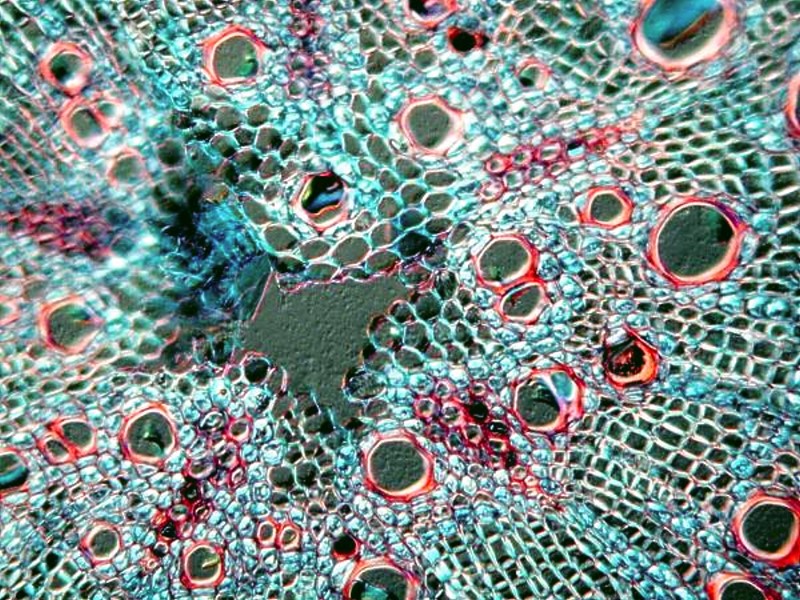
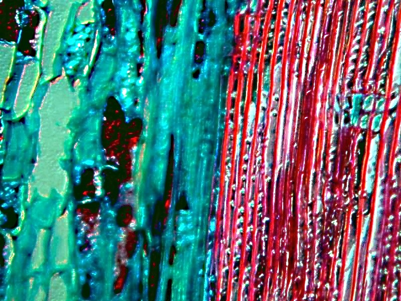
If we take a bit of stem and make a section, we see that it is, as we would expect, much denser and more like what we ordinarily think of as woody.
Sometimes, however, we may anticipate that a certain type of thing will look a certain way only to discover on examination that it is quite different than what we expected. This was for me certainly the case with a cross section of Pisum, the seed of a pea.
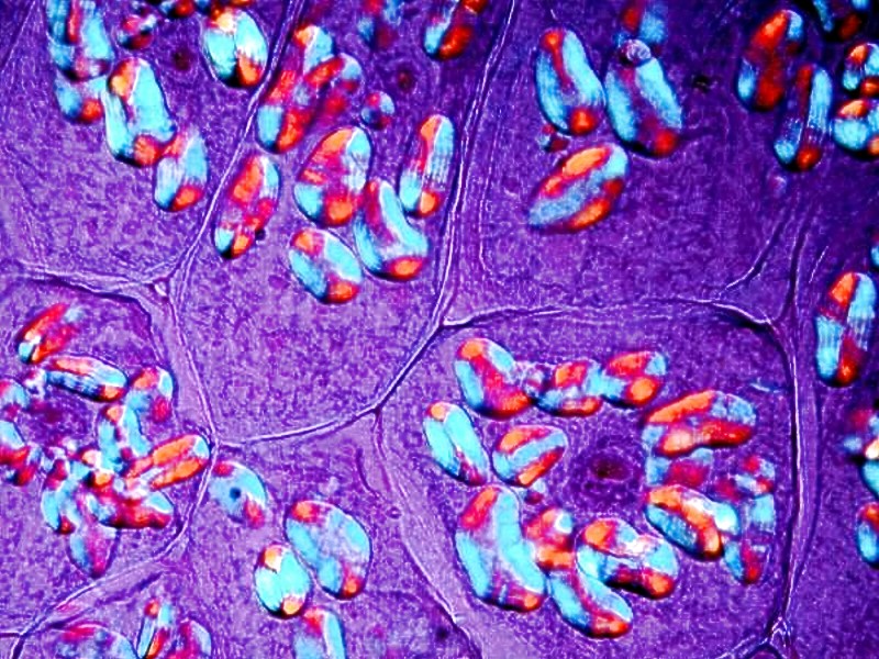
What stands out so vividly here are starch grains stained within the cells.
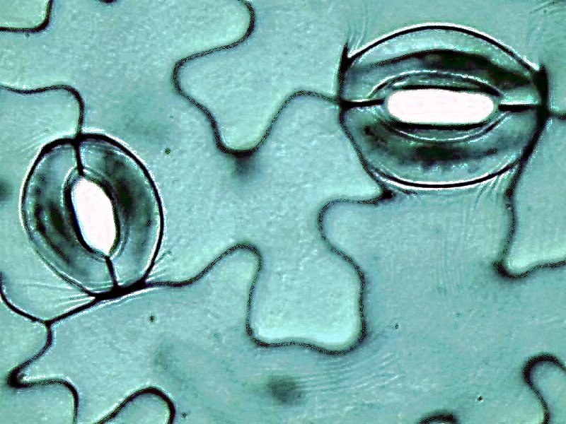
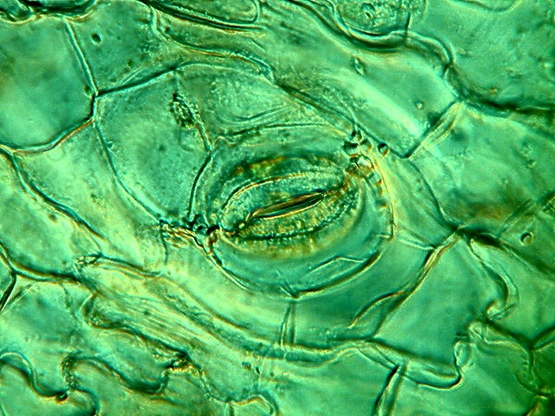
For those of you who are Hannibal Lector fans and like liver (frankly, I detest liver in any form), you may be familiar with the plant Vicia-Faba and here we have a section of leaf very nicely showing us the stomata through which respiration takes place. This is a section of the notorious Fava bean.
Those of you who have cats have no doubt discovered that there is one kind of plant which they will not nibble on and that is cacti. I have a small cactus from which I extracted a spine and discovered that it is quite a colorful feline deterrent.
A tiny section of tissue once again reveals stomata to us and we see the typical 2 curved cells enclosing the opening.
Another root tip is from a very popular, colorful little plant, namely, Ranunculus or buttercup.
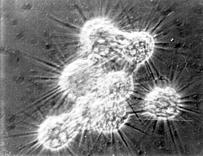
Very often when I am out during the collecting season and wandering around the ponds and lakes, I will take a sample of aquatic plant material, take it home, and spread it out on the lawn in the sun to dry. Then I put it in bottles or plastic bags until the winter. Some artesian water and a boiled grain or two of wheat and I get a culture started. Some years ago, I was collecting out at the Hutton Federal Game Reserve where there are 5 or 6 lakes connected by channels to maximize the distribution of the snow melt-off. These are very alkaline lakes and later in the summer, on the bushes surrounding the edges of the lakes there will be deposits of vegetation as the water recedes and both the bushes and the aquatic vegetation will be coated with a thick layer of white mineral deposits. I collected some of this material and then the following winter, before I had a digital camera and only a Polaroid microscope camera, got a photograph of a rather amazing organism. Admittedly, the image is not very good, but it is informative. The organism is an odd little amoeba called Raphidiophyrs. Here we have a cluster of 8 of them. They some times form these groupings when feeding, creating a “net” that few tiny critters will be able to escape from. After feeding for a while, they separate and continue on as solitary organisms.
Since it has now been quite a few years since I have encountered these bizarre critters, I am encouraged to find my packet of dried material to see if after all this time, I can still get some of these amoebae to appear.
Thereafter, I think I’ll take some samples from the Anthurium, Kalanchoe, Cyclamen, Amaryllis, and Geraniums which I have growing in a South window. That should carry me through until Spring which is scheduled for June 27th this year.
All comments to the author Richard Howey are welcomed.
Editor's note: Visit Richard Howey's new website at http://rhowey.googlepages.com/home where he plans to share aspects of his wide interests.
Microscopy UK Front
Page
Micscape
Magazine
Article
Library
Published in the March 2017 edition of Micscape Magazine.
Please report any Web problems or offer general comments to the Micscape Editor .
Micscape is the on-line monthly magazine of the Microscopy UK website at Microscopy-UK .
©
Onview.net Ltd, Microscopy-UK, and all contributors 1995
onwards. All rights reserved.
Main site is at
www.microscopy-uk.org.uk .