|
Galleries of Old Image Reprocessed Part 1
Richard L. Howey, Wyoming, USA |
Having taken photographs of many kinds of organisms and parts and structure thereof, using both photomicrography and photomacrography for over 40 years now, I ended up accumulating an overwhelming number of images. I confess that I am not the most organized person and, as a consequence, the arrangement of these thousands of images is bewildering even to me.
Last week, I started browsing through some of these old files and I had a wide range of reactions ranging from 1) Why did I ever keep that? It’s really bad, 2) Well, that’s interesting, I wonder what it is?, 3) Not bad, but is it really worth keeping? 4) Quite good, but it could do with some better processing now that I am more familiar with aspects of my graphics program. It is category 4 that I am going to be concerned with here and, I hope, in a series of subsequent galleries. My goal is two-fold; first, I would like to create a kind of record or mini-catalog of my work over the years and second, I hope that sharing these images will not only interest others, but perhaps inspire a few individuals to further investigate some of these fascinating aspects of the world of Nature.
Do try to think of this as simply a gallery and not an exposition with a theme. I will not include images which I have recently taken nor will I include any crystal images, at least not in the early galleries, since I have recently written a good deal about crystals. (Although, as it turns out, I will make one exception, as usual.) Also, I will keep my comments at a minimum, just enough to help orient you regarding the subject matter of the image. So, this is just a kind of ramble from one thing to another with no particular order.
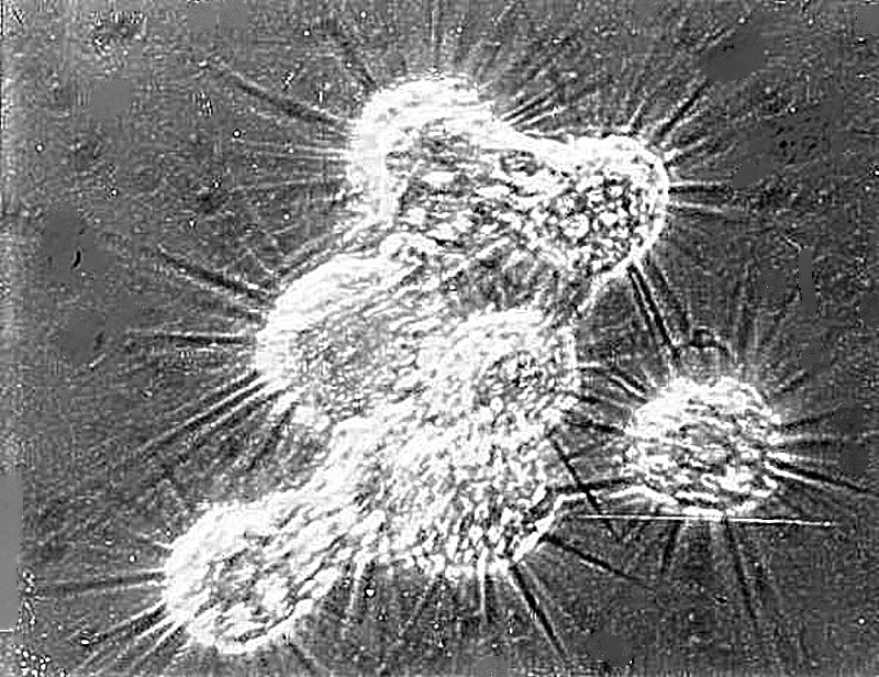
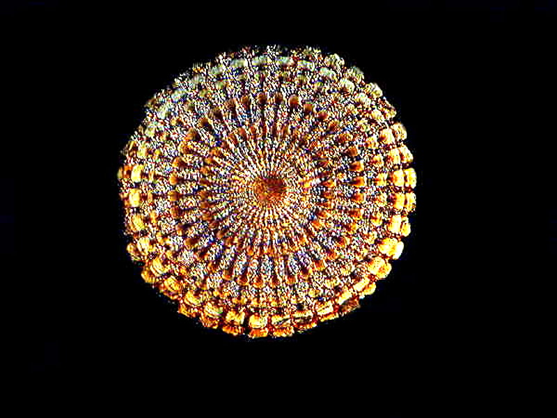
The first image is, as it happens, one of the first photomicrographs I ever took. It is a Polaroid taken with an awkward camera device designed for microscopic photography. It has a separate focusing tube with a small, circular ground glass screen. The film I used was a combination which produced a print within a minute or so and also a negative, which had to be carefully washed in water to remove any chemical residues. A rather cumbersome and somewhat messy procedure but, of course, nothing at all like the complexity of processing film. I must admit, I was never a fan of film cameras for photomicroscopy. (No, need to send me irascible, lengthy protests about its superiority. I have a couple of friends who are dedicated film users and they keep me on my toes.) The expense alone was always a major factor for me, plus the need to have ones own darkroom to get the best results. My first digital camera was a Nikon CoolPix 995 and it’s still the camera I use. Others have been recommended and I tried out a very fine JenOptik camera linked to a laptop, but I could never really get into it. I’m a tech dinosaur and a cantankerous old coot who is very stubborn. When I find something that works for me, I am very resistant to change. (Drives my friends and my wife crazy.)
I kept this image and present it here, because of the organism which is quite unusual, and I found it only once in a highly alkaline lake in the high prairie (about 7,000 feet). It is a cluster of Amoebae, named Raphidophyrs (although the taxonomists may have changed it by now). These little critters form clusters and create “bridges” with their pseudopodia, apparently as traps to capture prey for feeding–a remarkable example of cooperation at the most basic level. Well, so much for keeping my comments brief but, I promise, I’ll try harder as we proceed. (However, don’t make any bets on my having much success.)
For many years, I have been fascinated by echinoderms and also by calcareous structures and, quite wonderfully, echinoderms have lots and lots of calcareous structures. So, let’s look at a few.
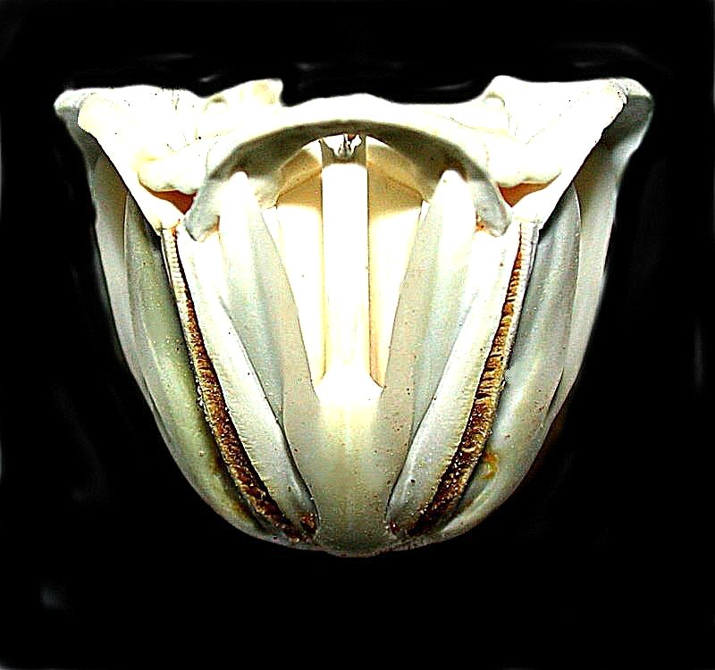
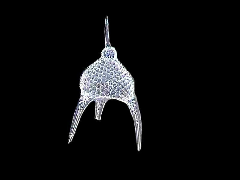
Sea urchins, the “pin cushions of the sea”, possess an incredible structure, called “Aristotle’s Lantern”. This is the “mouth” and usually consists of about 40 “plates” The image is a glimpse inside a lantern, which though relatively small, is the largest I have ever encountered. This is a view from the side.
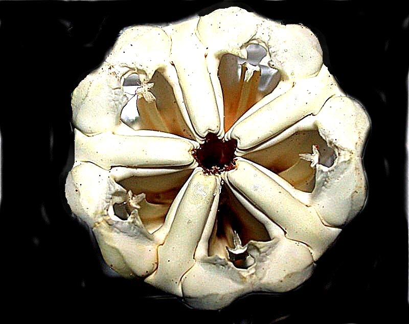
And here is a view from the top.
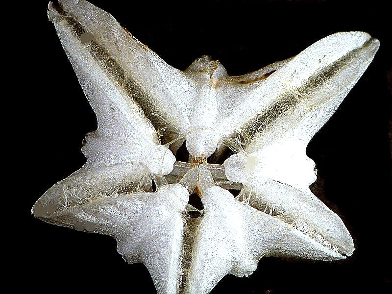
Equally remarkable is the “mouth” structure in sand dollars. This is a view from below and here you can clearly see the five teeth. Throughout the “lanterns”, pentagonal symmetry tends to be preserved.
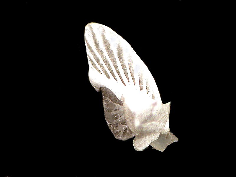
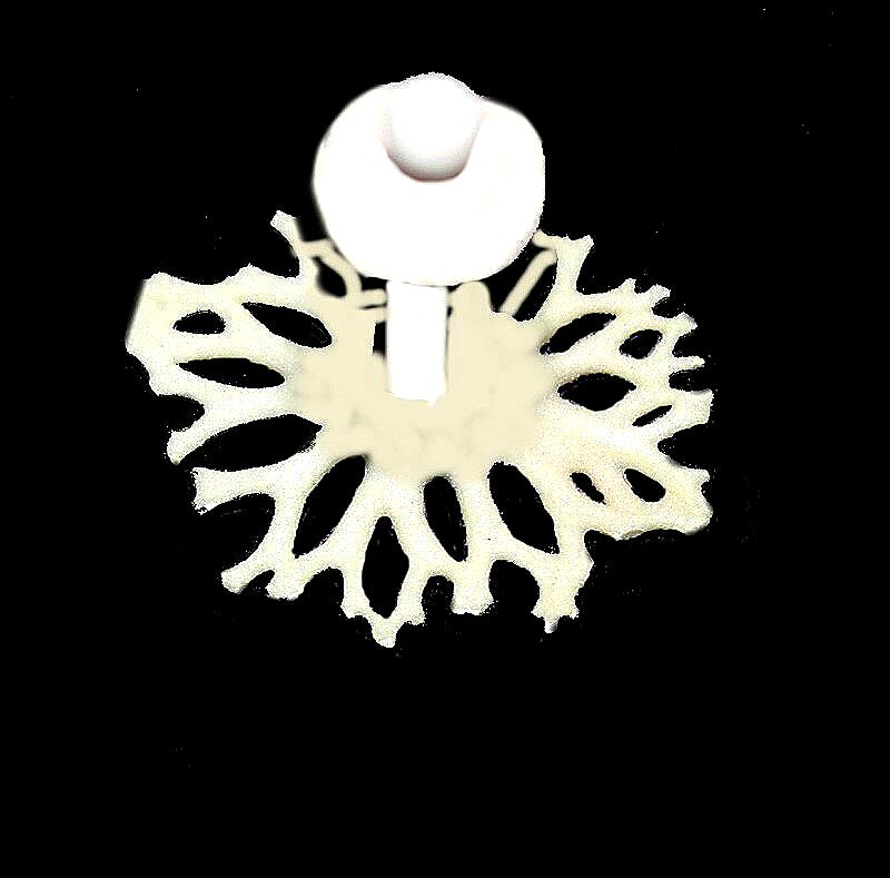
Even the parts of this structure are fascinating. Here is one which looks rather like some bizarre calcareous “wing” or perhaps an ultramodern, super high-rise hotel in Dubai.
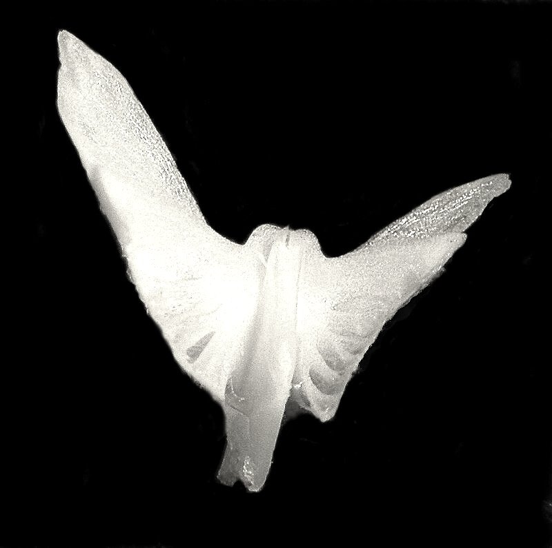
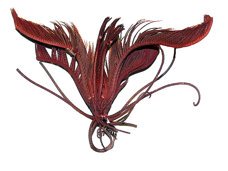
Below, you can see 2 of the “wings” fused in a larger structure which appears to be some extraordinary albino bird.
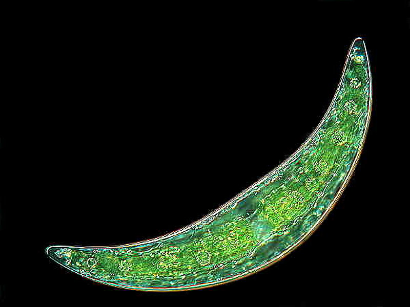
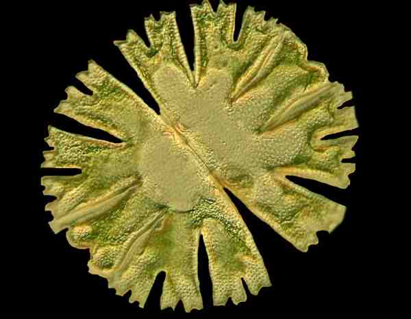
We’ll return to the echinoderms a bit later, but now I would like to turn to some truly microscopic organisms. For starters, two of my favorite desmids. These minute protists display remarkable diversity. I’ll show you Closterium which is crescent-shaped and Micrasterias which displays bilateral symmetry.
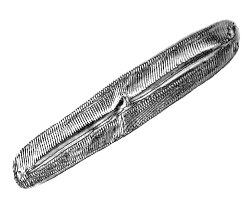
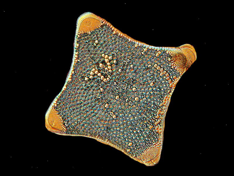
Some other great beauties are the diatoms, often described as “jewels of the sea”. However, it should be noted that they also occur in fresh and brackish water as well as soil. Traditionally, desmids and diatoms were regarded as algae and now recent taxonomists have decided to muddy up the waters, as it were, by calling them protists and creating new categories. Well, to that I say: Poppycock! Unnecessary obfuscation aimed at all of us amateurs. Anyway, there are two basic types of diatoms; the centric and the pennate. The pennate forms are bilaterally symmetrical and the centric diatoms are radially symmetrical. First, I’ll show 2 examples of pennate diatoms.
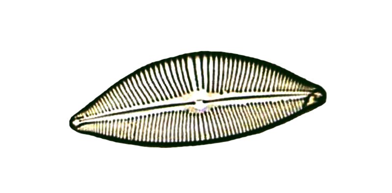
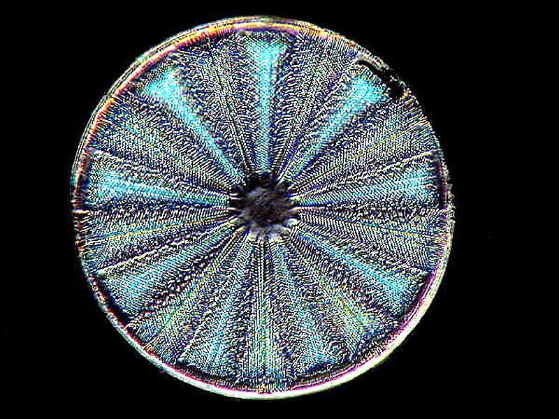
The centric diatoms, because of their radial symmetry are sometimes regarded as more beautiful, but that’s not quite fair. Diatoms are difficult to photograph, because they have so much fine structure. You can demonstrate this for yourself by going on Google and looking at SEM (Scanning Electron Microscope) images of diatoms. Further complicating things for the optical microscopist is the fact that the diatom frustule (shell) is largely composed of silica (glass) and creates special challenges to make the details accessible. However, using special contrast techniques such as Nomarski Differential Interference Contrast and adjusting the prisms so as to obtain what is called “optical staining”, one can get some rather spectacular results. I’ll show you another example of a pennate diatom, but this time using Nomarski and then an example of a centric diatom, also using that technique.
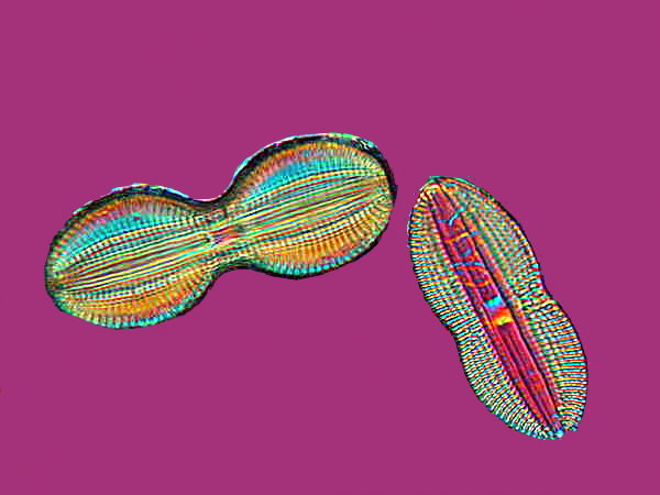
Of course, we should expect that Mother Nature will complicate things for us in our attempt to put things into order and so, I give you yet another diatom. Is it centric or pennate? Well, it isn’t centric, so I guess that settles it. Maybe.
Or perhaps a triangular one, just to keep things interesting.

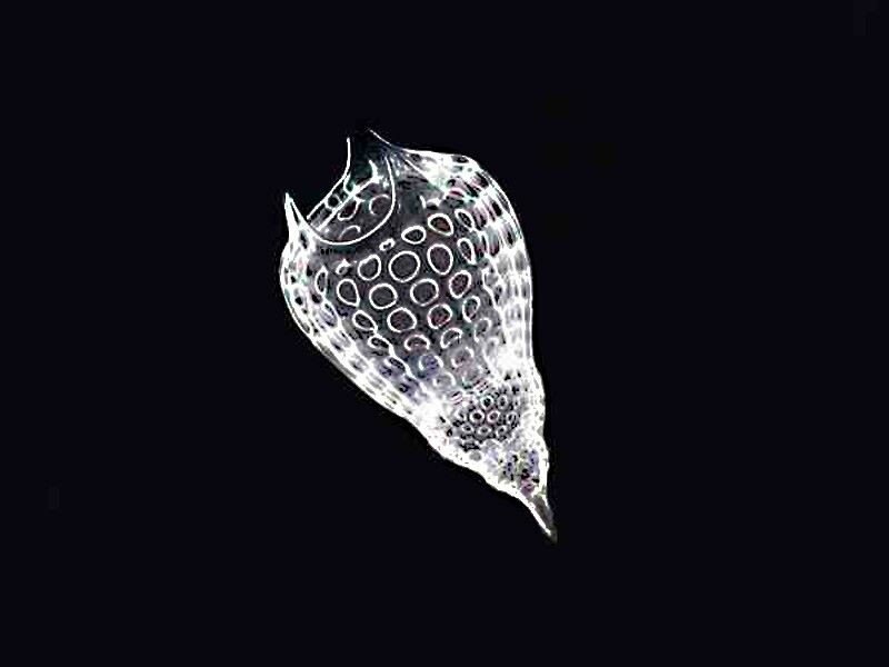
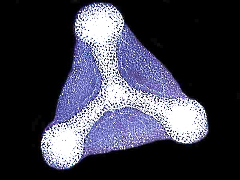
While we’re on things with glassy shells, there are some ameboid type organisms that are highly creative architects and create amazing “houses”. These are the radiolaria. I’ll give you 3 examples of their shells. The first 2 came from dredgings off Barbados and the third form is from the Pacific.
We have by no means exhausted calcareous things in the items for this first gallery, so let’s return to them. Let’s begin with a big item, Acanthaster planci, the infamous Crown-of-Thorns starfish which can get up to 12 to 14 inches in diameter from the center to the tips of the arms. This creature attacks coral reefs, digests the corals and regurgitates the indigestible calcareous bits. It’s possessed of a kind of toxin, so divers have to collect them with considerable care. There have been programs to remove them from designated areas, but they have to be removed by hand, lifted up into a boat above, and then either ground up or removed elsewhere for disposal. One can simply chop them into pieces and return them, because they will regenerate from the parts and one ends up with even more than one had in the beginning. Poison can’t be used, because that would kill off all the other reef flora and fauna as well.
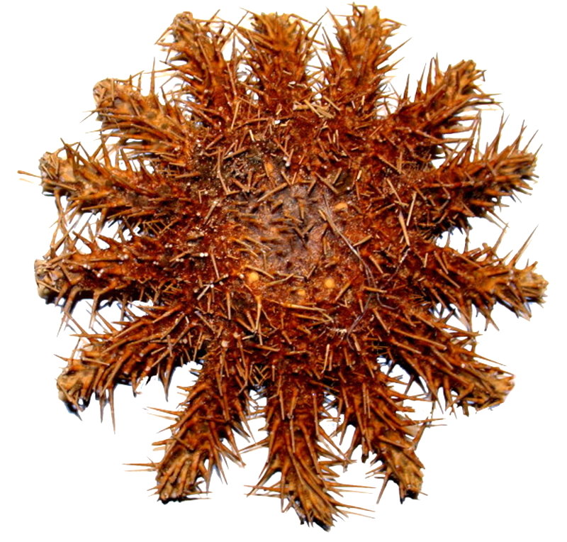
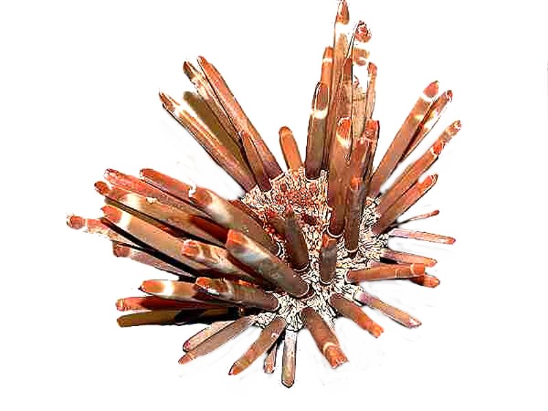
Another large echinoderm is the “slate pencil sea urchin”, Heterocentrotus mamillatus and probably apocrophally, in early days, the spines were used as chalk for writing on slate boards in classrooms. It is a tropical form found in the Indo-Pacific region and does have long, thick spines which have a sort of resemblance to brown sticks of chalk.
The spines of sea urchins are mostly composed of Calcium carbonate and consist of stacks and stacks of crystals. In the thick, pigmented spines, this cannot be easily observed. However, if one takes a relatively thin spine that is not heavily pigmented and places it in a solution of immersion or cedar wood oil, one can sometimes, with the help of the oil which has penetrated, see the columns or stack. Here is an example.
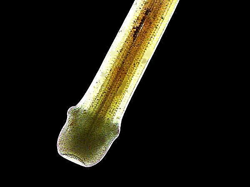
Spines in sea urchins come in all sorts of strange shapes and sizes and here I want to show you one of the most bizarre. It is a spine from a tropical species, Goniocidaris mikado, and I am showing you an upside down view. The ball and collar, which look rather like a tropical sun hat, form the ball portion of the ball and joint apparatus by means of which the spine is attached to the test (shell) of the urchin. This means that the frilly part at the bottom is actually projecting upward when attached.
The cross sections of sea urchin spines, when properly prepared can be some of the most beautiful specimens one can find. In this example, you can see concentric circles of crystals radiating out from the central core. This was photographed using Nomarski DIC.
Echinoderms are of such persistent fascination in large part, because they come in so many unexpected forms. Here is a specimen of a comatulid or “feather star”.
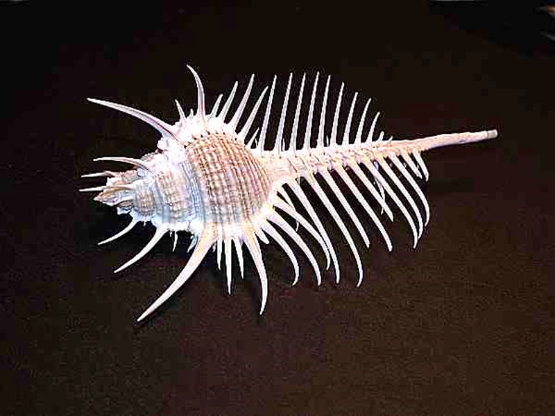
Mollusks are, of course, usually loaded with Calcium carbonate and I want here to consider a particularly lovely example. This is a Murex shell, commonly known as a “Venus Comb”.
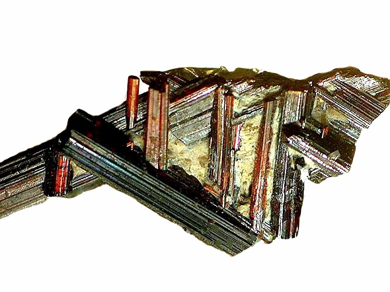
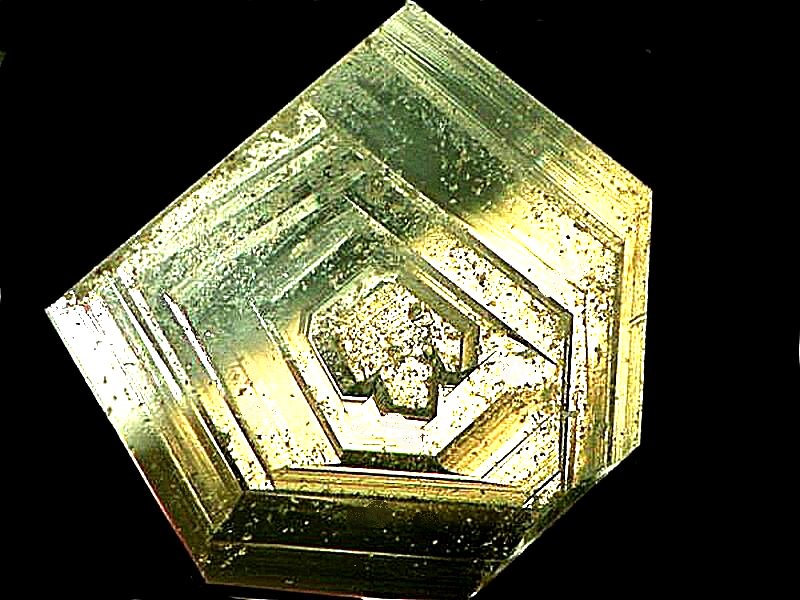
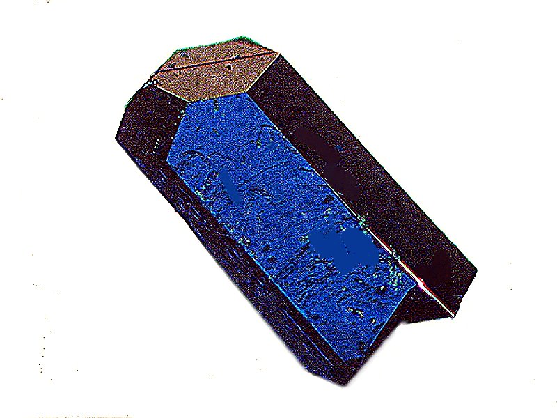
Well, as usual, I have rambled on too long, so I’ll finish this first gallery with non-biological images, namely, crystals-- with 3
examples.
The first consists of crystals of rutile in a matrix.
The second is a small chip of a mineral which I found in a small box of samples and which remains unidentified, but it has an intriguing structure and appearance.
The final example is a chemical crystal which formed on a slide and which I isolated both because of its form and its beautiful rich color. It is from a solution of Copper acetate.
I hope you enjoyed wandering through this gallery with me and I also hope that I find time to follow up with additional galleries. If you liked this one and would like to see additional ones, I would appreciate hearing from you.
All comments to the author Richard Howey
are
welcomed.
If email software is not linked to a browser, right click above link and use the copy email address feature to manually transfer.
Editor's note: Visit Richard Howey's new website at http://rhowey.googlepages.com/home where he plans to share aspects of his wide interests.
Microscopy UK Front
Page
Micscape
Magazine
Article
Library
Published in the March 2022 edition of Micscape Magazine.
Please report any Web problems or offer general comments to the Micscape Editor .
Micscape is the on-line monthly magazine of the Microscopy UK website at Microscopy-UK .
©
Onview.net Ltd, Microscopy-UK, and all contributors 1995
onwards. All rights reserved.
Main site is at
www.microscopy-uk.org.uk .