Not too long ago I bought an Intel PC Camera; and naturally, photomicrography was one of the first things I tried. My first attempts at hand-holding it to the eyepiece of my Bausch & Lomb monocular microscope were promising enough to encourage me to refine the technique.
Note: See the Image Notes below for size information and details. The video files are large (i.e. slow download). Click on the thumb-nail picture for the full image.
Steady Camera Support
The major problem all photomicrography has to overcome is supporting the camera. Almost any movement produces a blurred image, or misses the full image area. While hand-holding the Intel Camera gave me a few test images, I had to have a better solution.
 My next
test was crude but effective. The microscope was sitting on a
kitchen counter with overhead cabinets. I opened a door,
clamped a board to the door and clamped the camera to the
board. By adjusting the clamps, and swinging the door; I
could nicely align the camera over the eyepiece. Given the
limitations of the camera (and the door), this arrangement
gave excellent images.
My next
test was crude but effective. The microscope was sitting on a
kitchen counter with overhead cabinets. I opened a door,
clamped a board to the door and clamped the camera to the
board. By adjusting the clamps, and swinging the door; I
could nicely align the camera over the eyepiece. Given the
limitations of the camera (and the door), this arrangement
gave excellent images.
For a more permanent solution, I took some scrap PVC pipe, and mounted an upside down "J" of it to a board to provide a support that hangs over the microscope. Intel makes a 'laptop adapter' for clamping the camera to laptop screens. I purchased one of these adapters, and use it to attach the camera to the PVC. It works great. The only thing I might do different would be to use a larger diameter base pipe (2.25 inch?). The 5/8 inch pipe has some flex, so the camera tends to vibrate a bit if the set-up is touched. Then again, the board and door are so convenient I tend to ignore the stand.
Limitations
Image Size
The Intel Camera gives a true VGA image (640x480). While not as good as a 35mm photograph, or the mega-pixel digital cameras; I find the image quite acceptable (especially for the price). In fact, I borrowed and tested a Sony Mavica digital camera, and while the total image is bigger, the viewable image (due to vignetting) does not seem much different.
Capture Delay
 The one problem with the Intel
Camera (and all digital cameras for that matter) is the image
capture delay. From the time you press the shutter (or click
the mouse), the camera has to charge up the CCD and transfer
the results to memory. This means several seconds may pass
between when you want to take the picture, and when the
picture is actually taken. This may not be a problem with
prepared slides, but my enjoyment is live organisms like
daphnia, which rarely hold still. I list this as a
limitation, but it can easily be overcome (by using the video
mode).
The one problem with the Intel
Camera (and all digital cameras for that matter) is the image
capture delay. From the time you press the shutter (or click
the mouse), the camera has to charge up the CCD and transfer
the results to memory. This means several seconds may pass
between when you want to take the picture, and when the
picture is actually taken. This may not be a problem with
prepared slides, but my enjoyment is live organisms like
daphnia, which rarely hold still. I list this as a
limitation, but it can easily be overcome (by using the video
mode).
Features
USB Cable
Having the camera attached to your PC might be considered a limitation; but for photomicrography, this is an advantage. For general purposes, it is easier to view images on-screen than it is to look through the microscope eye-piece. For taking photographs, it is much easier to see the image on-screen than having to watch a small image on the camera back.
Video mode
 The Intel
Camera can be set up to take either snap-shots, or video.
While the standard video mode captures a smaller image than
the snap-shot; there is an option to take 640x480 video.
Though the high resolution video will quickly eat up memory
and disk space (a 200 frame video will be 2-3meg), it has
great advantages, and has become my standard technique.
The Intel
Camera can be set up to take either snap-shots, or video.
While the standard video mode captures a smaller image than
the snap-shot; there is an option to take 640x480 video.
Though the high resolution video will quickly eat up memory
and disk space (a 200 frame video will be 2-3meg), it has
great advantages, and has become my standard technique.
No matter what technique you use, you will always take more bad images than good ones. You always have to edit your pictures to find the good ones. However, the more pictures you take, the more chances you have of getting a good image. In addition to capturing action (heart beats, digestion, etc.), video mode is also a way to get rapid-fire still images. Rather than waiting for the right image to appear, then trying to capture it; I will run a stream of video, then go back and pull individual frames (which is a feature of the Intel software). Several times I have had a daphnia move, unexpectedly giving me a clearer exposure of an 'abdominal process' (used for taxonomy identification).
 Video can
also expose details that you don't see in a snap-shot. A 400x
image of a vorticella doesn't show much detail (like cilia),
but a video lets you see the water currents generated by the
waving cilia.
Video can
also expose details that you don't see in a snap-shot. A 400x
image of a vorticella doesn't show much detail (like cilia),
but a video lets you see the water currents generated by the
waving cilia.
Another nice trick, with specimens that are thicker than your depth of field; is to start a video, then focus through the full depth range of the object. Now go back and look at the sequence, image by image. This method will often show up details in individual images that you might miss otherwise.
Image Size and Editing
 If you are
interested in video images (not just using the video to
capture individual frames), you may not want the high
resolution due to the space it requires. Video editing
software (such as VidEdit) can give you the best of both
worlds; capture the video images at 640x480 frame size and
pull off selected frames, then resize the video to a smaller
format.
If you are
interested in video images (not just using the video to
capture individual frames), you may not want the high
resolution due to the space it requires. Video editing
software (such as VidEdit) can give you the best of both
worlds; capture the video images at 640x480 frame size and
pull off selected frames, then resize the video to a smaller
format.
Since I store the images as standard jpg files, I can manipulate them with any standard image editing software. There are a lot of possibilities here to adjust color, contrast or even 'sharpening' the images. This is a topic in its own right, both technically and ethically. (i.e. does altering an image change its scientific value?) [The images attached to this article have not been manipulated, though possibly cropped or shrunk to cut down on the size].
Auto light adjustment
A nice feature of the PC Camera is the automatic light adjustment; given a few seconds it will adjust for light intensity and give a good color balance. Unlike the expensive digital cameras, this cannot always be manually controlled. There may be some limitations with extreme contrast lighting, but so far the camera has kept up with my abilities.
Conclusion
 I want to be
objective about the Intel PC Camera, and present it's
limitations as well as it's features; but on the subjective
side I think it would be difficult to get much better. With
no specialized equipment, and at very low cost (other than
the PC), I can consistently obtain good images.
I want to be
objective about the Intel PC Camera, and present it's
limitations as well as it's features; but on the subjective
side I think it would be difficult to get much better. With
no specialized equipment, and at very low cost (other than
the PC), I can consistently obtain good images.
While 35mm photography would give better image quality, the cost of film and development make me hesitant to take a lot of pictures. With a digital camera, especially in video mode, I don't hesitate to click. I will take 200 images in a session and throw away 198 without concern. The net result is more good images.
For most of us, the goal of photomicrography is our own personal satisfaction. If I like the results, that is sufficient. By that standard, the Intel PC Camera is excellent value, I like what I get. Having said that, I don't want to minimize what can be done for so little effort. My excuse for getting the microscope was for teaching science to my children. The PC Camera makes it much easier for them to see, and we can share a common view. It is much easier to answer "what's that?" by pointing to the screen (or a captured image) rather than trying to understand a child's description of something only they saw. My patience for set-up and finding specimens exceeds that of my children, and can quickly bore them into disliking microscopes. With the camera, I can work by myself, and save the prime experiences to share with them when they are interested.
 However,
these are not just child quality images. The resolution and
quality is good enough that I have e-mailed some of them to
micro-invertebrate professionals when I needed help with
identification.
However,
these are not just child quality images. The resolution and
quality is good enough that I have e-mailed some of them to
micro-invertebrate professionals when I needed help with
identification.
If you already own a PC and a good microscope, and want to get into photomicrography; this is a great way to get started.
Image Notes:
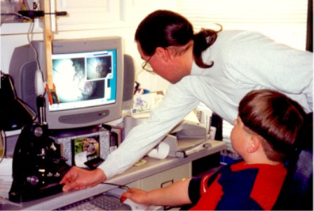
- Gregory and I in the 'lab': 35mm scanned photograph, 39kB jpg - other than using the Intel clamp for the camera, this is my typical set-up.
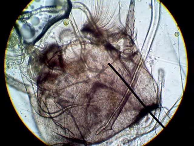
- Mosquitoe larvae: 400x, 145kB jpg, full 640-480 image.
- Vorticella: 400x, 1,004kB avi video, cropped - note the debris spinning in the cilia water current.
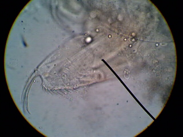
- Ceriodaphnia sp? abdominal process: 400x, 73KB, full 640x480 image.
- Ceriodaphnia sp?: 400x, 353kB avi video, reduced to 320x240 - pan through focus.
- Ceriodaphnia sp?: 100x, 27kB jpg, cropped 640x480.
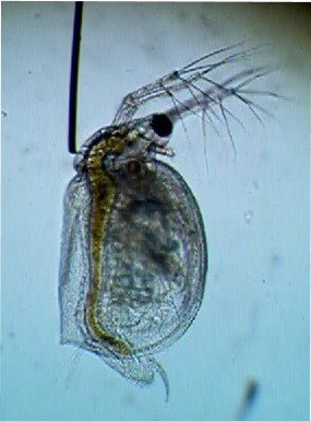
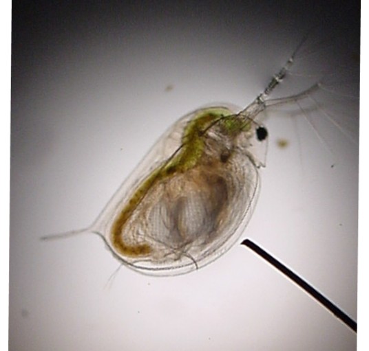
- Daphnia magna: 40x, 40kB jpg, cropped 640x480.
Notes:
- The Intel PC Camera usually costs about $69 (US). Requires a Pentium 166MHz or faster running Windows 95 or 98, and a USB port. While this is a great price for photomicrography, it should be noted that it can be used for other purposes. Intel's web site also has good technical information on digital cameras.
- The laptop adapter cost $10 (US) plus shipping (on-line, directly from Intel).
- My microscope is a Bausch & Lomb monocular, with standard Nikon optics (10x eyepiece, 4x, 10x and 40x objectives). It was originally owned by the Kansas State University, Veterinary Pathology Lab; I got it used for $100 (US). The black line in my pictures is due to the pointer eyepiece it came with.
- VidEdit is a Microsoft freeware product, a quick web-search should locate a download site.
Daphnia Web Sites:
Contact: Howard Webb