|
THE RED MANGROVE-TREE CRAB USING THE COMPUTER SCANNER AND THE INTELPLAY QX3 MICROSCOPE (As recommended by Micscape articles) by Hugh Mitchell-Tapping, Florida, USA |
Having read intriguing Micscape articles concerning the application of the scanner for recording small biota, such as leaves, flowers, bugs, etc., I decided to try the technique for myself using a red crab obtained from the mangroves on an island in Estero Bay in southwestern Florida. For microscopic examination of parts of the exoskeleton, I tried using the Intelplay QX3 microscope (now being sold in the US for less than $60). And here are the results.
The crab Aratus pisonii is a common mangal browser throughout south Florida and the Caribbean as reported in the literature. However, in January, February, and March 2001 many Estero Bay mangroves were searched without finding a single species. Normally, it can be found clinging to bushes, rocks, pilings, wharves and mangroves. In Estero Bay and other areas of Southwest Florida, the creature is a conspicuous resident of the red mangroves, but often confused with the abundant green crab of the same size that is found in the same environment. Mangrove-tree crabs tend to remain in the same general area all their lives. As reported in the literature, observations over a period of several months, 12 percent of the tagged individuals were recovered at or near their point of release.
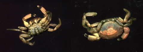
The common green crab found in the mangal tidal zone as recorded by a scanner:
Left lower view; right is top view. Actual size. Carapace diameter at widest point is 2.1cm.
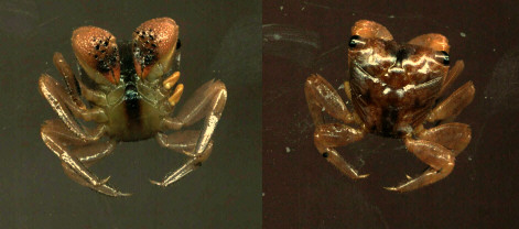
The scanner recordings of the red mangrove-tree crab found in the mangroves of Estero Bay: Left lower view; right is top view. Actual size. Carapace diameter at widest point is 2.1cm.Above water, predators of the mangrove-tree crab include birds, raccoons and the spotted mangrove crab. At low tide, the mangrove-tree crab explores the exposed mud bottom to feed on algae and small crustaceans. It also scavenges dead fishes and crustaceans, and consumes insects that it finds on the mud and in the trees. Apparently, however, one of its favorite foods is the leaf of the red mangrove. Laboratory-raised crabs have a preference for the leaf of the red mangrove over the black mangrove, even when they were originally caught on black mangroves.
The red mangrove-tree crab moves slowly with stealth when traveling over the mangrove branches and roots. The creature easily clings to even the smallest twig as it is equipped with legs that have needle-like tips.
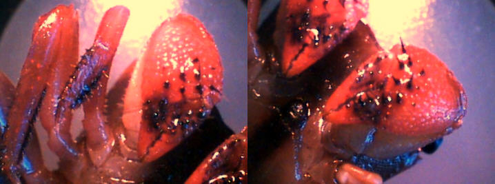
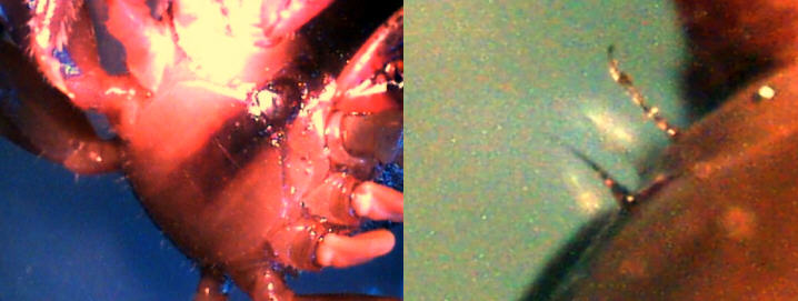
(left) The claws and a pincer of the red mangrove crab x10.
(right) The mouth area of the crab x10.
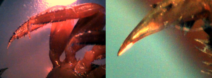
The claws of the red mangrove crab x10; x60.
(left) The underside view of the carapace x10. (right) The body hairs of the crab underside x60.
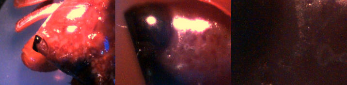
The eye of the red mangrove crab. x10; x60; x200.Due to its coloration, the crustacean is difficult to spot as its brown to mottled olive-green carapace color also helps conceal it. Most spend their time in the inner portions of the mangrove forest. During the day it hides by holding itself upside down on roots and branches. If the crab moves, it often gives itself away. When approached too quickly or closely by a human intruder or potential predator, the creature either scampers off or releases its grip and plunges to the waters below. But a watery escape route is not always perfect. Passing fishes and blue crabs sometimes snatch up this tasty handsome crustacean.
Females carrying eggs do, however, migrate to prop roots along the waters edge to release their young. Mating takes place throughout the year while the females shell is hard (many other crabs must wait for the female to shed her shell. mating then takes place while the females shell is still soft.). The male body and claws are much larger than the females. During mating the male holds the female upside down - belly-to-belly (abdomen) - with his claws and some of his legs. He clings to the mangroves roots or branches with his remaining legs. Within a day or two, the female attaches 3,000 to 27,000 (averaging about 11,000) fertilized eggs to her abdomen.
As the young (larvae) develop, the eggs pass through four color stages. The eggs swell and change from khaki to dark brown, light brown and finally gray. The eggs are ready to hatch in about 16 days. Hatching usually takes place at night, during a spring high tide (during a full or new moon). When ready, the female moves down the mangrove roots to the waters edge. With her body only partially submerged, she moves her abdomen back and forth as she dispatches the larvae into the water.
For about 30 days the larvae drift in the currents; they are part of a much larger population of ocean drifters known as zooplankton (animals). When first hatched, the crabs larvae somewhat resemble a mosquito. At that time, the larvae are known as zoeae. As they grow, they shed their exoskeleton (shell) about four times. The fifth and final larva resembles a crab. Called megalopa, the last larvae continue to drift along the coast until they metamorphose into minute crabs. Less than one percent of the larvae survive to become crabs. The highest predation pressure on the crab is while in the planktonic larval phase.
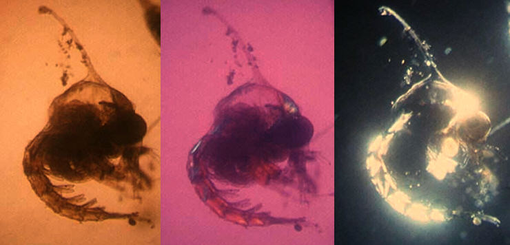
Estero Bay planktonic crab larvae x160; (a) under plain light, (b) crossed-polars, (c) incident light. The crab larva was taken in a trawl in the bay and examined under a regular microscope.Comments to the author Hugh Mitchell-Tapping are welcomed.
Editor's note: The author has written some fascinating web articles on other aspects of the fauna and flora of the Florida area. These are available on the Quekett Microscopy Club website which has many excellent resources and articles for the microscopist, together with details of club membership; see the links below.
A special reef
A unique tropical island beach
Dolphin's teeth