|
Snow Crystal Photography
by E.M. Kinsman, Rochester, New
York
|
This article is titled Snow Crystals,
even though many people would like to call the crystals snowflakes. Scientifically
the term snowflake relates to large clusters of dendritic flakes that fall
in clumps. The single crystals are called snow crystals. Ice crystals are
formed from freezing liquid ice. During this project I photographed both
snow crystals and snowflakes.
I would also like to state that this
short article is an introduction to an amazing topic that can easily absorb
a whole life of study. The many aspects of the "simple ice crystal" are
nothing but complex, intriguing, and fascinating. I encourage serious students
to look up the two references below as a starting point. It would take
several text books to cover all the aspects of just ice, not to mention
the complex nature of liquid water.
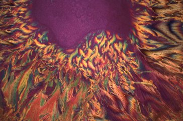
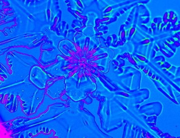
Polarized light shows different
ice crystals frozen from liquid water - I should mention this shot was
rejected from a prestigious weather photography competition as being "too
scientific". An honor I consider better than the winning prize!
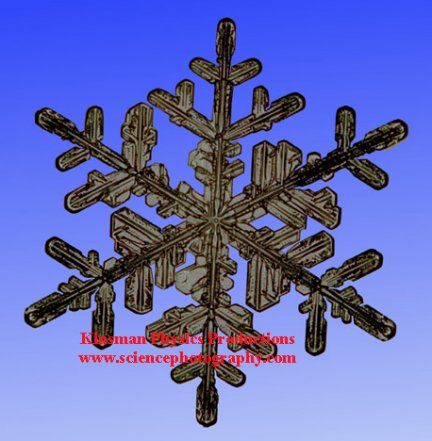
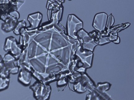
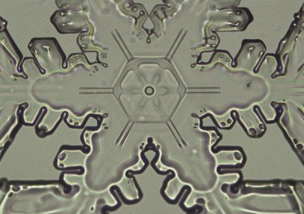
A snow crystal that has been
outlined in Photoshop®.
Dendritic form.
For the past 8 years I have been
experimenting with ways to document snowflakes. I have tried to preserve
them in optical ultraviolet cement, built a laser system to record the
diffraction patterns, and more recently used video techniques to make a
survey of the crystals.
It became obvious from the experiments
performed during the 1999-2000 season that to correctly record snowflakes,
a photographic system needed to be built that had the following parameters.
-
Ability to record images at highest
resolution possible (35 mm film)
-
Have auto exposure capability
-
Have a field of view from 1 mm to 6
mm
-
Quickly adjustable.
-
Have the ability to be both dark-field
and bright-field.
I chose to use the classic Olympus OM2N
camera on a B&L microscope body.
Although ice does rotate the polarization
field as many crystals do, snow crystals are too thin to use this property
of light to show the differences in the crystals. Polarized light only
has the effect of changing the color of the background. For this reason
I have designed the photographic apparatus to only use bright-field and
dark-field illumination, not polarization.
In many storms the precipitation
is often just about all identical and many times not worth photographing.
My goal on this project is to collect high resolution images that are pretty,
although I could not resist collecting some images of scientific value.
I found extreme close-ups using a 10X objective (62x at the film plane)
to be my favorite. If I wanted the whole snowflake I used a 5X objective
that gave about 8x on the film plane. Observant readers will realize that
the last two values are not in ratio to each other - this is due to the
microscope having an adjustable tube length. A highly useful characteristic
of this particular microscope.
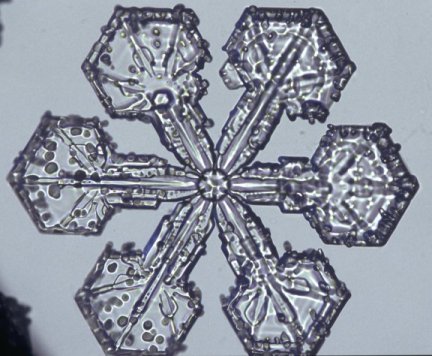
Large dendritic crystal covered
with rime. White light illumination.
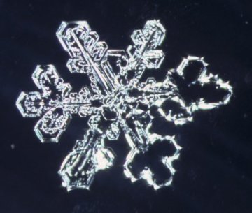
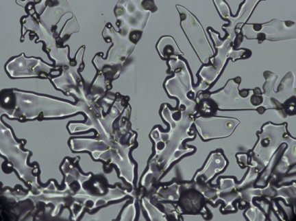
Dark-field illuminated dendritic
crystal.
Here in Rochester, NY we are just
a few miles from one of the great lakes (Lake Ontario) that greatly affects
our weather. One of the common forms of snow happens when the warmer lake's
air mixes with a cooler air mass. The resulting snow is called Lake Effect
Snow, it is characterized by rapidly growing crystals that are basically
formless spheres and elongated masses. The pretty dendritic forms are created
in airborne temperatures of approximately 13°C. Once the crystals can
form they fall through warmer air and build up ice under different crystal
orientations. The opposite is also true - where warmer ice can fall through
a cooler layer and an ice crystal starting in an ugly mass can start to
grow the perfect dendritic arms. Snowflakes can also fall through layers
of different relative humidity where they can both build up and lose mass.
Ice can also sublimate. Here in New York, I estimate that we get 15 good
storms when it is both cold enough to photograph and there is good snowflake
development.
Due to my proximity to the Great
Lakes, there are a number of snowflake types that are either very rare,
or just do not form in this location. These include the very cold forms,
and the top-hat shapes.
You can basically think about photographing
a snowflake as being identical to optically testing a small piece of glass
or micro-lens for defects.
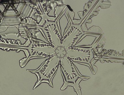
Dendritic, 10x objective.
Dendritic, 10x objective.
Image used for false color tests
below.
Rarely do snow crystals exist as
a flat object, flakes are very three-dimensional objects and due to their
thickness some of the snow crystals will be out of focus. Some forms of
dendritic flakes go in all directions and are impossible to get a picture
of, unless you hack off some of the protrusions.
I only photograph snow crystals when
the ground temperature is at or below 25°F. Warmer temperatures cause
the snowflakes to quickly melt around my body's microclimate of warmer
air and higher humidity. The illumination system of the microscope, even
though it has an infrared filter to block out heat energy, it radiates
enough heat to warm up the snowflake a few degrees and cause melting.
The process of snow crystals photography
is quite simple. I use a sheet of masonite to collect snow crystals. The
sheet of masonite is placed just outside the garage door, while the microscope
and camera are in the garage. The microscope is kept in the garage to keep
it cool, while the camera is kept in a plastic container and moved to the
garage when there is good flake development. When the photography is done,
the camera is placed into the plastic box and moved into the house. The
plastic box keeps water vapor from the warm house condensing on the camera.
The camera system can be used if the microscope is moved into a warmer
environment.
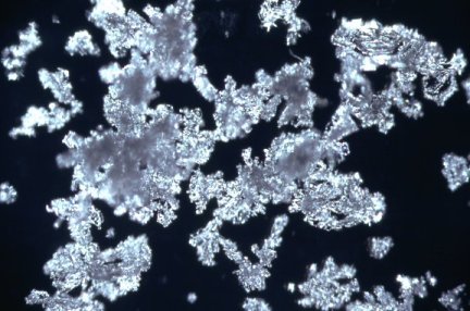
Good snow crystals are picked up
with a thin pin taped to the end of a pencil. The flakes are transferred
to a pre-cleaned microscope slide. I typically try to pick out the best
dendritic flakes that fall on the sheet of wood. If the flakes fall on
the microscope slides the chances of getting a nice flake are relatively
few. The majority of flakes that fall will collide with other flakes and
loose arms, or will grow unevenly. Since the snowflakes have an electrostatic
charge, they often clump together and form falling masses. Some times the
masses can form more interesting images than solitary flakes. Collecting
flakes in a snowstorm is often a challenge, any snow is accompanied by
huge collections of broken flakes, bits, and little particles or dust,
as well as micro crystals. All of these particles will end up on the slide.
Once a snowflake is photographed
it can be cleared off the microscope slide with an artist's paintbrush,
and the slide can be reused a few times. I like to photograph snow anytime
the flakes are good, but the cooler temperature required, mandate that
I photograph late at night. With good crystal development and a quick setup
you can take about 30 images an hour. Many times the rates are nowhere
near this number. For every 30 images taken I would estimate that you might
get 2 good images. You cannot really tell how good a flake is until you
get it under the microscope. Even broken flakes can have some interesting
parts on them, so I have a 10x objective that I swing into place to see
if I cannot get an interesting image out of a bad flake.
I have a lamp post in my front yard
that I can quickly look at night to see if it is snowing and what type
of snow is falling. Dendritic flat flakes reflect light so if you look
a few degrees away from the light and see little reflections then there
is good snow falling. Hard snowstorms have too many broken flakes and bits
to bother going out and photographing. The most desirable storms are when
it is just barely snowing and there is no clumping of the flakes yet. Many
times a storm can have windows of different types of flakes. Some of these
windows of good flakes will only last a few minutes, so I collect my ½
square meter of masonite wood with the good flakes and move it into the
garage (I also keep the garage door open to equalize the temperature),
then I can take my time to photograph the good flakes while a polycrystalline
mess falls just a few feet away. Of course the collected flakes will change
depending on the humidity conditions on the ground (grow larger, smaller,
condense).
If you observe a snowstorm and decide
that the flakes are not desirable, just wait 20 min and look again. The
snow forms can change quickly and you must be ready to act quickly. This
waiting and hoping for good snowflakes can lead to sleepless nights.
I am quite surprised at the lack
of snowflake photographers. After corresponding with numerous interested
photographers, several important variables to make this process work have
come to my attention.
The photographer must meet the following
criteria:
-
Live somewhere there is a lot of snow
-
Be very interested in microscopy
-
Have the required equipment
-
Have the required time
-
Have a covered place to work cold
-
Like watching the weather
The first point eliminates 50% of the
photographers, while the second point eliminates another 49.99% while the
last four points eliminate just about everyone else. I would be interested
to hear from anyone else actively involved in snowflake photography.
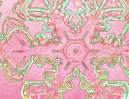
An image is converted to black
and white, then false colors are applied to highlight details.
Another false color image.
Future Experiments
The idea of thinking of snowflake
photography as a form of optical testing is of particular interest. There
are a number of both optical and digital techniques that can be employed
to document snowflakes.
I am also intrigued by the idea
of growing heavy water (deuterium oxide) crystals. Since D2O
freezes at 3°C in theory I should be able to grow frost underwater.
In this case thicker crystals can be photographed by using polarized light,
schlieren photography, or a modification of optical lens testing called
Ronchi Testing.
I am also interested in photographing
more snowflakes with a much smaller F# so as to yield a much greater depth
of focus.
Comments to the author Ted
Kinsman are welcomed.
For additional snowflake / snow crystal
images check out the author's web site, Kinsman Physics Productions: www.sciencephotography.com
References
Field Guide to Snow Crystals
Edward R. LaChape
University of Washington Press 1969
This is a nice little text showing
the changes in snow crystals due to time, humidity, and evaporation. The
results are applied to the field of predicting unstable snow fields or
avalanche prediction.
Snow Crystals: Natural and Artificial
Ukichiro Nakaya
Harvard University Press 1954
This is the definitive textbook
on snow crystals. Nakaya reproduced just about every natural form in his
cold laboratory. A wonderful researched book by a brilliant scientist.
This is a must read for any serious snow crystal researcher.
Microscopy
UK Front Page
Micscape
Magazine
Article
Library