 |
| The
Binocular Head
A few thoughts about binocular vision and user adjustments in compound microscopes regarding observing pleasure as well as reducing eye fatigue By Paul James (uk) |
Binocular microscopes have been in use for approximately 140 years now, and though the principles involved have not changed a great deal, design and technical advances have improved this most desirable observing facility.The binocular compound microscope divides a single image from the objective into two equally intense portions which our minds fuse into one, which is devoid of any impression of stereoscopy. It is true that some spatial perception can be sensed, but this is small and varies between binocular designs, and may even be psychological in origin?
|
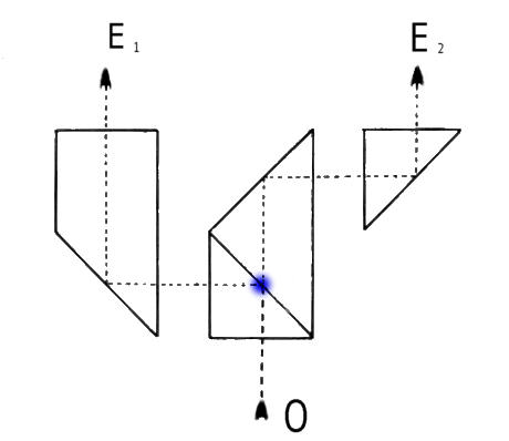
The principle of this image division is shown above, although prismatic designs vary between makers. Notice the light from the objective at O meets a semi-transparent metallic film which allows 50% through to the eyepiece at E2, and another 50% to be reflected to reach the eyepiece at E1. The essential element of design allows 2 identical images with effectively identical path lengths to emerge toward the eyepieces. Note that the vast majority of binocular head designs in modern times, provide parallel * emergence of light paths, which encourages a state of infinity focus in our eyes, and a more restful disposition for prolonged observation.* Early English binocular compound stands tended to have converging eyetubes.
Inter-ocular separation and dioptric corrections
Since the distance between our eyes varies from one individual to another, some form of provision has to be made for variations in this interpupillary distance, as well as dioptric differences between eyes in individuals too. The former is not easily accomplished, and again maker's have various ways of achieving this. I'm not concerned here about the individual merits of the different systems of adjustment, but only the actual methodology and subjective assessment of these adjustments.
There is no doubt in my mind that accurate adjustment of both is absolutely essential if the best imagery is to be enjoyed by the observer. Some observers may make adjustments rather quickly without a great deal of thought about what might be going on, but to provide the most restful imagery for our eyes and to reduce muscular fatigue to the minimum, a little more care is necessary. I will relate a simple method of achieving this collimation of binocular/eyepiece to our eyes which I use, which works extremely well for me.
A Method of attaining Interpupillary Distance Adjustment (IDA)
Place a simple self contained specimen, preferably of small size in the field centre with a x 10 objective in brightfield, and with the substage iris reduced to its lowest limit.
Before proceeding, remember that your eyes must be concentrating on the specimen, and only your peripheral vision used to assess the following :-
Now to check whether the interpupillary distance setting is accurate for your eyes, simply withdraw your head from the eyepieces by about 20-30 mm (varies with the eyepiece type). You will see two field circles of light overlapping each other to appear as one, or at least you should do if your IDA is correct.
If your IDA is not quite accurately related to your eyes, then you will see something similar to the 'split field circles' shown below at A:-
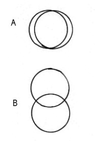 |
When you have either closed or opened the eyetubes correctly, the two circles will merge perfectly. Another test is to rotate your head slightly left or right, and this will separate the two circle fields as shown in B above depending on the degree of rotation. If your IDA is accurate then these two circles will be vertically in line or plumb.This method is not foolproof, and its ability to help adjust the IDA accurately, depends on the observer's ability to concentrate on the subject in the field, and keep their eyes equidistant from each eyepiece.* However with some practice this adjustment can be accomplished more quickly than writing this sentence!
* It may help to use two cardboard tubes cut to length to keep the eyes at the same distance if you find this difficult?
Dioptric adjustment
When you are satisfied with the IDA, you can adjust the focussing of the adjustable eyepiece tube to bring about identical focus for each eye. Possible the best method is to focus the instrument on a tiny dust speck using the fixed focus eyepiece first, then follow by adjusting the setting of the variable eyetube last. Making sure that the eyes are relaxed is the difficulty, and it is probably better that we use a piece of card to mask each eye as required.
Other concerns regarding collimation
Though the binocular head should be collimated, and the vast majority will be, it is entirely possible that it is slightly out of alignment, or that one or both eyepieces may have their optical axis slightly askew to their bodies.
Eyepieces
To check these, look at a slide of strewn radiolarians or diatoms etc., with a x10 objective after adjusting the IDA. Observe the detail of the specimens around the periphery of the field, with each eye in succession. If there is any discrepancy in collimation, you will find subtle variations of positioning of the subjects in relation to the field boundary, between the two images.
Reduce this by rotating one eyepiece a few degrees, then compare the two images as before. Repeat this until you reduce any discrepancy to the minimum. This is well worth doing thoroughly, and when you are satisfied that the images are as near identical as it is possible to get, mark each eyepiece L and R very clearly, so their correct placement orientation will be preserved permanently.
Binocular collimation
If the above method of revolving eyepieces does not eradicate these discrepancies, then it might suggest that the binocular head prismatic assembly itself is misaligned. More likely is that the eyepiece tube(s) have been moved away from their original seating, since most manufacturers embed their prisms in cages which are virtually immovable.
I cannot recommend the casual alteration of eyepiece tube positioning etc., but advise the owner to consult a technician if this needs to be done.
Conclusion
The involvement of both eyes in microscopy is a real joy, and especially if using quality optics which are correctly collimated. Apart from the obvious pleasure we experience whilst observing, the importance of the working conditions to which our eye's are subjected to cannot be over emphasised. Therefore if anyone finds observation through their binocular microscope less easy than viewing a mid distance scene, they should pay some attention to their instrument's optical integrity. You can buy a different microscope, but not your sight.
|
|
Please report any Web problems
or offer general comments to the Micscape
Editor,
via the contact on current
Micscape Index.
Micscape is the on-line monthly
magazine of the Microscopy UK web
site at Microscopy-UK
WIDTH=1