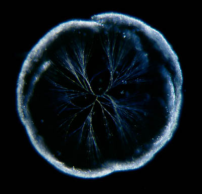
| The
trouble with this organism is that the colony is easily mistaken for a
piece of decaying matter. But if you examine it closer it appears to be
delicate flat structures made by tiny flagellated protists. The branches
are flat sheets formed by thin tubes of mucus. The BI-flagellated builders
of the structures are sometimes visible, their flagella protruding from
the tubes.
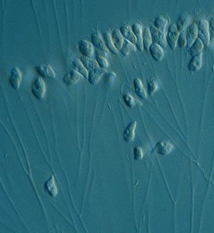
I must
admit that I always get confused by all these little flagellated protists.
Many unrelated groups look very similar and you need much more patience
than I have to figure out what is what. But sometimes the structures they
form make it a bit easier to identify them.
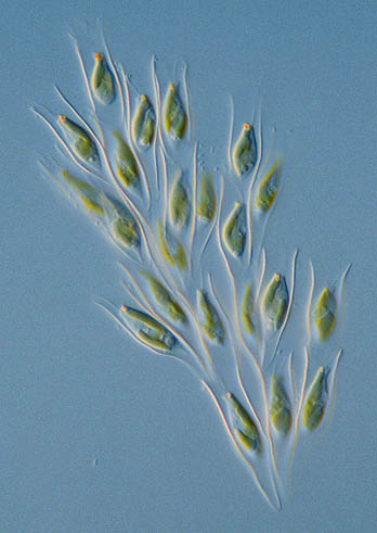
Note:
I call the organisms in this article 'protists' instead of 'protozoa'.
'Protozoa' is an informal name used for many unrelated unicellular organisms
that don't use photosynthesis. They are heterotrophic (get their food from
organic substances produced by other organisms) Algae is used for the more
plant-like unicellular organisms. These are phototrophic (they use light
as source of energy). So in this case you could call Pseudodendromonas
and
Rhipidodendron protozoa. Or is Rhipidodendronphototrophic?
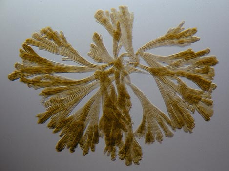
Now I am confused. I give up these tiny problematic organisms. Next time
I'll write about something big, like aquatic insect larvae!
As
long as they don't bite! |