 |
 |
| Butterfly
and Moth Scales
Some simple notes for beginners By Paul James (UK) |
A
Word of Advice :- Please do not capture and kill a butterfly or moth just
to observe its wings etc. Dead examples can often be found. When you do
find one try to handle it carefully so its structures are preserved, and
keep it in a container, away from sunlight and dust for it will keep for
a very long time.
|
|
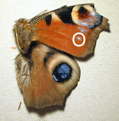
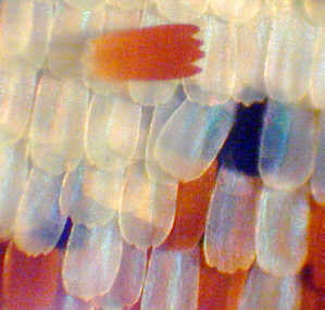
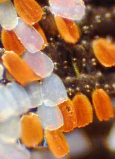
Only two main wings remain on the right side, and of course all the colourful patterns of this pretty creature are nearly all preserved. I have put a white 'O' over the part of the wing that I have photographed through the microscope using a hand held digicam. The microscope is a simple one that can be bought secondhand for a few pounds.
Demonstrating that we can get
results using this simple and easy technique will hopefully encourage the
beginner to combine photography with microscopy.
 |
 |
 |
 |
 |
 |
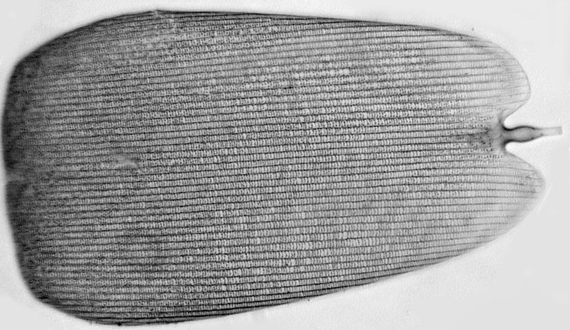 |
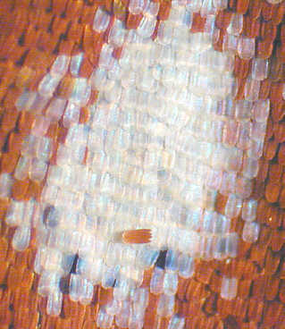
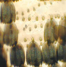
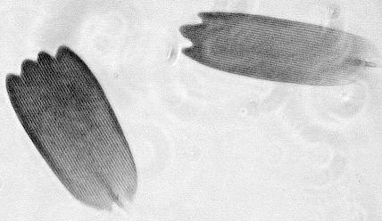
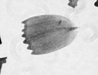
This image has been greyscaled to reduce download time. It shows the intricate detail of the butterfly's scales which are very thin, yet immensely stiff for its weight. It is a non lobed example from a prepared slide of the Red Admiral butterfly. The stalk is clearly shown. The markings are present on most scales and some actually split up the light that reflects off them into different colours, causing the butterfly to appear even more colourful.
Finally, after observing these rather delicate structures, some questions might cross your mind....."Why are these scales necessary on the wing's surface?"......"Why are they coloured?"......... and perhaps after seeing them for yourself you might wonder how all this could possibly be created inside the pupa (chrysalis), in a matter of a few days or weeks.
Good questions, and even if you do not know
the answers, it does not prevent you from appreciating one of nature's
most beautiful creatures. It is far more important however that we protect
their environment, and if you are interested in butterfly and moth conservation
write to 'Butterfly Conservation' PO Box 222, Dedham,
Colchester, Essex CO7 6EY.
| All comments to the author Paul James are welcome |
Please report any Web problems or offer general comments to the Micscape Editor.
Micscape is the on-line monthly
magazine of the Microscopy UK web
site at http://www.microscopy-uk.org.uk