|
Observations on a Common Cold
by Joe Schuller (Frederick, Maryland, USA)
|
Recently I made the unexpected discovery of ciliates living in my nose! I was three days into a typical end-of-winter cold, and feeling miserable. My attempts to examine some pond water under the microscope were being frustrated by a profusely running nose. In a sudden flash of inspiration, I allowed a drop of the mucus to fall on a clean slide, and gently placed a coverslip over the drop.
At 40X the mucus appeared full of particulate matter. At increasing magnification it became apparent that much of the particulate matter was actually cells of a variety of shapes and sizes. Some were large, block-shaped cells with a distinct nucleus; others were smaller and round or amorphous. I also observed red blood cells, although my mucus was not obviously bloody.
While scanning this cellular mess at 400X I detected movement. Centering the field on the movement and switching to 1000X, I observed an egg-shaped cell, with a thick tuft of waving cilia projecting from one end. My initial thought was that I had contaminated my slide with a protozoan from the pond water I had been observing. However, the cilia of this cell appeared longer, and moved more slowly, than those I have seen on freshwater protozoa. With an abundant supply of sample material, I repeated my observations on several more slides. Although I spotted only a few more live ciliated cells in my samples, I did observe a number of apparently dead ciliated cells mixed in with other cell types and cellular debris.
I photographed some of the ciliated cells using a Fuji FinePix S3000 digital camera held by hand to the eyepiece of an Observer IV microscope. Because these preparations were not stained, the unprocessed images are unimpressive (see Figures A, C, and D). However, more detail is revealed after enhancing the contrast and brightness of the images using Photoshop (see Figures B, D, and F). The tint of these images is due to a blue filter that was in place when the photos were taken.
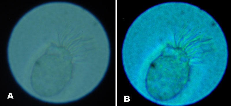
Figure A/B: An oblong ciliated epithelial cell at 1000X, (A) before and (B) after brightness/contrast enhancement.
In addition to the tuft of cilia projecting from one end of the cell, it is possible to make out some intracellular
or cell surface details after image enhancement.
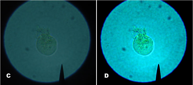
Figure C/D: A rounded ciliated epithelial cell at 400X, (C) before and (D) after image enhancement.
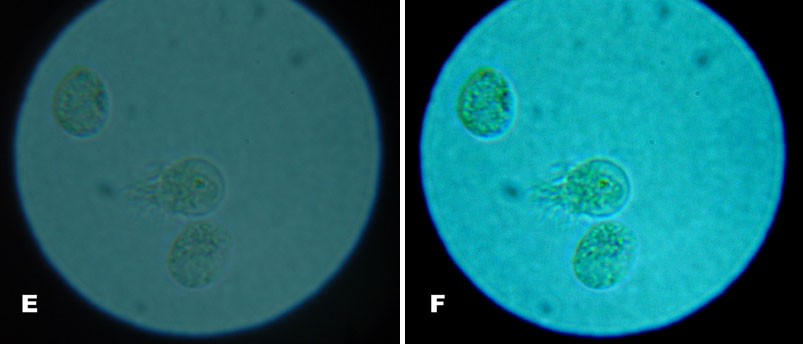
Figure E/F: A ciliated epithelial cell flanked by two cells that do not appear to have cilia,
(E) before and (F) after image enhancement. (400X)
Where did these cells come from? Was I infected with an exotic protozoan as the result of spending too much time peering at pond water? Turning to the internet for answers, I was soon reassured. I learned that the nasal lining includes goblet cells which produce mucus, and ciliated epithelial cells which move the mucus and any trapped particles toward the back of the throat. The ciliated epithelial cells are susceptible to infection by certain viruses, including those that cause colds. Impaired ciliary activity due to the infection can result in the accumulation of mucus in the nasal passages. Meanwhile, immune cells alerted to the viral intruder begin to invade the infected area. Some of the immune cells release inflammatory signals that cause swelling of the nasal passages. These events contribute to the stuffy, runny nose that accompanies a cold. Depending on the virus, the infected ciliated epithelial cells may be shed from the nasal lining and expelled in nasal secretions, along with red blood cells, white blood cells, and other immune system cells.
Alas, my cold symptoms improved too quickly to follow up on my initial observations. Future sick-day projects may include attempting to video record the ciliary movement of the secreted epithelial cells and using histological stains to try to identify other cell types present in mucus. At the next sign of a sniffle, take the opportunity to look for the fascinating ciliates living right under (or in) our noses. It might even take your mind off your cold.
Resources:
Chapters on the common cold and ciliated cells appear in Medical Microbiology and Molecular Biology of the Cell, both freely available through the "Books" database at the National Center for Biotechnology Information website (www.ncbi.nlm.nih.gov).
Long PH, Bliss EA, Carpenter HM. Studies Upon The Nasal Secretions. I. The Cellular Content Of The Nasal Secretions In Acute Disease Of The Respiratory Tract. J Clin Invest. 1933 Nov; 12(6): 1127-1134. This is a freely available journal article from PubMed Central (www.pubmedcentral.nih.gov) that describes the cellular content of nasal secretions sampled from subjects suffering from common colds. The authors describe "curious globular ciliated cells which resemble bursting grenades" and "[i]n many preparations the cilia were highly motile, in others non-motile." Leukocytes, macrophages, and red blood cells were also frequently observed in the samples.
Reed SE, Boyde A., Organ Cultures of Respiratory Epithelium Infected with Rhinovirus or Parainfluenza Virus Studied in a Scanning Electron Microscope. Infect Immun. 1972 Jul; 6(1): 68-76. Another freely available journal article from the PubMed Central website. This article contains spectacular scanning electron microscopy images of ciliated epithelial cells from normal and virus-infected epithelial tissue. Infected epithelium has numerous ciliated cells in the process of being "extruded" from the epithelial surface (see Figs. 2 and 3). These extruded cells bear a striking similarity to those I photographed for this article.
Understanding Colds, at www.commoncold.org.
Ciliated Epithelial Cells, at the University of Leicester.
Comments to the author,
Joe
Schuller, are welcomed.