|
The Marvels of Crustacea By Annette and Lewis Hein, Casper, Wyoming, USA |
Recently my parents bought a piece of land west of Casper, Wyoming. It and the surrounding properties have many ponds, streams, and springs. They are rich in protozoa, rotifers, crustacea, algae, and other invertebrates. The crustacea are particularly interesting to me so I have been focusing on them over the past few months.
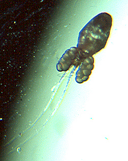
None, so far, is longer than a centimeter, except Gammarus shrimp, which are common in the streams and ponds. The youngest shrimp are about ˝ centimeter long, but they grow up to 3 centimeters. The digestive tract is visible as a black tube
I have tried to culture crustacea, with limited success and lots of failures. I intend to keep trying, though, and if any of you have suggestions, please e-mail them to me.
My only success with cladocera was a total accident. I had taken a pond sample which seemed to have nothing in it. I decided to throw it out, but it was in an out-of-the-way corner and I forgot it. Then, one day, I found it again. I took it off the windowsill...and suddenly saw that it was full of little moving creatures. As I stood staring at it I thought, This was the sample I could have certified empty.
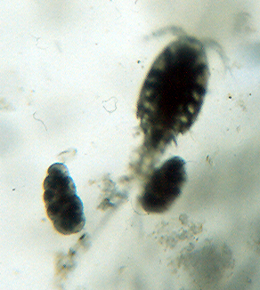
I think the explanation was that a few organisms had come into the sample and then multiplied. Over the next week I studied this culture. I found two kinds of Chydorus, a small round water flea. Also, I found some ostracods, about 1˝ millimeter long.
This happened in early September, and now, in November, the culture is still plentiful. It has adults and young ones of all kinds, including Cyclops.
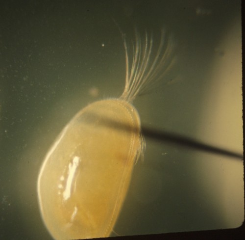
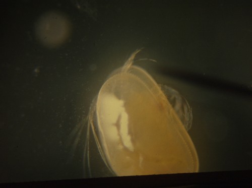
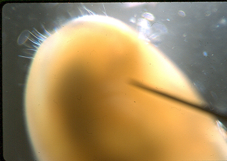
I have also observed one kind of Chydorus in detail. The adults are about 2 millimeters long. They have yellow legs, a short beak, and a striated shell. The shape depends on whether you view it from the top or the side.
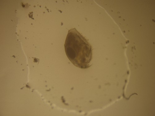
The adults are apparently all females. They often have eggs or up to six young ones in the brood pouch behind the body. When they have enough water they swim with the hinged part of the shell uppermost.
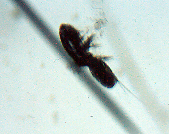
Another thing I have discovered is how Cyclops turn from a small brown egg into a full sized adult. So far I have only observed the nauplius, metanauplius, and some copepodid stages. The Cyclops hatches as a nauplius larva then, after about 4 days, turns into a metanauplius. In the copepodid stages it looks like a Cyclops but is not the size of an adult and has fewer legs. The nauplius larva move by means of what later (I think) becomes the first and second antennae. Since these larvae move so fast—and with the typical jerky motion of copepods—I have not been able to observe much more than that. I found the names of the stages in a Barnes biology textbook.
I have also worked with ostracods. They are easy to culture. I take away their natural enemies, the insect larvae with ant-like jaws, put in some silt and algae from wherever the ostracods came from, and leave them alone for about a month. They multiply quickly.
One nice thing about observing crustacea is that they are quite transparent (except ostracods) and so I can see what is going on inside of them. One of my favorites is the digestive system of Chydorus sphaericus. It has a looped gut, and now and then I will see a piece of the food break off and slide along an invisible spiral for a few twists.
The crustacea are a most interesting group, and play a major part in food chains. They also have many facets to study and after they are dead, their shells remain among the bottom sand to make a home for algae.
EQUIPMENT:
The camera is an old ca. 1980 Olympus OM2-n. I used it with a T20 flash, a T32 flash, a remote cord, slave, and cable release. The microscope we used is an old Wolfe Student microscope. It has a 10x eyepiece, and 4x, 10x, and 40x objectives. We do not know precisely how old it is, but we think our parents bought it around 1999.
BIO:
Annette Hein is 13 years old, and Lewis Hein is 11. They have been working together on microscopy for about 6 years.
Comments to the
authors
Annette
and Lewis Hein are welcomed.
Published in the May 2006 edition of Micscape.
Please report any Web problems or offer general comments to the Micscape Editor .
Micscape is the on-line monthly magazine of the Microscopy UK web site at Microscopy-UK
© Onview.net Ltd, Microscopy-UK, and all contributors 1995
onwards. All rights reserved.
Main site is
at www.microscopy-uk.org.uk
with full mirror
at www.microscopy-uk.net
.