|
|
A
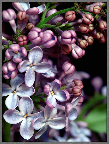
Close-up View of the Wildflower (Syringa vulgaris) |
|
|
A
Close-up View of the Wildflower (Syringa vulgaris) |
How slowly through the lilac-scented air
descends the tranquil moon.
Henry Wadsworth Longfellow
(The Spanish Student)
When lilacs last in the dooryard
bloom'd
And the great star early droop'd in
the western sky in the night,
I mourn'd, and yet shall mourn with
ever-returning spring.
Walt
Whitman
(Written at the death of Lincoln)
April is the cruelest month, breeding
Lilacs out of the dead land, mixing
Memory and desire, stirring
Dull roots with spring rain.
Spring has truly arrived when the first lilac flowers fill the air with
their unmistakable scent. This wonderful plant is usually found
as a multi-stemmed shrub with an irregular, rounded outline. Part
of its usefulness to the landscaper and gardener comes from the fact
that it is particularly hardy, being able to withstand severe
environmental conditions.
Originating in eastern Europe, lilacs were brought to North America by
pioneers in the early 1600s. From the beginning, their
appearance and scent proved extremely popular. So popular in
fact, that during the 1800s, plant scientists began developing hybrids
(cultivars) which improved upon the lowly common lilacs
characteristics. In some of these cultivars however, larger and
more colourful blooms were obtained at the expense of the strength of
the scent.
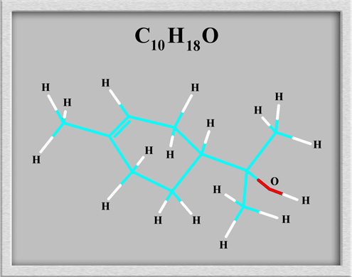
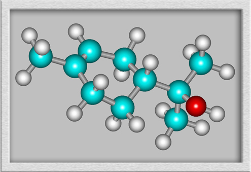
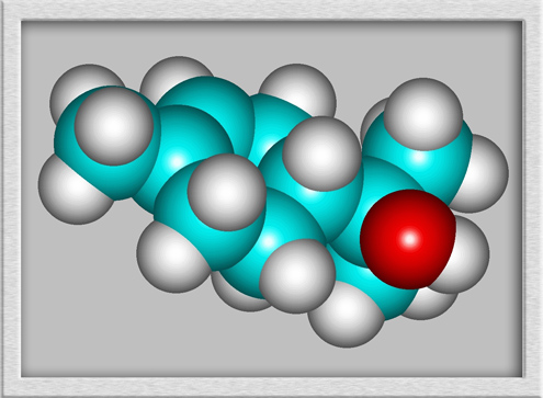
Modern chemistry has found that the scent associated with lilacs is due
to a number of organic molecules manufactured by the plant. The
most important of these is alpha-terpineol, an organic (carbon
containing) alcohol. Historically, this compound was obtained by
removing the essential oils from lilac flowers, and was used to produce
scented soaps, bath preparations and household products. This is
still done today, but the alpha-terpineol is produced synthetically in
the laboratory. (The true, and synthetic molecules are
identical.) In smaller amounts, the compound may be used to
formulate synthetic flavours such as nutmeg, orange, peach and other
florals.
The structural formula, ball and stick model, and molecular shape of
alpha-terpineol are shown below. (HyperChem Pro was used to
produce the illustrations. A technique called geometry optimization was used to
find a low energy (stable) shape. Other stable configurations
exist.)
The name lilac is derived from
the Arabic word layak and the Persian word nilak referring to the
colour blue, and from the Sanskrit word for purple. The genus
name Syringa
comes from the Greek word for pipe, and refers to the tubular bottom
of a lilac flower. The species name vulgaris
translates to the more modern common.
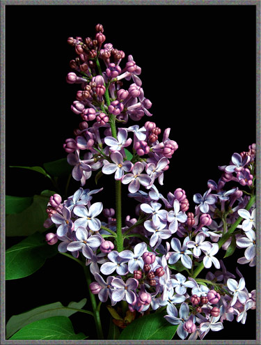
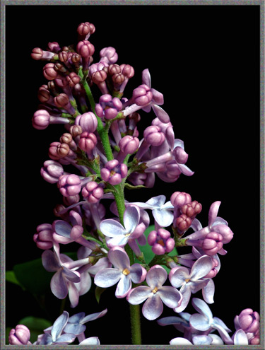
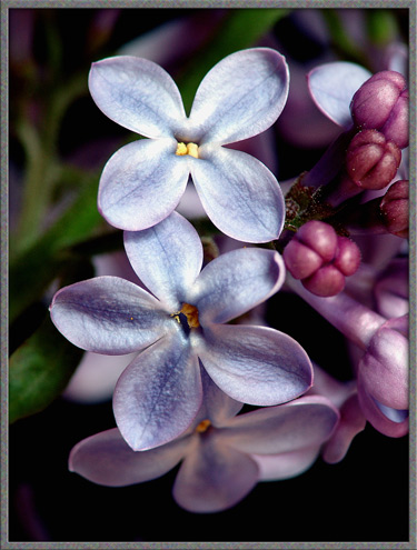
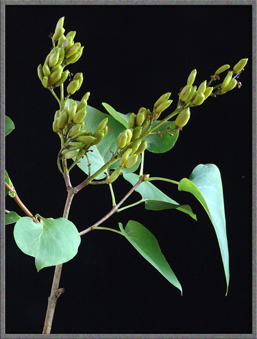
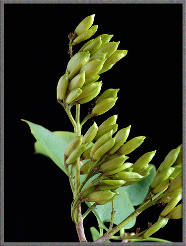
On a lilac bush, the flowers appear in spikes. Each spike has a
central stem with the secondary (and sometimes tertiary) branches
ending in flowers. The flower clusters are called thryses and may be up to 20
centimetres in length. As can be seen below, the flowers bloom
from bottom to top in each thryse.
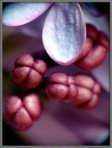
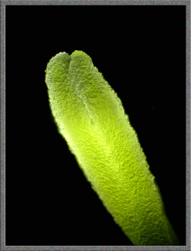
The buds of the lilac plant are almost as striking as the flowers
themselves. They are roughly club-shaped, with the bulbous end
being divided into four compartments. Two buds are attached at
their ends to a single terminal branch.
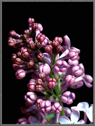
Most of the buds, when examined at higher magnification, appear to be
dusted with tiny crystalline specks.
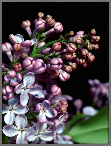
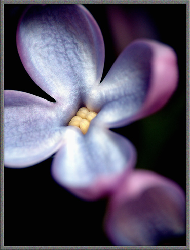
As a bud begins to bloom, each of its four compartments opens up to
form a petal. The interior of each petal tends to be a lighter
shade of purple than the exterior. (In very sunny weather, the
colour of the flowers may fade.)
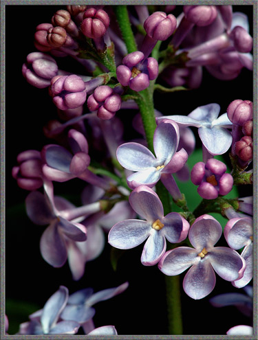
Notice that the open flower has a relatively long tube connecting it to
the branch. It is this tube that gives the plant its genus name Syringa.
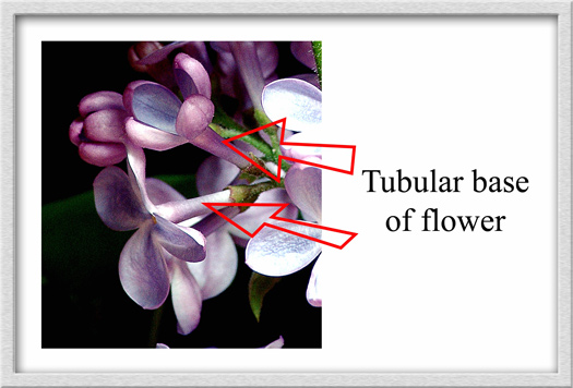
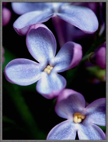
Compared
with many other flowers, the lilac bloom is remarkably simple in
structure. Individual flowers are from 0.5 to 1 centimetre in
diameter and have four petals that are fused together where they meet
the long tubular base. As often happens in nature, mistakes in
DNA replication can produce anomalies such as the flower shown below,
which has five petals!
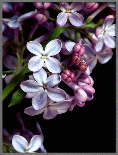
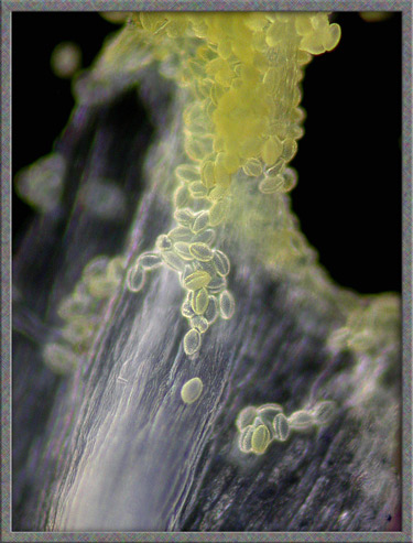
A closer look reveals four bright yellow anthers (male, pollen producing
structures) packed closely together at the point where the tube widens
out to form the petals. The stigma
(female, pollen accepting structure) is held by the style in a position
beneath the anthers and is not visible.
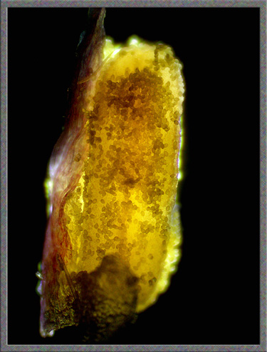
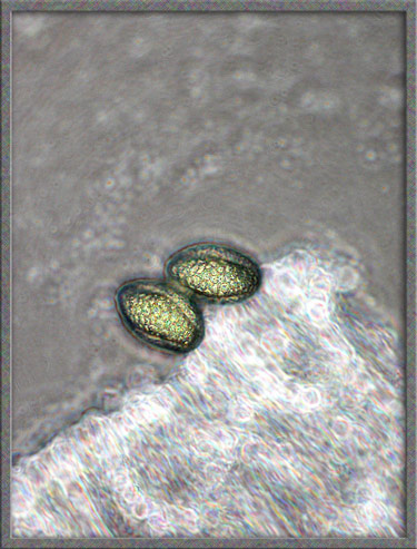
Under the microscope, one of the anthers appears encrusted with pollen.
The single green stigma is divided at its tip into two parts.
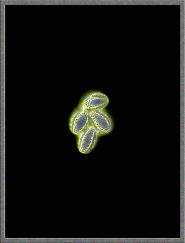
Lilac pollen appear egg-shaped and have several longitudinal grooves on
their surface. Many small dimples pockmark the surface of each
grain.
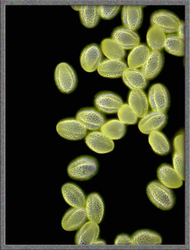
Phase-contrast illumination at a higher magnification resolves more of
the surface detail.

Once
insects have fertilized the flowers, many pale green fruit form on the
branches. At a later stage, the fruit dry out to form capsules
containing two seeds each. (Notice the characteristic
heart-shaped leaves.)
My parents home was built on land which had been a large garden
containing many lilac bushes. During construction, the lilacs at
the edge of the property were left standing. This all happened
sixty years ago, and those same plants, and their descendants were the
source of the lilac flowers used in this article. Lilacs are not
only beautiful to look at, and wonderful to smell, but they are also
very long-lived!
Photographic Equipment
The photographs in the article were taken with an eight megapixel Sony
CyberShot DSC-F 828 equipped with achromatic close-up lenses (Nikon 5T,
6T, Sony VCL-M3358, and shorter focal length achromat) used singly or
in combination. The lenses screw into the 58 mm filter threads of the
camera lens. (These produce a magnification of from 0.5X to 10X
for a 4x6 inch image.) Still higher magnifications were obtained
by using a macro coupler (which has two male threads) to attach a reversed 50 mm focal length f 1.4
Olympus SLR lens to the F 828. (The magnification here is about
14X for a 4x6 inch image.) The photomicrographs were taken with a Leitz
SM-Pol microscope (using a dark ground condenser), and the Coolpix
4500.
References
The following references have been
found to be valuable in the identification of wildflowers, and they are
also a good source of information about them.
Published in the May
2006 edition of Micscape.
Please report any Web problems or
offer general comments to the Micscape
Editor.
Micscape is the on-line monthly magazine
of the Microscopy UK web
site at Microscopy-UK
© Onview.net Ltd, Microscopy-UK, and all contributors 1996 onwards. All rights reserved. Main site is at www.microscopy-uk.org.uk with full mirror at www.microscopy-uk.net .