This article can also be downloaded in Acrobat pdf format here (file size ca. 3Mbyte prepared by the author).
This format is easier to print if desired.
Leitz Orthoplan
Universal Largefield Research Microscope
A brief illustrated history
Mike Andre, USA
This
article can also be downloaded in Acrobat pdf format here
(file size ca. 3Mbyte prepared by the author).
This format is easier to print if desired.
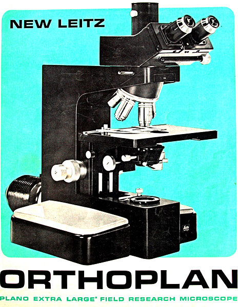
Leitz introduced their flagship large field research stand, Orthoplan, ca. 1966, which continued in production essentially unchanged until 1991, a run of over 25 years, attesting to the capabilities designed in from the outset; it was just as viable in 1991 as when first introduced and remains a premier research stand used at universities and other research facilities around the world today. From one of the original sales E. Leitz NY flyers: “The ORTHOPLAN is a new member of the family of Leitz Research Microscopes. It is the first instrument designed for a field of view of 28mm; it allows more than twice the field area of a conventional microscope with widefield eyepieces.” The Orthoplan was designed to be the epitome of functionality and flexibility with ability to switch from one form of illumination to another quickly and easily, setting the standard for quality and capability, costs be damned! Likely this is the primary reason for its eventual discontinuance – the cost to produce these units was economically unacceptable.
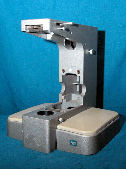
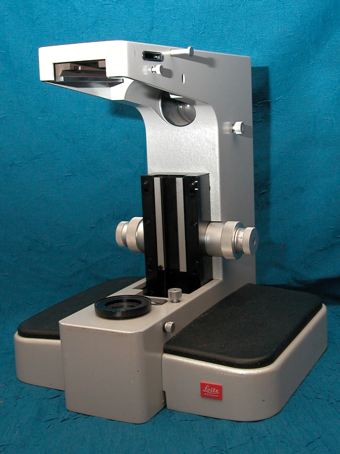
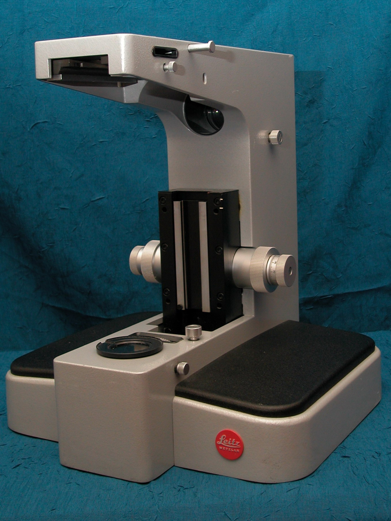
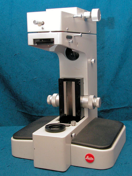
The initial fifty Orthoplans were made in all black; these are very rare and rarely seen (or even known) today, almost all surely in the hands of collectors. General production was 'hammer tone gray' followed by ‘light silver gray' and finally the off-white/ivory version. The hammer tone gray units had light gray handrests that tended to turn yellow-gray with age; all later models came with textured black handrests. Another variation was the very early models had a square blue Leitz logo, followed by square red and ultimately the round red logo seen on the later versions.
|
First standard (after black) issue – Hammer tone Gray. Yellowing of the armrests is evident in this shot. If anyone knows how to remove 30 year-old cellophane tape – please advise! (This stand is used for parts.) |
Later production in 'Silver Gray' – note the square red Leitz logo. Note the change in the armrests from gray to textured black, which were used on all subsequent production Orthoplans. |
|
Later variant of 'Silver Gray' with round red Leitz Logo |
Orthoplan in final Off-white/Ivory color. This unit has a Variotube mounted. |
|
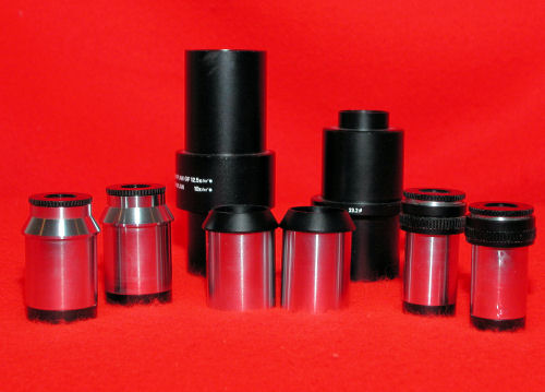
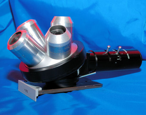
A
comparison of GW (30mm) eyepieces on the left versus GF (23.2mm)
eyepieces on the right. At the back are two photo tubes for use in
the trinocular port of the body tube; center are adapter sleeves
that allow the GF (23.3mm) eyepieces to be used in the FSA body tube.
|
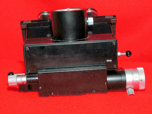
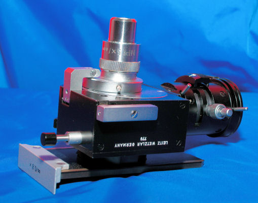
Left side view of FSA-50 body tube. The large silver knob on the bottom is the analyzer control drum that can be rotated 360° with an accuracy of 0.1°. |
The two silver knobs on left of the black knob are for centration of the Bertrand lens. The black knob controls the pin-hole stop. |
On the left are the controls for the Bertrand lens: black knob controls the Bertrand lens, the silver knob is for focusing. (See note below) |
It is quite
easy to open the back cover (be careful as there is one spring under
the cover which is easy to lose if one is not aware of it!) and move
the lens by hand to determine if it needs to be cleaned of dried
grease and relubricated. This is a good job for a microscope
technician, as the risk of damaging an FSA-50 easily outweighs the
cost of having someone who knows what they are doing work on it.]
|
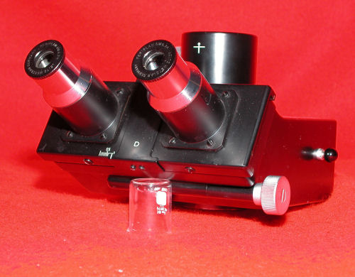
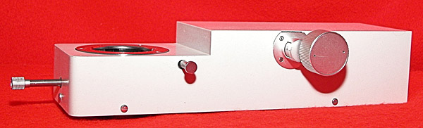
This is an FSA-55; the knob on the right controls the light either 100% to eyepiece or 100% to the trinocular tube. (The 10ml beaker is for photo positioning only!) |
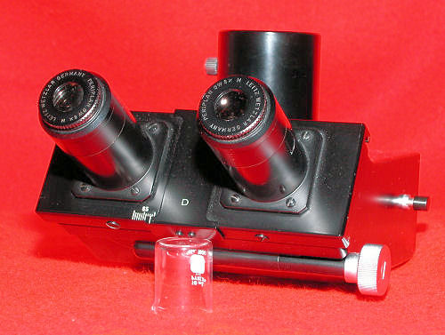
An FSA, the only difference between it and the FSA-55 is this model provided choice of 100% to eyepieces or 80% to trinocular and 20% to the eyepieces. |
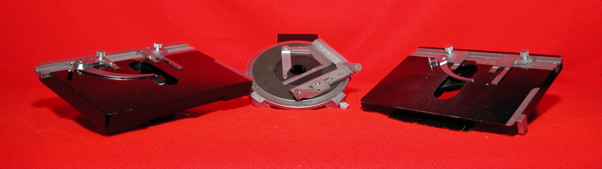
Here are
three of the many stages available for the Orthoplan; on the left is
a large mechanical stage with ball bearings, while the one on the
right is the standard unit. In the middle is a 150mm Ø
rotating stage #837 with scales and verniers reading to 0.1°,
click stop at 45° intervals, locking device and interchange
carrier, fitted with attachable low-profile mechanical stage #42 with
graduated vernier to 0.1°.
|
|
|
|---|---|
|
|
|
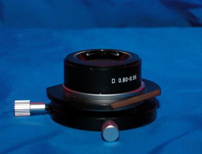
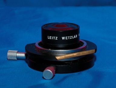
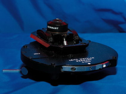 Phase contrast condenser 402A with 0.90 NA top lens |
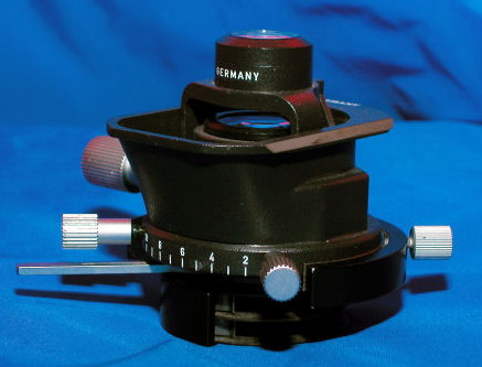 Condenser with rotatable film polarizer at bottom; 0.90 NA |
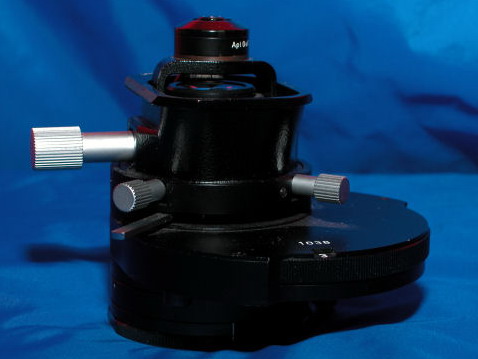 Nomarski DIC Condenser with Wollaston prisms, rotatable film polarizer at bottom; 1.25 aplanatic top lens |
|---|
At
least fourteen different condensers, four different lamphouses each
capable of using halogen, HBO or XBO lamps, four mirrorhouses, at
least 7 different nosepieces with varying mags from 0.8X to 1.25X,
three or more different bodytubes, I don't know how many stages......
I think you get the idea!
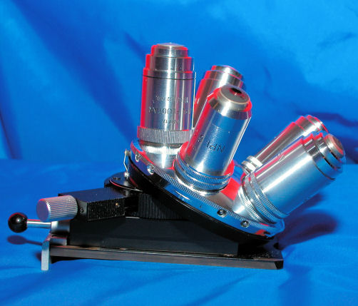 Centerable pol nosepiece with two filter slots, swing-in auxiliary lens for conoscopic image of small object detail. |
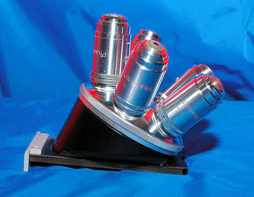 Standard 1X quintuple nosepiece with NPL Phaco lenses. |
|---|---|
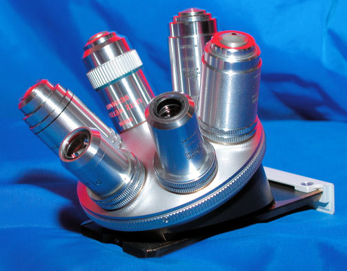 Standard 1X sextuple nosepiece with Plan and Plan-apo lenses. |
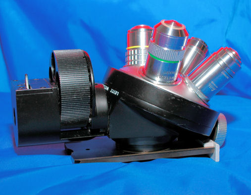 Epi-fluorescence after Ploem nosepiece. This uses standard 170 TL objectives. |
 Pol vertical illuminator infinity corrected optical system, plane glass plate and compensating prism and auxiliary lens |
 Incident light for BF/DF 0.8X quadruple nosepiece with Plan BF/DF lenses |
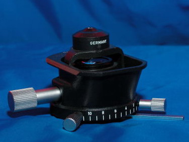
The Variotube allows one to change magnification from 1X - 3.2X by rotating the large silver knob. The knob on the front controls the built-in Bertrand lens – by pushing/pulling the lens is moved in/out of the optical path, what is interesting is the Bertrand lens is focused by rotating the same knob!
Leitz supplied equipment for the Orthoplans allowing Interference Contrast in at least the following methods, (there may have been more):
DIC after Nomarski (Original Leitz France version, only available a very short time) Smith T System Jamin-Lebedoff Interference Contrast R Françon Pol-interference for Incident light All these bits & bobs were made to the highest levels of quality and performance – it's been said many times, “if it's Leitz and it moves it probably has ball bearings!” An example of the level of quality built into every part for these instruments is the rotating objective carrier on the original epi-fluorescence nosepieces; the turret revolved on forty-eight 3mm steel bearings held in a race formed by the nosepiece and the turret; a machine screw holds the turret to the body on a race set on ten 1mm bearings. Overkill? Absolutely! Will it last a long time? Absolutely! This is typical of the engineering, build quality and attention to detail demonstrated in every element of the Orthoplan. Although there were numerous improvements and changes made over the ~25 years this model was produced, all of the various parts will fit all of the Orthoplans. As one can easily see, amassing a 'complete' collection of Orthoplans and allied equipment is a daunting task.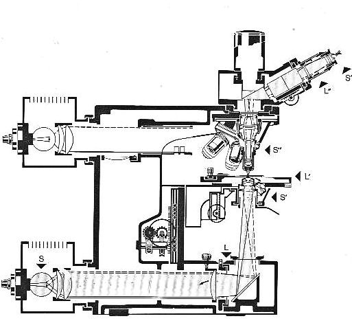
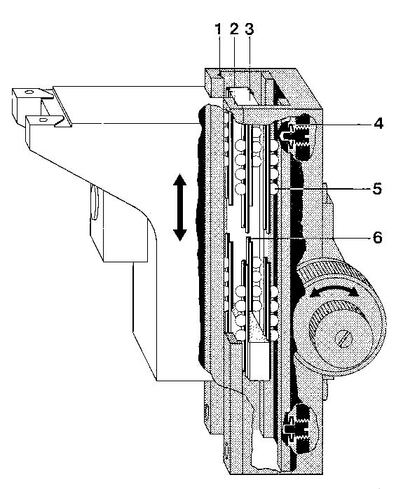
Schematic
of light paths and main conponents
Comments / questions
to the
author
Mike
Andre are welcomed.
Editor's note: This article can be downloaded in Acrobat pdf format here (file size ca. 3Mbyte prepared by the author). This format is easier to print if desired.
Published in the May 2006 edition of Micscape.
Please report any Web problems or offer general comments to the Micscape Editor .
Micscape is the on-line monthly magazine of the Microscopy UK web site at Microscopy-UK
© Onview.net Ltd, Microscopy-UK, and all contributors 1995
onwards. All rights reserved.
Main site is
at www.microscopy-uk.org.uk
with full mirror
at www.microscopy-uk.net
.