|
|
A Gallery of Beta-Alanine & dl-Alpha-Alanine Photomicrographs (using
polarized light illumination) |
|
|
A Gallery of Beta-Alanine & dl-Alpha-Alanine Photomicrographs (using
polarized light illumination) |
This article shows images of the two
isomers of the amino acid alanine.
Amino acids are the building
blocks from which proteins are formed. Proteins then, consist of a series
of amino acids linked together in a particular order specified by a
genes DNA sequence. These proteins may act as structural
components in a cell, as signaling molecules, or as enzymes (catalysts)
which speed up chemical reactions in the cell.
There are twenty amino acids which
can combine to form the 50 000 to 100 000 different proteins in the
human body. Nine of the twenty amino acids must be obtained from
foods, since the body cannot synthesize them. The other eleven
amino acids, to which group alanine belongs, can be produced by the
body by using the aforementioned nine. In the body, alanine is
synthesized in muscle cells.
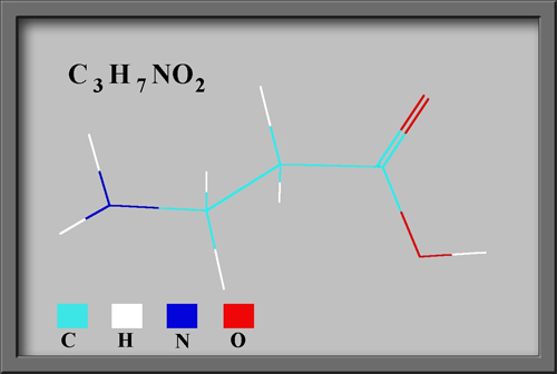
Beta-Alanine
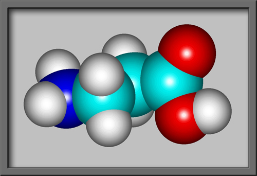
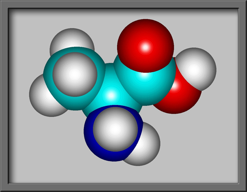
The structural formula and
molecular shape can be seen below. (Both illustrations were
produced by HyperChem
software.)
Since the colourless crystalline
solid has high solubility in water, I was able to produce an
evaporation specimen by dissolving a small quantity in distilled water,
and then placing a couple of drops of the solution on a microscope
slide. After the crystals that formed were completely dry, a drop
of Permount was applied over
them, and a cover-glass added. This produces a specimen that
lasts for decades! Note that the two compounds discussed in this
article may act as skin or eye irritants.
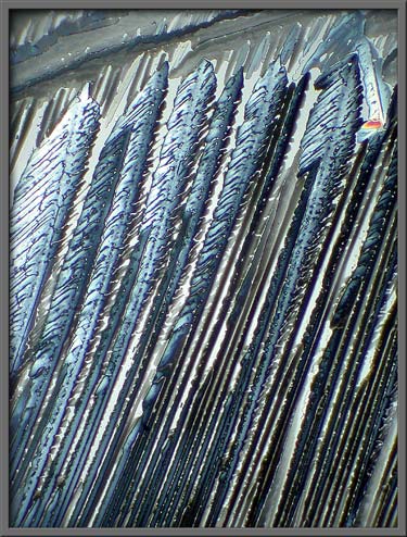
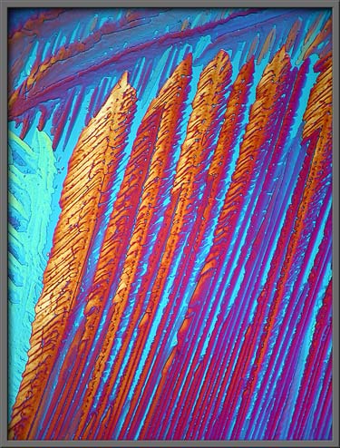
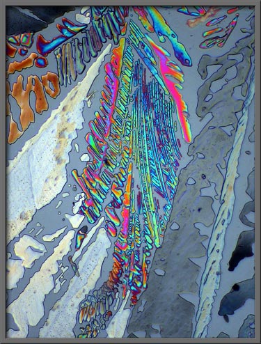
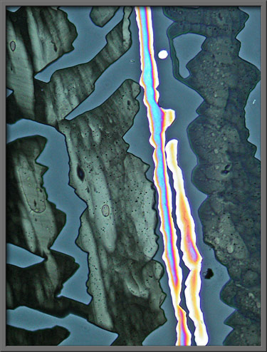
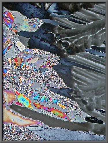
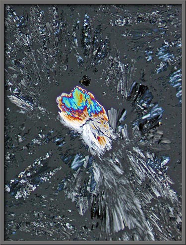
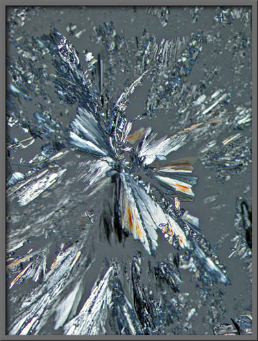
The first image in the article, and
the one below, show crystal structures that formed on the slide during
evaporation of the solvent. The background is gray, instead of
the expected black when using crossed polarizers, because two
quarter-wave plates were utilized to produce elliptically polarized
light.
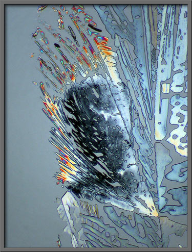
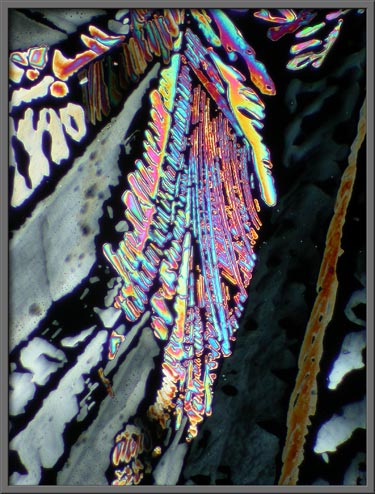
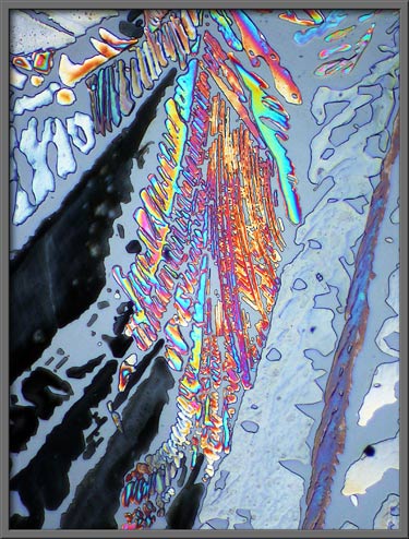
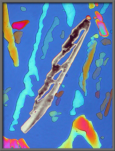
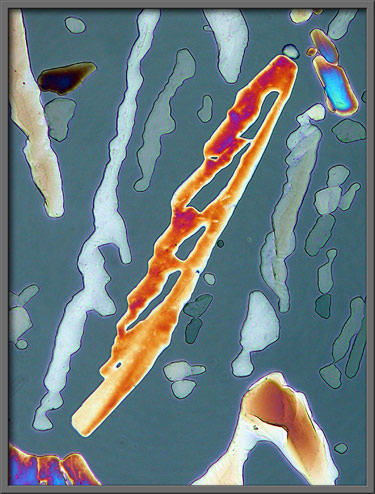
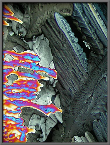
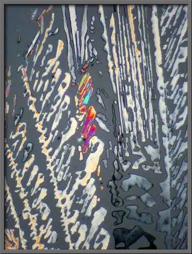
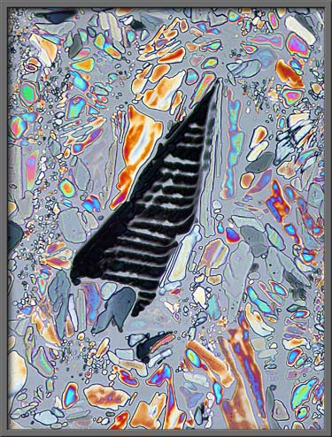
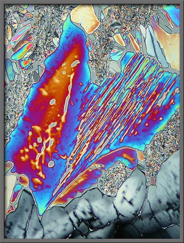
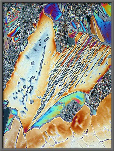
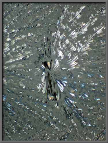
The use of such plates, (called
compensators), can dramatically change the appearance of a particular
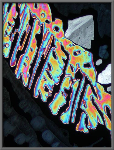
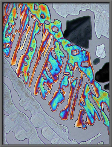
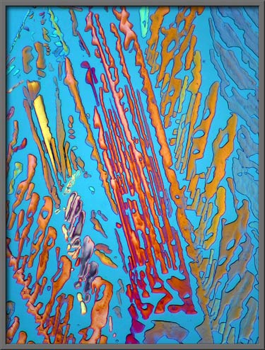
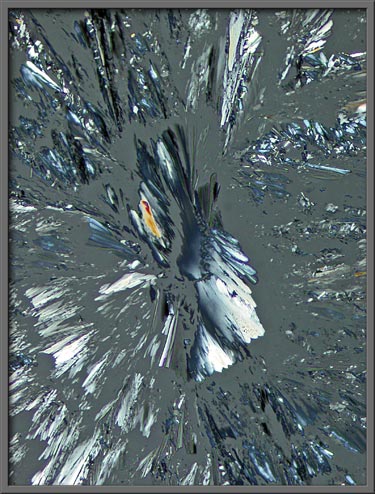
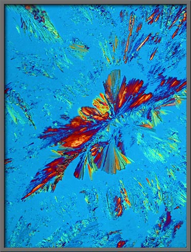
visual field. For example, consider the three images that
follow. The first uses two lambda/4 compensators, the second
lambda/4 and lambda compensators, and the third uses no compensators.

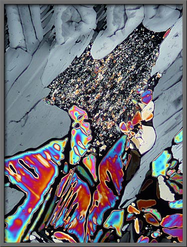
Another example follows. No
compensators were used when producing the first image, while two
lambda/4 compensators formed the second and third images. The
subtle difference in the last two images was caused by rotation of one
of the compensators.
The use of these techniques can
dramatically alter the appearance of a field.
Lambda/4 and lambda compensators
were used in the three images below. Rotation of the lambda/4
compensator resulted in the different background colour in each
photomicrograph.
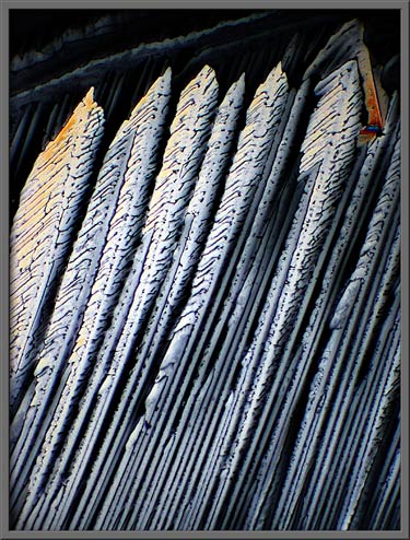
If the crystals formed by
evaporation are thicker than the ideal, under polarized light, they
may appear as shades of gray or brown, as in the four examples below.
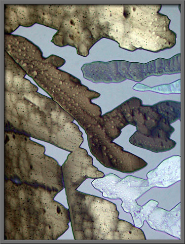
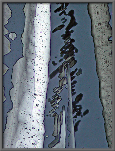
All but the second image below
utilized elliptically polarized light instead of the normal plane
polarized variety.
dl-Alpha-Alanine
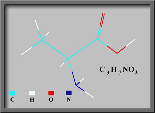
Alpha and beta-alanine are isomers. This means that they
have the same formula, C3H7NO2, but
the molecular structures are different. If you compare the
molecular shapes, it is evident that the NO2 group in
alpha-alanine is attached to the central carbon, whereas in
beta-alanine, it is attached to the end carbon. This results in
different chemical properties for the two compounds. (For
example, alpha-alanine has a melting temperature of 314 degrees
Celsius, while beta-alanine melts at 196 degrees Celsius.) It is
not surprising then, that the crystals formed by alpha-alanine in
evaporation specimens bear no resemblance to those of the beta compound.
Some organic molecules have a
mirror image twin that is structurally different. The twin
molecules are referred to as the right-hand (d) form, and the left-hand (l) form. The dl in the name above tells the user
that the bottle contains both the left, and right hand forms of the
molecule.
By comparing the images below with
the previous ones, it is clear that alpha-alanine is less photogenic
than its beta sibling. The first four images were taken using two
lambda/4 compensators, and the last image used lambda/4 and lambda
compensators.
I would enjoy photomicrographing
the other nineteen amino acids. Unfortunately, finding pure
samples is a problem!
Photomicrographic
Equipment
The images in the article were
photographed using a Nikon Coolpix 4500 camera attached to a Leitz
SM-Pol polarizing microscope. Crossed polars were used in all polarized
light images. Compensators, ( lambda and lambda/4 plates ), were
utilized to alter the appearance in some cases. A 2.5x, 6.3x, 16x
or 25x flat-field objective formed the original image and a 10x
Periplan eyepiece projected the image to the camera lens.
All
comments to the author Brian
Johnston are welcomed.
Published in the May
2007 edition of Micscape.
Please report any Web problems or
offer general comments to the Micscape
Editor.
Micscape is the on-line monthly magazine
of the Microscopy UK web
site at Microscopy-UK
© Onview.net Ltd, Microscopy-UK, and all contributors 1995 onwards. All rights reserved. Main site is at www.microscopy-uk.org.uk with full mirror at www.microscopy-uk.net .