|
|
|
Preservation of Contrast in the
Compound Microscope (2)
Concluding part : A simple comparison between images of widely ranging contrast By Paul James |
If it seems reasonable to try to eradicate any contrast diluting light generated along the optical train of the compound microscope in order to render a more faithful image of a given specimen, then it would seem logical that the resulting image should reveal evidence of this within its tonal range and ultimate resolving capability of a given objective. So in order to try to demonstrate this premise I reasoned that one photographic image taken on the same microscope, whose contrast preservation was intrinsically sound might compare more favourably with another photographic image generated with identical lighting, but with artifically reduced contrast. This method of using the same microscope would be free of a host of subtle problems if using two different stands, when the only principal purpose of the experiment was to keep to the contrast issue and nothing else. The stand used was a Zeiss Photomicroscope using the trinocular port to transfer images to a 5 megapixel digicam.
Methodology
So in this comparative test using the same stand, the optical glassware was identical for both images. Since the contrast preservation was intrinsically sound when used in its normal mode I reasoned that I would need to lower this for the second photomicrograph to a level that might be present in a somewhat inferior or poorly adjusted stand. After thinking about this for a while I decided to leave the Kohler setup exactly as it was for best imagery in COL for both photo images and to reduce the contrast preservation for the second image by a singularly simple but very effective means of using a frosted/ground glass screen between the lamphouse and substage condenser. Such drastic means of destroying the ascending cone's integrity from the lamphouse/condenser unit might seem a little OTT but at least there could be no doubting that the contrast preservation would be compromised severely in the second image. The whole point of the exercise was to see if the quality of the images so produced varied and to what extent.
Results
One of the vital elements of comparative photography in this situation is to maintain identical settings for camera and 'scope aside the inclusion of the ground glass screen for one of the exposures of course. The only cause for concern was the minute variation of focus caused by the camera's auto focus (Minolta F300). The discrepancy is very slight and although not noticeable in ordinary terrestial photography where depth of field is desirable, it nevertheless affects the interpretation of resolution between the images in a situation where the depth of field, especially when using COL is at a premium, and thus the white or black dot focus effect becomes more difficult to maintain indentically between both images. Post processing in software was performed to highlight the detail rather than the tonal differences in the images, so that greyscaling/sizing/image intensity were altered accordingly to try to emulate the 'live' views. Visually, the diffused imagery looked much dimmer and with lower contrast as would be expected, but clearly the resolving capability of the objective did not seem to be affected. The camera compensated for the duller image with a longer exposure.
|
|
|
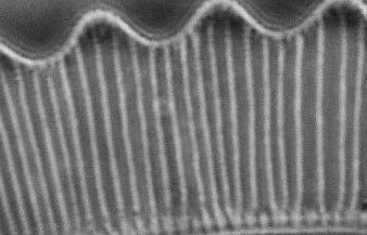
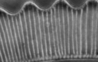
The two images above appear to be very similar despite the fractional discrepancy of camera focus. The diffusion screen was used in the second photo (right). The amplification of this Eutonia diatom is deliberately excessive which was chosen with dotting just beyond the objective's ability to grasp and reveal with clarity.
In Brightfield there was similar variation in tonality between the pair of images, but the resolution was markedly inferior to the COL rendering on this subject, and so the contrast appeared to be more diluted than in the case with COL.
Conclusion
My understanding of the ramifications of this simple experiment is that the rendering of detail is first and foremost a product of the objective's pedigree and aperture value in conjunction with a suitably appropriate substage and lighting technique. The ease with which the eye and the camera perceive this, is a function of the degree of contrast present in the emerging image from the eyepiece. It therefore seems to me that contrast preservation in its widest sense in microscopy, whilst being fundamentally essential, is not necessarily the ultimate gaol for observers. The range of specimens suitable for compound transmission microscopy is so vast that there must surely exist many situations where contrast taming, especially in lower powers is desirable, but when dealing with subject matter requiring the ultimate resolution and amplification of a given specimen, contrast preservation is absolutely essential for high fidelity image reproduction.
Author's Note : My ability to furnish decent photo's for this article has been made difficult owing to some problems concerning the camera and the fine focus control of the Photomic.
| All comments welcome by the author Paul James |
Microscopy UK Front Page
Micscape
Magazine
Article
Library
Please report any Web problems or offer general comments to the Micscape Editor.
Micscape is the on-line monthly
magazine of the Microscopy UK web
site at
Microscopy-UK