|
Nineteenth Century British Microscopy and Natural History: Part 5 by Richard L. Howey, Wyoming, USA |
Visit the Micscape Library to read other parts in the series.
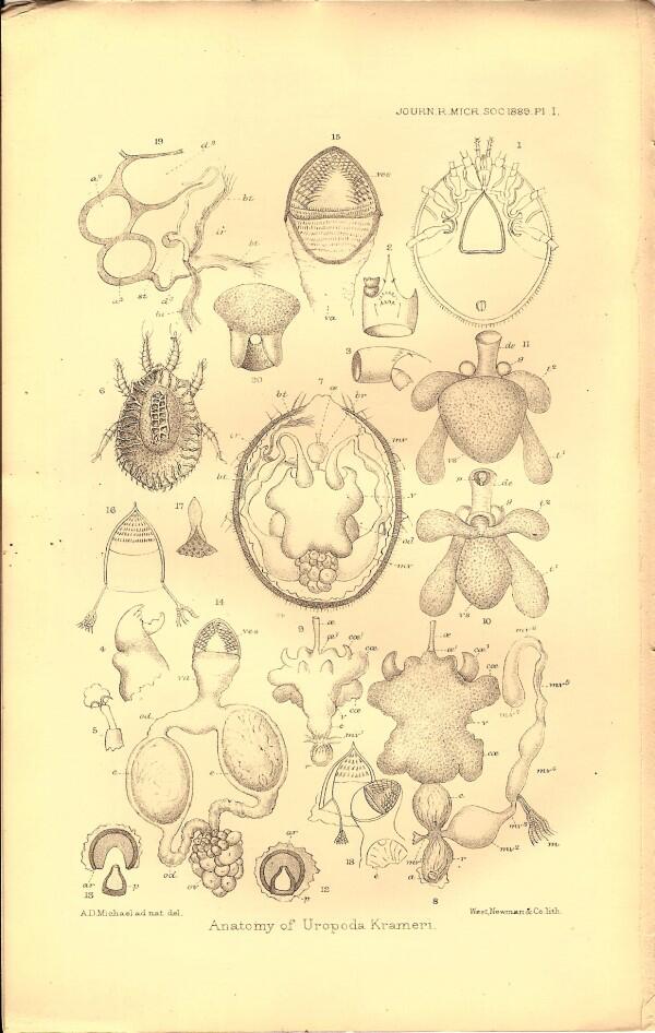
This time I want to examine the February issue of 1889 of the Journal of the Royal Microscopical Society. Like the other issues, it is also filled with fascinating investigations. The first major paper is a 13 page, careful examination of the mite Uropoda krameri accompanied by a splendid plate displaying its internal anatomy.
The author Albert D. Michael remarks:
“During the summer of 1888, when staying at a farmhouse in Derbyshire, I found Uropoda Krameri (Canerstrini) in great abundance on the floors and walls of an old barn used for storing hay. This seemed to be a favourable opportunity for ascertaining something about the internal anatomy.” An abundance of mites in an old hay barn–this is what distinguishes the naturalist from ordinary folk, who might ask: “Who in his or her right mind would want to muck about with a bunch of mites?” Well, as it turns out there are hundreds of specialists around the world who do. It’s the people who aren’t interested in such things whom I find strange. Curiosity is such a precious gift and it should be encouraged on every possible occasion.
The second major paper is titled: “List of Desmids from Massachusetts, U.S.A.” by Wm. West, who was well know for his studies of desmids and algae. The article is accompanied by two very interesting plates which I will reproduce for you here.
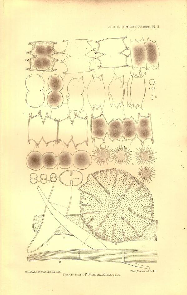
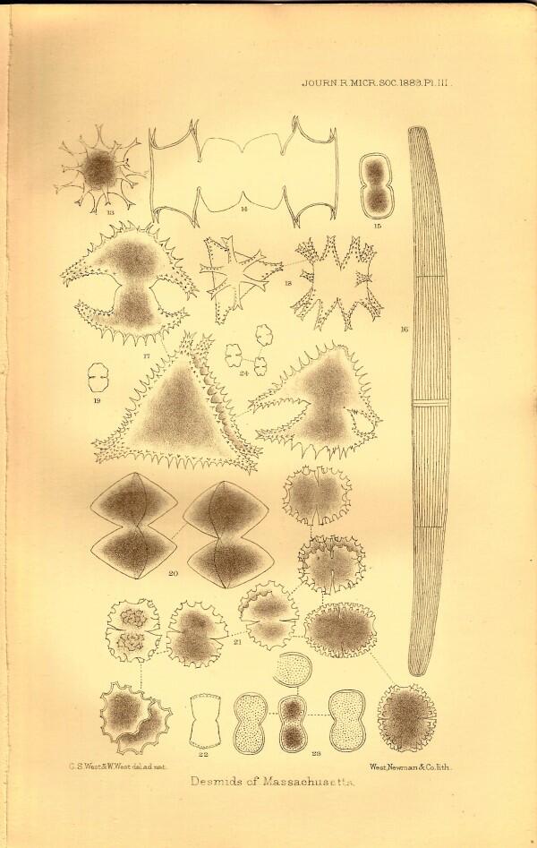
Desmids are among the loveliest micro-organisms and now with the techniques of modern photography, we can see them in great detail and in color. For those of you who are interested in algae, there is a superb book by Hilda Canter-Lund & John W.G. Lund called Freshwater Algae which is unfortunately no longer available, but might be found in a good university library. It is well worth seeking out.
This issue atypically has a third major article “Reproduction and Multiplication of Diatoms” which is 6 pages in length and written by the Abb Count F. Costracane, Hon. F.R.M.S. This issue of reproduction in diatoms is one of great interest to diatomists even today and this paper should still be of interest to the specialist.
In the summaries of researches, there are, as we have come to expect, many strange and interesting things. We learn that Mr. A.H.S. Lucas “considers that the effect of the surrounding during the time of the formation of the shell, upon the mental or nervous constitution of the bird, is a very important factor in determining the colouring of the eggs.” I’m not an ornithological oologist and so have no expertise in this matter, but my intuitive response to this theory is one of considerable skepticism.
Remember, this is the 1880s and Lamarckianism was by no means dead and, in certain circles, it was a mark of distinction to attack Darwin. In the previous article in this series, I mentioned how Victorian titles often tended to be quite elaborate and detailed. However, the Germans are second to none in this regard. Professor G.H.T. Eimer produced a work (this was just the first volume) with the grand title: Die Entstehung der Arten auf Grund von Vererben erworbener Eigenschaften, nach den Gestezen organischen Wachsens. Ein Beitrag zur einheitliche Auffassung der Lebewelt. Whew!
Eimer is discussing the origin of species through the inheritance of acquired characteristics. The reviewer informs us that:
“In his introduction, Prof. Eimer criticizes the Darwinian postulate of indefinite variations; emphasizes the deficiencies of an aetiology which does not discuss the primal conditions of variation and maintains that the utilitarian principle, which does not explain the origin of new qualities, only partially at most accounts for their increase and dominance.”
Incisive points, which explains why no one has heard of Darwin today and the dominant theory of the origin of species is Eimerism. Yes, I know, a cheap shot and a logical fallacy; but Darwin was a genius and Eimer was a drudge.
There is in addition another summary of a paper devoted to evolution, this one by a cleric, who evidently was a believer in the long scholarly tradition of obscurantism; that is, the view that if you make things so obscure and complex that they are virtually impossible to understand, then they will be regarded as profound and the author will escape criticism for propagating nonsense. The reviewer’s opening paragraph reads as follows:
“In an elaborate paper the Rev. J.T. Gulick follows up some previous communications, in which he has maintained that ‘separation without a difference of external circumstances is a condition sufficient to insure divergence in type.’ The abundance of technical and unique terminology, combined with the intrinsic complexity of the inquiry, renders it very difficult to present a brief summary without injustice to the patient author.”
The reviewer adds in his last paragraph:
“As an analysis of the conditions of association and isolation the memoir possesses great interest, not a little spoilt by the elaborate and ugly terminology. The author certain cannot be charged with depreciating the complexity of the inquiry.”
Talk about a high-level diplomatic response! This reviewer must have broken his back bending over backward to avoid offering offense.
A bit later on we find a review of slightly over 5 pages regarding a monograph devoted to a single species of a colonial, sessile (attached) tunicate. At this time, tunicates were still grouped with mollusks, yet in spite of this taxonomic deficiency, it was already recognized that the tunic consisted largely of cellulose compounds which are, of course, quite rare in animals, although common in plants.
This lengthy review is followed by 2 short ones of posthumous papers of Dr. M.L. Joilet on 2 other types of tunicates–salps and pyrosomes. These remarkable organisms are planktonic and thus free-swimming. (See Micscape articles on tunicates). These are elegant, transparent creatures which look like they were made of blown glass. Both occur in aggregates formed by budding. Pyrosoma means ‘fire-body” in Greek and this name derives from the fact that these organisms are bioluminescent. (See 'The JelliesZone' website.) There is an old account of a fishing trawler having brought up a large pyrosome–4 foot long, if my memory serves me correctly–and while it was still dark, a sailor inscribed his name on its surface with his finger and then watched his name “magically” appear in the cold, blue-white light of bioluminescence. These are wondrous creatures and deserve much wider recognition and study.
A bit ago, we noted the remarkable diplomacy of a reviewer with regard to the “theories” of the Rev. Gulick, but not all of the reviewers were so deferential.
There is a summary of an article titled: “Notes on some Rotifera from the neighbourhood of Geneva” by W.E.F. Weber. The reviewer of this article is clearly a no-nonsense sort of person who, from the morphological details he presents and the other experts whom he cites, is a highly knowledgeable specialist. He remarks: “...these ‘Notes’ are written throughout with an assumption of authority which is by no means warranted by M. Weber’s observations, figures, or descriptions.” Weber claims to have discovered four new species of rotifers and the reviewer takes them one by one and demolishes his claims. Weber also scorns the accounts of other specialists. The reviewer comments:
“The very memoirs he quotes from, and of which he gives a list, ought to have preserved him from such a blunder; were it not that M. Weber appears to have no doubt that, when an observer differs from him, the person in error cannot be himself.
The following is an amusing instance of this curious belief in his own infallibility. M. Weber fails to find the contractile vesicle in the male of Hydatina senta, so he dismisses all observers who have seen it by saying, ‘Cohn, Leydig, Daday, and Hudson have seen it with the eye of faith!’”
I find it reassuring that there were crusty curmudgeons like myself back in the 19th Century who had a low tolerance for pompous pretensions, but I do sometimes worry about where such curmudgeons will come from in the future.
This issue of the journal is a particularly rich one with interesting summaries of researches on bryozoa, starfish, sea urchins, sea cucumbers, jellyfish, and hydroids. In addition, there are a number of summaries regarding protozoa and the one which I found most interesting was a brief account of the species which Prof. L. Maggi found on mosses. There were 21 species; 17 amoebae, 1 flagellate, and 3 ciliates. I should note that the sections on botany are also of wide variety and interest.
In the section devoted to microscopes, accessories, and other apparatus, there are a number of items that are too irresistible not to show you. In fact, there are 6 instruments which I wish to show you; 4 in some detail–the other two are minor curiosities. The first curiosity is a relative of the modern otoscope and was know as Czapski’s Ear-(Tympanum) Microscope. This has an ingenious design worthy of Torquemada, but then I have an inclination to regard almost all medical instruments as devices of torture. Here’s a scan to show you what it looks like.
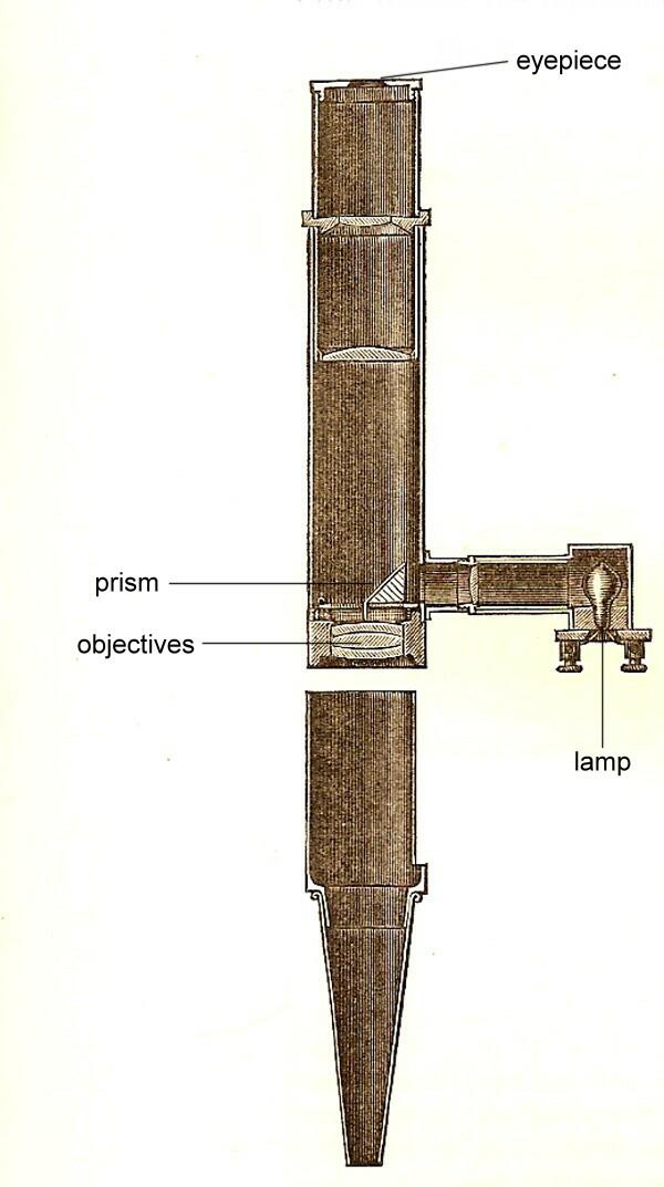
I have added 4 labels to help clarify the description. When placed into the ear, it allowed an examination of the eardrum at a magnification of 6 to 8 times. The prism is a half-reflecting one, so that the light from the illumination source goes down through the objective lens, bounces off the inner parts of the ear and comes back up through the objective to the eyepiece and to the eye of the examining physician. The design as shown was for a small electric incandescent lamp (also referred to as a glow-lamp) in the side tube. The reviewer informs us: “Instead of a glow-lamp a gas or petroleum lamp can be used, placed at the side.” I don’t find this at all comforting since the reviewer also remarks:
“The illumination is of course most intense over the whole field of view when the lamp is as near as possible to the prism, but regard for the ear and cheek of the patient places a certain limit to the approach of a hot source of light.”
I should bloody well hope so! Frankly, I wouldn’t want this instrument of the Inquisition anywhere near me.
Have you ever seen a Kitsch microscope? Well, feast your eyes on this beauty.
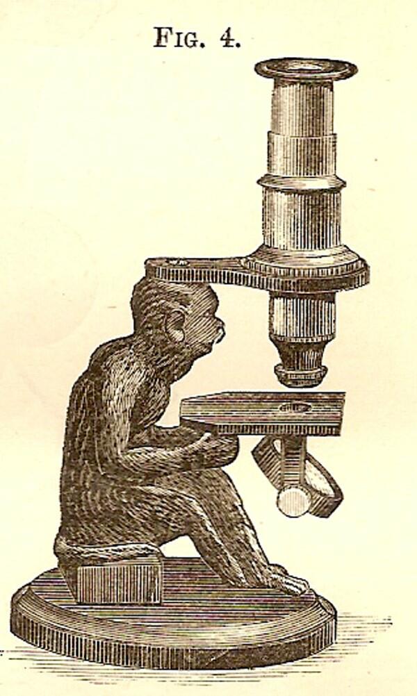
This is know as Moreau’s Monkey Microscope and was not inspired by the short novel of H.G. Wells The Island of Dr. Moreau wasn’t published until 1896. However, Wells’ novel did inspire 3 movies, the most recent staring Marlon Brando and immortalizing one of his all-time worst performances. However, it is just the tatty sort of little microscope that would have been right at home in the film.
The reviewer’s description of the microscope is only 2 sentence and I’ll provide them to you here.
“This Microscope (fig. 4), by M. Moreau of Paris was exhibited at the December meeting of the Society. In its design Art as well as Science has been drawn on, for instead of an ordinary base and pillar a figure of a monkey is introduced which holds in its hands the stage and mirror, while the cross-arm carrying the body-tube and socket is screwed to the top of its head.”
It seems that Kitsch is inescapable.
The next instrument, Reichert’s Petrological Microscope is quite remarkable.
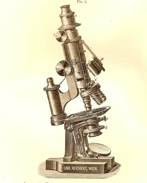
The reviewer informs us that this instrument has two special modifications. “The first is the introduction into the body-tube of a second analyser c, which is supported on a hinged arm so that it can be rapidly inserted and removed.”
The second innovation is 3 apertures in the body tube: o, o', and a. Into a and o' quartz wedges can be inserted and o is for the reception of a lens s,
“...which magnifies the axial image sketched by the objective, and which conducts the rays of the objective, so that if we will pass from the observation in parallel light to that in convergent light, it is but necessary to place the corresponding objective and the lens s without changing the eye-piece, which can be exactly adjusted on the object by the aid of a rackwork of the draw-rube b.”
Here is a microscope of extraordinary sophistication manufactured at least 119 years ago! Imagine the machining skills which were necessary to produce such a precision microscope. Reichert had a long and venerable history of manufacturing superbly engineered instruments culminating in the Zetopan which ultimately was the company’s undoing. The demand for these microscopes became so extensive worldwide that the company could not meet all of the orders and maintain their high quality standards. Eventually–a sad day for microscopy–Reichert was bought up by Leica.
Next we encounter Hughes’ Patent Oxyhydrogen Microscope.
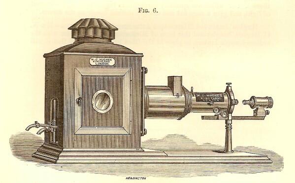
From this image, you might think that this is a rather prosaic piece of equipment, but you would be seriously mistaken.
“This instrument (fig. 6) has been designed and constructed by Mr. W.C. Hughes with a view to enable scientists, teachers, and lanternists to display on the screen in a clear and well-defined manner the minutiae of anatomical and geological sections, preparations of insects and vegetable tissues, and general microscopic objects either by ordinary or polarized light.”
One of the intriguing facets of Victorian society was that there existed a sizeable general public with an interest in natural history and for whom lectures and slide shows on microscopical subjects were a special treat. So, having apparatus which could project large images onto a screen with clarity and detail was a powerful attraction both in classrooms and in lectures halls for the general public. How impressive were these images? Let’s allow our reviewer to speak.
“To obtain this maximum of illumination Mr. Hughes has designed a special chamber jet, with which sufficient light can be obtained to magnify transparent subjects to 1900 diameters, which has hitherto been unattainable; thus a flea, which is about 1/10 in. total length, will be thrown on the screen 16 feet long, and every hair distinctly defined and nearly as brilliantly as a picture shown by the Pamphengos lantern. The proboscis of the blow-fly can, with various powers be projected from 8 to 16 feet long, and the all the details of an insect’s eye in section can be shown most perfectly; the circulation of the blood in the foot of a frog is easily displayed, and the wonders of pond life made manifest without the slightest difficulty or trouble. With the electric light no limit can be put on the magnifying power of the instrument, although, for all ordinary purposes, the lime light is all that is needed to obtain the results above mentioned. Every precaution has ben taken to arrest the passage of heat to the objects by means of non-conductors, and the results obtained have met with the approval of all those who have seen its perfect performances.”
A 16 foot flea–imagine what a sensation that would be for a Victorian audience. Unfortunately today, unless it was interactive and the viewer could shoot at it with an electronic laser pistol, it wouldn’t stir much excitement.
The Pamphengos lantern used oil as a source of fuel. One could not always be sure that a given venue would be able to provide electricity in 1889. Also, you will recall that Hughes refers to this device as an Oxyhydrogen lantern which links to the reference to lime light. Lime is Calcium oxide and jets directed oxygen and hydrogen to “candles” of Calcium oxide which would burn with an intense white heat without quickly melting. It is from this rather dangerous bit of ingenuity that we get the phrase about someone seeking out the limelight.
Mr. Hughes was not, however, content with this straightforward sort of projection instrument; he created the “Special Scientist Optical Lantern.”
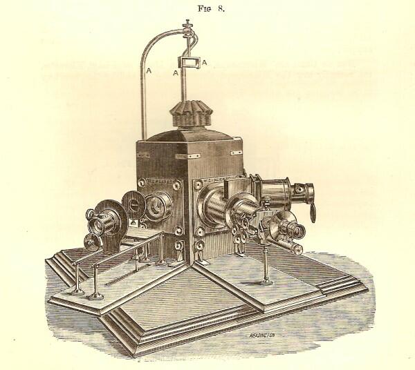
This remarkable piece of paraphernalia Hughes felt supplied “a want long felt by the professional lecturer both in the classroom and in the theater, namely, that of rapidly throwing upon the screen (a) the general view of an object (b) the microscopic portion of the same enlarged, and (c) in the matter of chemistry and physics, the experiment in actual operation.
The central body was made of mahogany and housed the illumination source. It could also be rotated to bring one of the three condenser systems into position behind the projection microscope. I strongly suspect that most of these instruments were in the possession of institutions and societies, not individuals.
Another ingenious, but rather eccentric-looking microscope was the Duc de Chaulnes’ Microscope.
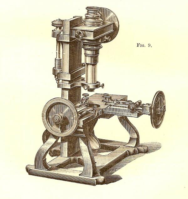
This was an instrument dedicated to the verification of micrometric measurements. It seems as though Victorian microscopists and instrument makers relished the challenges of solving complex optical problems by creating elaborate pieces of apparatus. Consider Engelmann’s Microspectrometer.
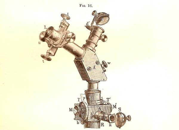
Look at all of the knobs, mirrors, and eyepieces–what microscopist worth his or her salt wouldn’t have drooled at the prospect of having one of these? What does it do? The reviewer kindly tells us.
“Originally devised for the quantitative determination of the absorption of different colours through living plant-cells, the apparatus is serviceable for quantitative microspectral analysis generally, and can be used with advantage for most microspectrometrical researches in place of the ordinary larger apparatus.”
This clearly was designed for special applications and was not something that the casual amateur microscopist would acquire. There are no indications of price regarding any of these instruments, but most of them I suspect were quite beyond the means of even most professional researchers of the time.
While many of the instruments and accessories were beautifully made and had metal parts of lacquered brass, others were more like Rube Goldberg concoctions, such as, Dr. Garbini’s small Steam-generator for Microscopical Technique.
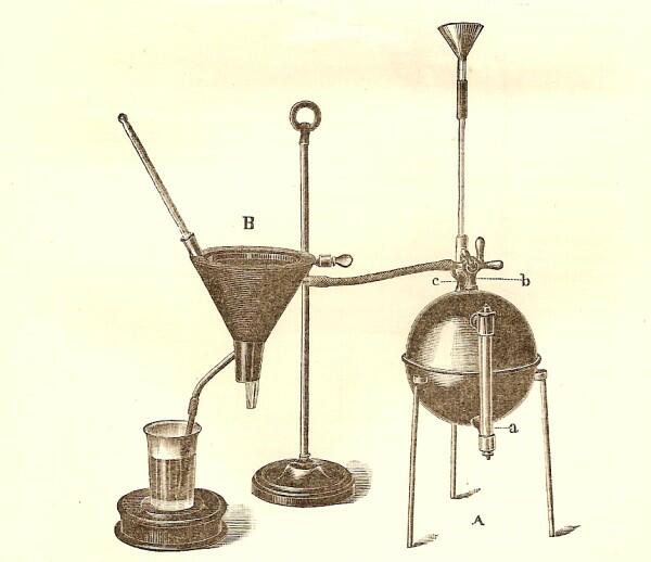
This odd piece of equipment was used for melting paraffin and gelatin for embedding tissue to section. One can certainly not deny the ingenuity of the Victorians in the production of scientific paraphernalia.
All comments to the author Richard Howey are welcomed.
Editor's note: Visit Richard Howey's new website at http://rhowey.googlepages.com/home where he plans to share aspects of his wide interests.
Microscopy UK Front
Page
Micscape
Magazine
Article
Library
Please report any Web problems or offer general comments to the Micscape Editor .
Micscape is the on-line monthly magazine of the Microscopy UK website at Microscopy-UK .