Forays into fluorescence - 2
A selection of prepared unstained slides studied in visible light fluorescence
by David Walker, UK
The author is currently exploring fluorescence microscopy as a hobbyist and the basic questions asked may be similar to those of others venturing into this area; so it is hoped these occasional 'forays' may be of interest. In parallel to simple autofluorescent studies of mosses and algae the author was also intrigued as to what prepared slides responded to fluorescence. The notes and images below are based on fluorescence studies of subjects in the author's fairly typical slide collection, which consists of both of old and more recent slides of a wide variety of subjects.
Sources of fluorescence - From background reading, primarily Rost (ref. 1), likely sources of fluorescence applicable to an 'active' prepared slide are summarised below:
Autofluorescence
- certain substances in plants, animals. Rost notes that aromatic molecules are 'in general'
responsible for the autofuorescence and that studies are 'best done' with fresh
material (ref. 1).
Autofluorescence - certain
chemicals,
minerals, rock sections, manmade objects etc.
Fluorescence of other materials -
certain mountants, reagents or their impurities present in the preparation.
Rost notes that Canada Balsam, one of the commonest mountants of older slides is
one of the 'worst offenders' for autofluorescence (ref. 2a).
Induced
fluorescence. Apparently some subjects
can react with certain reagents, neither which fluoresce, to undergo a
chemical change that induces fluorescence. 'Aldehyde and acid-induced fluorescence' is
one of a number of types which Rost discusses in detail (ref. 1).
Fluorescence of stains - some of the conventional
stains used to enhance selected subject
features for e.g transmitted light studies may also be fluorochromes e.g.
Eosin derivatives.
Fluorochromes (dyes
that fluoresce) intentionally added to subject - slides prepared specifically
for fluorescent studies may be rare in the hobbyist's collection. The author
only has one example, the triply fluorochrome stained 'Bovine Pulmonary Artery
Epithelial Cells, BPAE' slide courtesy of Molecular
Expressions. It has proved to be an invaluable reference slide because
its imagery has been explained and illustrated in detail on the web, allowing
comparisons of results obtained with own equipment.
Given the wide variety of protocols used to prepare slides, which is often not stated, arguably a 'try it and see' approach' is worthwhile to ensure some potentially interesting slides are not overlooked. This is the approach the author used! Presumably, the activity of prepared slides may vary considerably if the fluorescent components either degrade with age or were removed / altered during slide preparation. Also because of the uncertainty in the subject preparation, no comment is made on the likely source of fluorescence, although a number are likely contain material known to fluoresce like skin, bone, teeth (ref. 1).
The author's ongoing survey of his own slide collection was intended to get a feel as to what was worth studying in more detail; especially leads into fresh subjects worth investigating. Slides possessed known to contain stains that fluoresce e.g. plant sections and histology slides are not shown below; this seems to be a large subset which other contributors are studying and who may share their results in due course.
Transmitted v epi fluorescence - the non-standard slide thickness of many older slides prevented the setting up of darkfield for transmitted fluorescence studies with suitable filters on the author's Zeiss stand. Also to avoid damaging Victorian papered slides, water or oil condenser immersion was not desirable. The author therefore used exclusively in this article epi-fluorescence with a Ploemopak head on a Leitz Diaplan stand shared with my brother.
Lighting - for more convenient camera exposures, a 100W mercury arc lamp was used, but many 'active' subjects fluoresced brightly, permitting a quartz halogen lamp to be used for both visible and photo light studies. The quartz halogen lamp typically gave exposures 4 stops slower than for the mercury arc (in rough agreement with published relative intensities of both lamps, ref. 6). Few of the author's slides responded well to UV activation using the mercury lamp; a number of subjects successful in visible light excitation gave a washed out autofluorescence of the mount and subject in UV.
Filter cubes - see Footnote.
Selected Results
(All
images captured with a Nikon D300, 'neutral colour' setting, auto white balance. No
post capture colour adjustment.
Some level adjustment and resize only, slight
resharpening after resize where needed.)
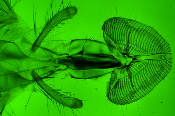
Fluorescence of the mount or other components - over half of the slides, both older and more recent, in the author's collection seemed unsuitable for this reason. Thus under the activation wavelength the mount was noticeably fluorescing instead of, or in addition to, the subject. This often resulted in a lack of contrast and subject definition, but in some cases, where the mount emitted strongly but the subject didn't, the result was a quite interesting 'backlit' effect, as the example below illustrates. The mount of the blowfly below isn't stated but likely to be Canada Balsam for this vintage, noted by Rost for its autofluorescence. Although photographed with a 100W mercury arc, using a 100W tungsten halogen lamp as epi source gives a bright fluorescing slide mount readily visible to the eye and for photography.
Blowfly tongue, Victorian
slide, unnamed (Watson style). Zeiss 2.5x NA0.08 objective. 'Zeiss blue' cube
excitation. The mouthparts are not fluorescing but the mount is, so
gives a contrasty 'other worldly' look to the subject not quite the same is
if a green filter was used in transmitted visible. Exposure 3 sec, ISO200.
Although subject brightness in epi studies
is proportional to the fourth power of the NA (ref. 4), the low NA of this objective
gave bright visible fluorescence imagery (and was used for many other subjects).
In the author's old slides however, the degree of autofluorescence of the suspected balsam mount seemed to vary widely. Thus the example below was weak compared with the subject itself allowing a contrasty image of the fluorescing subject to be taken.
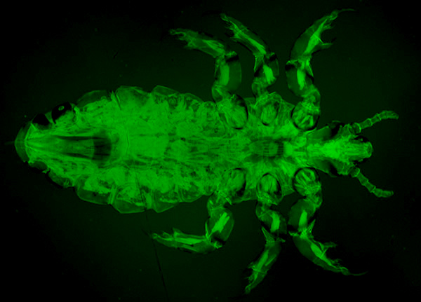
Human louse, Victorian
mount by C. M. Topping.
Left: 'Zeiss blue' cube. Zeiss 2.5x NA0.08 objective.
The internal structure
is clearly seen in this splendid mount. Exposure 8 sec, ISO200.
Right: Leitz
green cube. Leitz 6.3x NA0.20 objective. Exposure 2 sec, ISO200.
This subject
and a number of others shared here seemed to have a broad emission with both
blue and green excitation.
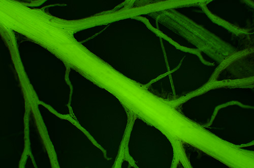
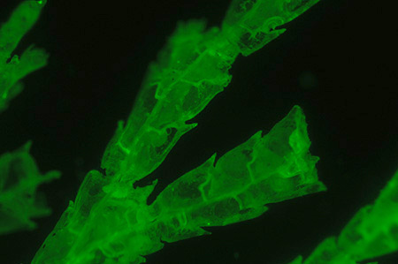
Plants
Some of the most successful subjects to date were old slides of untreated parts of plants in deep cells designed for incident light studies. I'd always regarded these slides to be rather drab in incident visible light but was pleasantly surprised by the interesting fluorescence imagery they offered. The absence of any mountant and competing autofluorescence possibly aided the viewing. This has prompted the author to keep an eye out for similar growths on fresh plant material.
'Gymnogramma peruviana'
(Silver fern) slide dated 1881. Zeiss 2.5x NA0.08 objective.
Left: Incident
visible light using external side lighting from Russian lamp.
Right: Fluorescence
using 'Zeiss blue' excitation. Although the spore releasing structures are
clearly seen in brightfield, the fluorescence introduces an attractive tonality
and highlights these structures.
Exposure 4 secs ISO200.
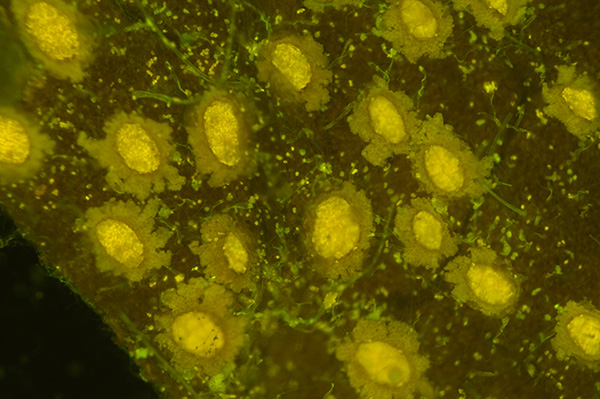
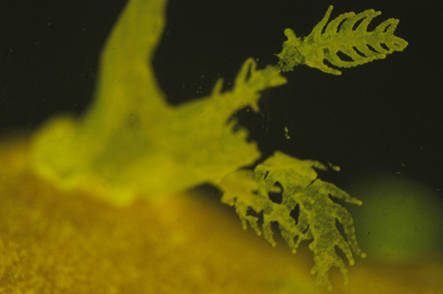
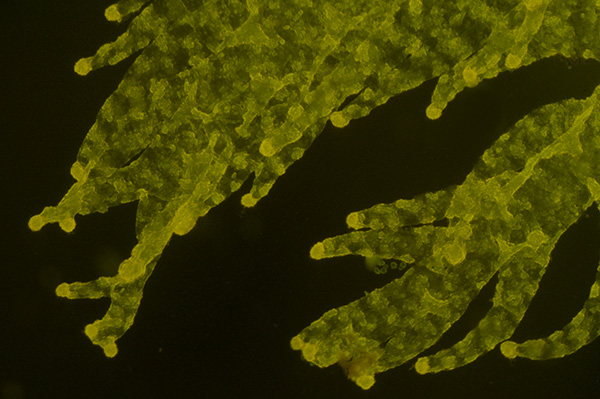
'Microfungi, cluster
cups' 'Aecidium epilobii' on leaf. Darlaston Victorian slide. Zeiss 2.5x NA0.08
objective.
Left:
Incident visible light using external side lighting from Russian lamp. The slide
suffers from fungal growth in the deep cell.
Right: Fluorescence using 'Zeiss
blue' excitation. Visible light shows the structures well but fluorescence gives
a complementary very different view of the fungal structures. The green hyphae
are the additional fungal growth. Exposure 2 sec ISO400.
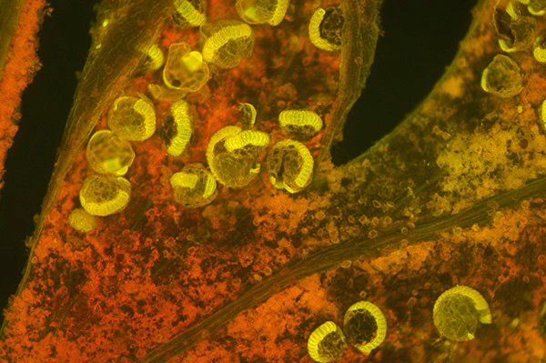
'Raphides in yellow
water lily, Nuphar lutea'. Victorian slide. Zeiss 2.5x NA0.08 objective.
Left: Darkfield
with green filter. Darkfield is non selective in showing both the structure
and debris in the field. Exposure 5 sec ISO400.
Right: 'Chroma blue' excitation.
In fluorescence the debris is not active giving a cleaner image. Some internal
detail is also more clearly seen. Exposure 5 sec ISO400. The subject also fluoresced
in green light to emit red.
Raphides are clusters of needle shaped calcium
oxalate crystals found in some plants.
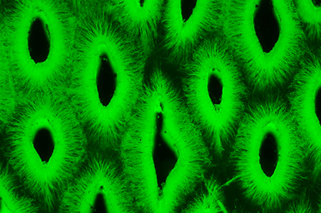
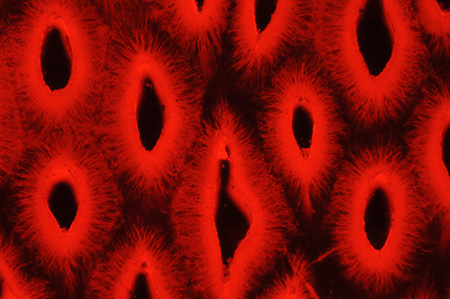
'Dissected box leaf, middle cuticle', Victorian unnamed slide. 'Zeiss blue' excitation. Leitz 6.3x NA0.2 objective. Exposure 1 sec, ISO200.
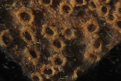
'Animals'
'Anthozoa. Corallium
rubrum'. A sea fan, common name Red 'coral' or Precious 'coral' (ref. 3). Victorian
mount by 'Flatters, Milbourne & McKechnie Ltd'. To the eye it is a pink
twiglike skeleton about 3 mm thick supporting the tiny polyps.
Left:
Fluorescence 'Zeiss blue' excitation, Zeiss 2.5x NA0.08 objective.
Right: Same subject
with Leitz 6.3x NA0.2 objective. Exposure 1 sec ISO400.

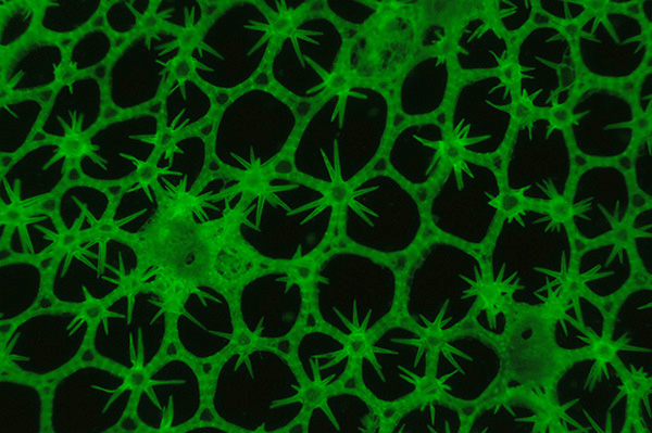
'Corallina. Sertularia
pluma'. Victorian unnamed, apparently, dry mount. Zeiss 2.5x NA0.08 objective.
Left:
Darkfield in white light. Exposure 1.6 secs ISO200.
Right: Fluorescence with
'Chroma blue' excitation. Exposure 15 secs ISO200.
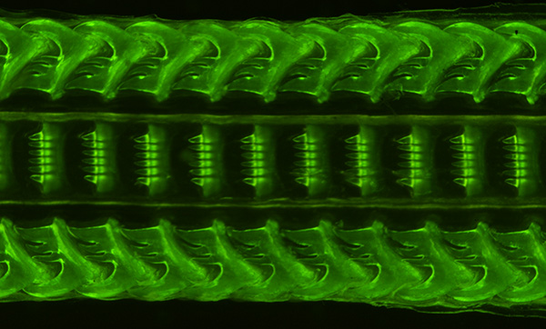
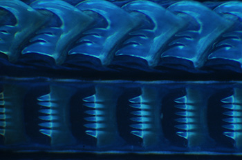
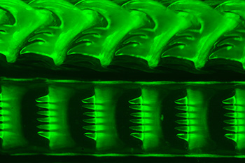
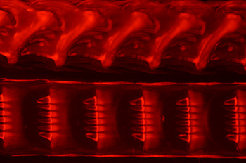
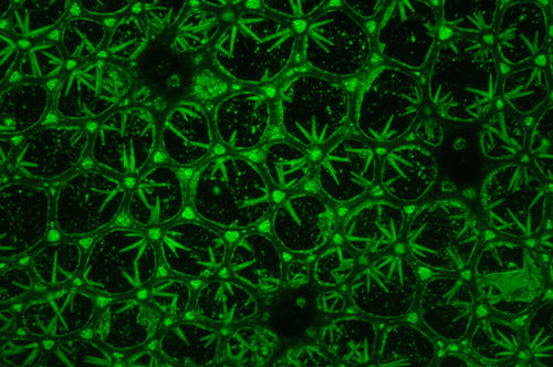
'Palate of whelk'.
Victorian dry mounted slide by 'J&TJ'.
Upper image: Zeiss 2.5x NA0.08
objective. 'Zeiss blue' excitation. Exposure 2 secs ISO200.
Second row:
Zeiss 6.3x NA0.16 objective. All exposed at 1.6 secs ISO400. Left: 'Leitz UV',
middle - 'Chroma blue', right - 'Leitz green'.
This appears to be a dry mounted
slide with black backing and gave contrastier results than the same subject
mounted by Wheeler for transmitted, presumably in Canada Balsam. This type of
subject, chitin? possibly modified by its processing, seemed to emit across
a broad spectrum of excitation wavelengths, as shown above. The author searched
for an emission spectrum for chitin and some of the other subjects above but
was unsuccessful.
Flurescence gives a surface detail view compared with transmitted
studies of similar subjects.
'Tooth of Myliobates.
Eagle ray. Transverse section.' Victorian unnamed and possibly a dry mount.
Zeiss 6.3x NA0.16 objective.
Left: 'Chroma blue' excitation. Exposure 0.62
secs ISO400.
Right. 'Leitz green' excitation. Exposure 0.62 secs ISO400.
This
was a very bright emitter, I'm unsure what the tooth material is for an eagle
ray.
Commments to date
For
the hobbyist, like the author, an interest in fluorescence studies is
likely to extend into the potential aesthetics of the imagery as well as the
information the technique may provide. As a beginner in this area, one
aspect that appeals is the element of surprise; some of the most interesting
subjects were slides I'd regarded as less so in normal studies.
In the author's slide collection it was mainly the older, probably dry mounted,
deep cell
slides that were most successful. i.e. a potentially competing fluorescing
mount was absent. This bodes well for studying fresh unprepared materials found in
nature e.g. microfungi.
Secondhand epi-fluorescent heads are readily available for many stands a hobbyist may use, eg for the Zeiss Standard and Leitz Labolux / Dialux etc. To date the author has found the UV excitation of a mercury lamp the least used for the prepared slides studied; blue and green excitation were much more useful. The intensity of the mercury lamp was helpful for short camera exposures and clear visual studies, but many of the subjects were readily visible using a quartz halogen lamp. If a sensitive camera like a modern DSLR is available, epi-fluorescence imagery with a 100W quartz halogen lamp and an epi head seems practical for subjects with brighter emissions with visible light excitation.
Comments to the author David Walker are welcomed.
Footnote: The filter cubes used in
a Leitz Ploemopak on a Diaplan were:
'A' - Leitz cube for UV excitation.
Present and tried, but few of the author's prepared slides gave a usable image; often because
of extensive none specific fluorescence of mount and subject.
'N2'
- Leitz cube for green excitation.
'Chroma blue' - a specialist blue excitation cube
in the head when purchased (labelled '41001 HQ:F'), with transmission
limited to 510 - 560 nm, green (ref. 5). This cube differs from a typical Leitz blue cube e.g. the
'I3' where
all light greater than 510 nm is transmitted.
'Zeiss blue' - for a wider
emission with blue excitation, a blank cube
was purchased and fitted with a ca. 510 nm dichromatic mirror by the specialist seller.
Zeiss 3mm thick blue excitation filters (2xBG12, BG38) and barrier filter ('Zeiss
50') used
in their III RS fluorescence head were used, which
conveniently are 18 mm diameter which also fit the Leitz. This approach
allowed a suitable cube to be built for a quarter of the cost of a commercial
Leitz cube.
(Safety note: If building cubes, the specs of the mirrors and
filters must be correct and undamaged to ensure no UV pass through, especially
if using a mercury lamp. If not activating in the UV, a UV blocking filter in
front of the lamp is a worthwhile additional precaution.)
Acknowledgements Thank you to Chroma (USA) for providing detailed information and spectra on the 'Chroma blue' cube fitted to the stand.
References
1) F. W. D. Rost, 'Fluorescence microscopy', vols. I and II, CUP, 1992. Obtainable by interlibrary loan or a local university library may have it and can also be 'dipped into' on Google Books.
2a) Above ref., Vol. II, page 177.
3) 'Seashores and shallow seas of Britain and Europe' by A J Campbell. Hamlyn Guide 1994.
4) B. Herman, 'Fluorescence microscopy', Bios Scientific Publishers, RMS Handbook No. 40, 2nd edition 1998, p. 19.
5) Chroma USA, personal communication.
6) Leitz Wetzlar, K. Koch, 'Fluorescence microscopy. Instrument, methods, applications', 1972, graph on p.8. The four stop difference remarked on was estimated for the general emission level of the mercury arc, not the specific wavelength peaks. Downloadable from www.science-info.net. Includes an excellent practical introduction to fluorescence microscopy.
Published in the April 2009 edition of Micscape.
Please report any Web problems or offer general comments to the Micscape Editor .
Micscape is the on-line monthly magazine of the Microscopy UK web site at Microscopy-UK
©
Onview.net Ltd, Microscopy-UK, and all contributors 1995
onwards. All rights reserved.
Main site is at
www.microscopy-uk.org.uk
with full mirror
at
www.microscopy-uk.net
.