Robert Pavlis, Girard, Kansas USA
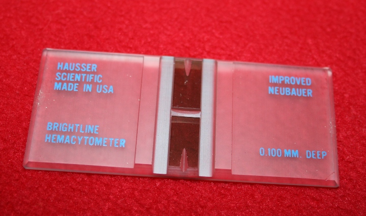
Over 100 years ago Louis-Charles Malassez invented an ingenious device to determine the concentration of particles in liquid suspensions--the haemocytometer. The device was originally designed to determine the concentration of blood cells, but it is commonly used for determining the number of any kind of particles in a suspension. Thus they are frequently used in both biological and physical science laboratories whenever there is a need to determine particle concentration.
An haemocytometer is really a very special extra thick glass slide that is graduated in a special way. Its surface is ground to a special shape so that there are two flat area that are etched with a special grids, and on either side of the etched areas there are slightly raised areas of glass that serve as supports that can hold a special thick cover slip 100 microns above the surface of the grids. (The reason that there are two grid areas on them is that when they are used for counting blood cells, one of the areas is used for leukocytes and the other for erythrocytes.)
Almost all haemocytometers are ruled in the same way. The rulings divide up the chamber between the cover slip and the slide into areas of known volume. The usual grid is as follows:
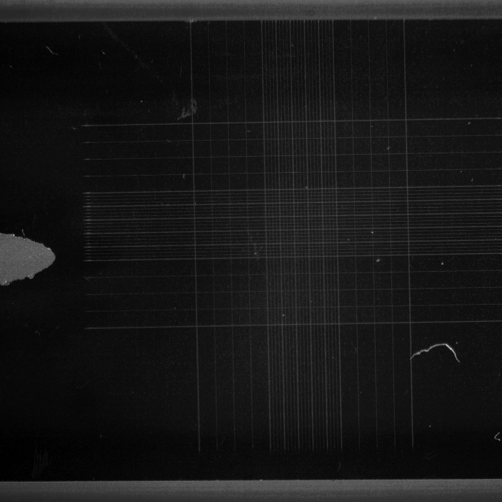
The image shown below shows one of the entire ruled areas on the haemocytometer shown in the first image. (I used my LOMO MBS-10 to obtain this image.)
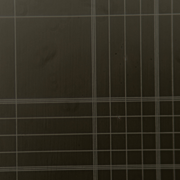
The image just below was obtained of the same haemocytometer using my Leitz Orthoplan microscope. The large square to the upper left is one of the 6.25 nL chambers. The smaller ones in the lower right are the 250 pL ones.
The next image shows basically the view that is seen when small particles like erythrocytes are being counted. (These are the 250 pL squares.)
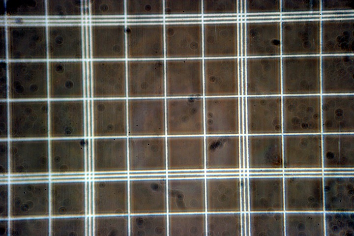
Different sized squares on the haemocytometer can be used for different concentrations of particles. (When used for counting erythrocytes in blood the small 250 pL squares are used. The blood is first diluted with saline solution to reduce the concentration to countable levels.) To get accurate concentration values, one needs to count many squares and average them because the number of particles in each square is subject to probability fluctuations involving the Poisson distribution.
If possible, one should try to count enough squares so that the total number of objects counted is at least fifty and preferably several hundred or more. (Some types of living cells may not be easily visible without using phase contrast.)
For particle counts to be accurate they must be mixed thoroughly in the liquid in which they are suspended. It is possible to check the distribution of particles by comparing the actual distribution of particles to the theoretical distribution given by a famous formula developed by the famous French mathematician, Simon-Denis Poisson.
The Poisson distribution formula is f(x)=(λx . e-λ)/x!
f(x) is the probability of exactly x occurrences when λ is the average number of occurrences. Although this expression is a continuous function, only integer values of x have physical significance because particles must either be in a given chamber or not.
The Poisson distribution formula is an important consistency check. If one should count the cells in 125 chambers and determine an average, the number of chambers with specific counts should not deviate too far from the Poisson distribution. When there are large deviations it indicates the material placed in the haemocytometer chambers was not homogeneous and that the determination should be repeated.
In this era of computers it is very easy to write a simple computer program that takes the data obtained by using an haemocytometer and then produces both the average number of particles per chamber and a Poisson distribution for that average to use as a check. A while back I wrote a program to do this. I think that it is correct, it certainly compiles correctly on any standard C compiler. This takes a data file with each line having the number of objects in a cell.
Haemocytometers are obviously very precision devices. Precision devices tend to be very expensive. However, because they are made in fairly large numbers, they are mass produced, so they sell for fairly reasonable prices. In fact they generally sell for less than simple stage micro-meters! Sometimes they are sold with special pipettes for diluting blood samples for determination of blood cell concentration.
When measuring particle concentration it is often necessary to dilute samples. There are special dilution pipettes for blood, as mentioned above. For general particle counting, however, it is better to have available standard serological pipettes. Chemists, for some truly bizarre reason, seem to like pipettes that are not calibrated to their tip. Biologists (and sensible chemists!) use the far superior ones that are calibrated to the tip. When a sample is found to have too many particles to count, it is a simple matter to dilute it!
Because haemocytometers are fairly thick they cannot generally be used on inverted microscopes. Because haemocytometers are often used to count living cells phase contrast may be required to find them! The chamber thickness and the extra thick cover slip tend to preclude using high power oil immersion objectives, though 40X and 60X dry objectives usually do not have any problems. Sometimes, however, there are problems with phase contrast systems because the glass thickness. Special thinner "phase haemocytometers" are available for phase microscopes that have these problems.
The prices of haemocytometers tends to be rather variable. In US dollar terms it is possible to find them priced anywhere from about $12 to perhaps $200. (The thinner ones specifically made for phase contrast tend to cost much more than the ordinary ones.)
Hospital laboratories tend to use flow cytometry to determine blood cell concentration. These devices are dramatically more expensive than traditional haemocytometers, but they require less labour to operate.
There is an important fact that many people fail to recognise: ONE CAN USE HAEMOCYTOMETERS AS STAGE MICRO-METERS TO CALIBRATE RETICLE OCULARS AND FILAR MICRO-METERS.
All comments to the authors via Robert Pavlis are welcomed.
Microscopy UK Front Page
Micscape Magazine
Article Library
Published in the May 2010 edition of Micscape Magazine.
Please report any Web problems or offer general comments to the Micscape Editor.
Micscape is the on-line monthly magazine of the Microscopy UK website at Microscopy-UK
© Onview.net Ltd, Microscopy-UK, and all contributors 1995 onwards. All rights reserved. Main site is at www.microscopy-uk.org.uk with full mirror at www.microscopy-uk.net .