Victorian Rambles - Part II
Three 'injected' microscope slide preparations.
by David Walker, UK
Old microscope slides can be enjoyed on many levels, and this occasional series is a random dip into the author's and brother's modest collection to illustrate some hopefully interesting slide subjects and brief aspects of their history. Different lighting techniques are explored to show their relative merits where appropriate.
Injected microscope slide preparations are those where vessels in the subject matter have been injected with a medium to better present the structure. There is a wealth of resources on the history of such slides prepared specifically for microscopy and also injected preparations for anatomical studies, the latter which date back to the 17th century (1-3). My brother and I only have three injected slides in our slide collection, none of which were specifically bought but were found amongst purchases of small general selections of slides.
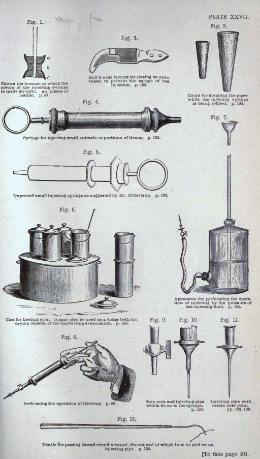
A number of the 19th century 'classic' microscopy books (many available on www.archive.org) have a section on preparing injected microslides. Lionel Beale in his book 'How to Work with the Microscope' (4), first published 1857 and in later editions, has an extensive coverage of procedures for injecting various subjects, including suggested recipes for injecting media; a book plate is shown below.
|
Plate XXVII from L S Beale's 'How to Work with the Microscope', fifth edition, 1880. Click image for larger version.
The plate shows the variety of apparatus typically used for injecting the slide subjects.
(Screen capture from a scan of the book on www.archive.org.)
|

|
The 'Micrographic Dictionary' by Griffith and Henfrey (published 1855 and later editions) has an extensive entry for ''INJECTION' providing injection procedures and a variety of formulae for coloured injection media. Three images from Plate 31 are shown below with the original captions.
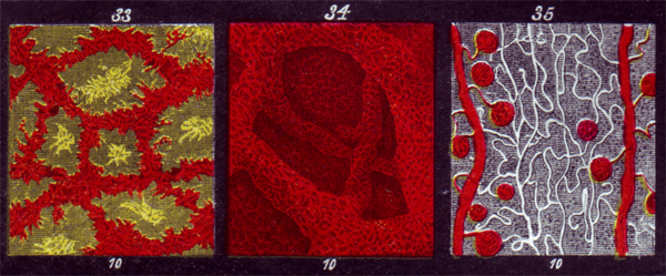
Crop of Plate 31 from the 'Micrographic Dictionary' by Griffith and Henfrey - three images from the plate showing injected preparations,
(scan from author's copy of fifth edition, 1875).
The 'Micrographic Dictionary' also describes 'self-injection' where an unfortunate subject such as a live frog was injected using the heart to pump the injection medium around the 'vascular system'.
Although such books did discuss in some detail the methodology of making slides and are referred to in later works such as those by Carpenter, it's interesting to note that in 'The Microscope and its Revelations' by Carpenter (eighth edition, 1901, revised by Dallinger) the following remark is made under 'Injected Preparations' (page 1061):
"The art of making successful preparations of this kind is one in which perfection can usually be attained only by long practice and by attention to a great number of minute particulars ; and better specimens may be obtained , therefore, from those who have made it a business to produce them than are likely to be prepared by amateurs for themselves."
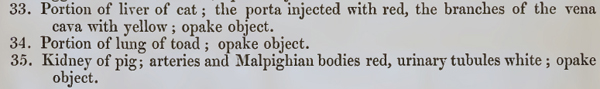
Three examples of injected preparations:
The three Victorian slides discussed. Only the middle example is named, by 'Hett 1850'. The right hand slide is double injected and the label reads 'Section of Human Kidney' and 'Artery Red Vein Blue injected'. The left hand slide uses a mountant, now partly oxidised, probably Canada Balsam. The other two have entirely colourless backgrounds but are believed to be Balsam mounts, see below.
Slide by 'Hett 1850': Alexander Hett is one of the notable 19th century makers of injected slides; Bracegirdle has an entry for him in his invaluable book 'Microscopical Mounts and Mounters' (3). (
Update May 16th, 2011: Brian Stevenson has just completed a fascinating illustrated essay on Hett and his work for his microscopist.net website which he has also kindly offered to share in next month's Micscape.)
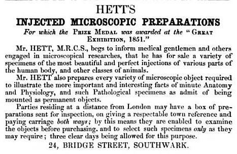
An advert by Hett in the back of John Quekett's 'Practical Treatise on the Use of the Microscope' published in 1852 edition. 'M.R.C.S.' indicates that he was a 'Member of the Royal College of Surgeons'. (Screen capture from a scan of the book on www.archive.org.)
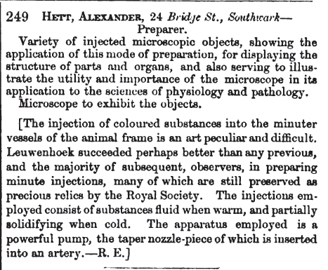
As the Quekett advert notes, Hett exhibited at the 1851 Great Exhibition in London where he states he was awarded the 'Prize Medal'; a transcription of the entry from the 'descriptive catalogue' is shown above (screen capture from Google Books). He also exhibited at the 'Paris Universal Exhibition' of 1855, catalogue entry on Google Books.
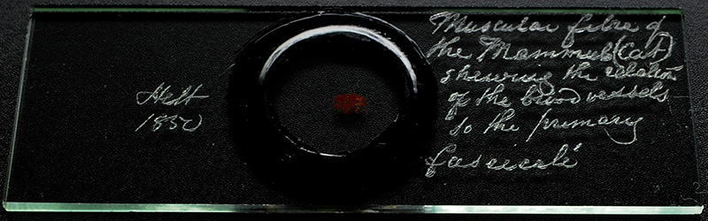
Image of author's slide with oblique top lighting to better show the diamond writing which reads: 'Hett 1850' and 'Muscular fibre of the Mammal (Cat) shewing the relation of the blood vessels to the primary fascicalé'.
The subject measures ca. 3.6 x 2.4 mm with ringed circular coverslip with no visible thickness to the preparation in side view. By inspection, it's uncertain if the subject has been mounted dry or in a mountant, as the slide remains perfectly clear, colourless and with no evidence of bubbles. However, both
the subject and background show green autofluorescence with a deep blue incident excitation light (shown below, 2.5X objective), a feature seen by the author for known 19th century Canada Balsam mounts.
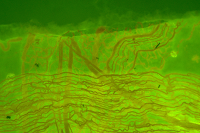
Green autofluorescence with deep blue excitation suggests a balsam mount for the Hett slide. Zeiss 2.5x objective.
This presentation of the subject, allowing in principle both transmitted and incident light studies, seems to differ from those by Hett seen to date described and illustrated in references, which show deep cell fluid mounts with an opaque black square background (3). The author would be interested to hear from readers who have come across other examples of this type of Hett mount.
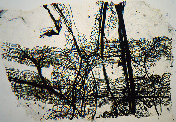
Transmitted light, Zeiss 2.5X objective. The vessels are opaque with little evidence of the three dimensional structure.
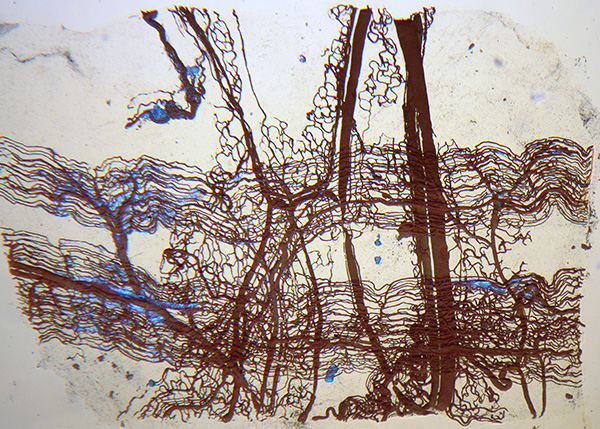
Transmitted and incident light, Zeiss 2.5X objective. The vessels now show some colour and three dimensional structure.
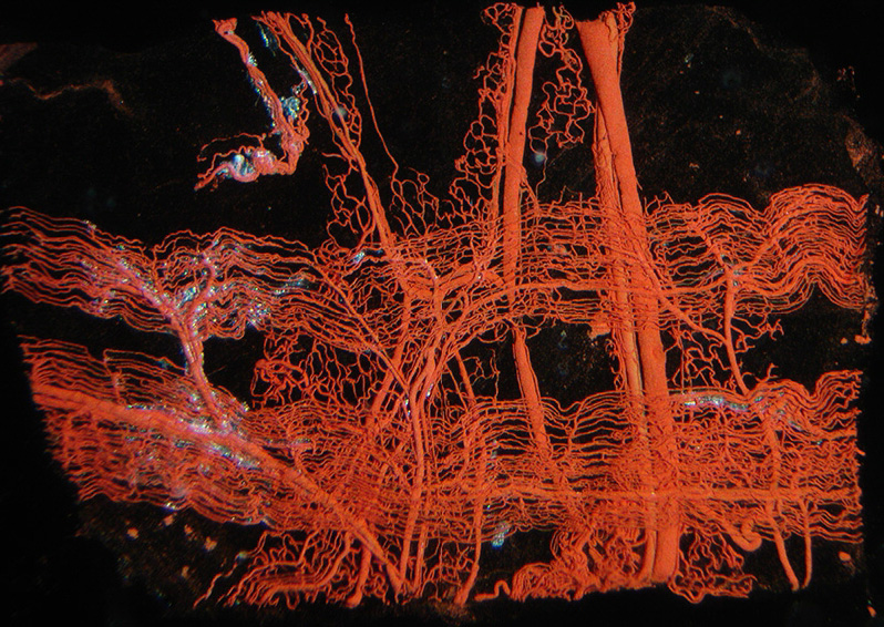
Incident light only, Zeiss 2.5X objective. This seems to be the most successful lighting with vessels now showing more colour and three dimensional structure. The latter is most impressive visually and demonstrates how effective the injection technique could be for showing complex three dimensional structure.
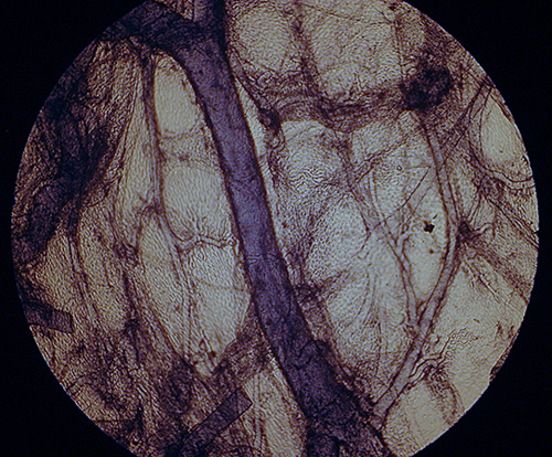
Injected lung wall of frog: This slide by an unnamed mounter was one of four conventional histology slides in a selection of slides purchased. The subject measures ca. 5.5 x 5 mm and is in a now partly oxidised balsam mount. Unlike the Hett mount, this mount works best in transmitted light but doesn't have the striking three dimensionality of the Hett slide. The finer vessels though are attractively rendered. Carpenter in 'The Microscope and its Revelations') has a short illustrated entry on the injected lung and lung wall of a frog (eighth edition, revised by Dallinger, page 1063).
Zeiss 2.5x objective, transmitted light.
Near infra red light gives better transmission through the denser thicker vessels, but is at the expense of contrast and detail on the finer vessels.
Zeiss 2.5x objective, Hoya R72 filter on field lens with 100W quartz halogen lamp, Sony S75 consumer digicam, home modified by author for exclusively near IR imagery (see this Micscape article for details).
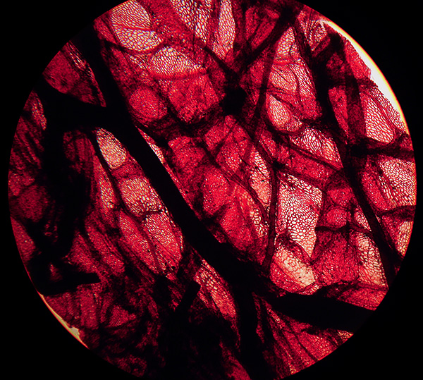
Injected human kidney: This is also an unnamed mount, the subject measuring ca. 16 x 12 mm. The mount is very clear but green autofluorescence in deep blue excitation light again suggests a Balsam mount. The handwriting has some distinctive capitals but did not seem to match those of other known makers of injected mounts of the time e.g. by Topping, Cole or Suter. Any comments on the likely preparer are welcomed.
Update May 14th 2011: Thank you to Howard Lynk who kindly comments that this slide is 'almost certainly' by Amos Topping from comparison with his own slide collection. He notes that there is a 'very similar example' by Topping on his 'Victorian Microscope Slides' website, (2nd image row from bottom, 3rd from left) showing Amos Topping attributed slides. Amos, the son of Charles Topping, were both noted preparers of slides, see entries for father and son in ref. 3.
This is an attractive dual injected specimen and delineates the arteries and veins and other structures well, and best
viewed in transmitted lighting. There is relatively little three dimensional information cf the Hett slide. The exact dyes used are uncertain, but the 19th century textbooks describe dyes both in soluble forms and as suspended fine solids in a medium; dyes mentioned include carmine (crimson) and Prussian blue.
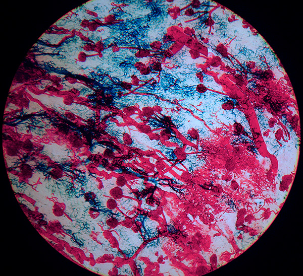
Zeiss 2.5x objective, transmitted light.
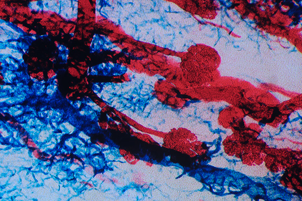
Zeiss 6.5x objective, transmitted light, slight oblique.
These type of slides are a fascinating genre of the microscope slide maker's craft. Old microscope slides of many categories seem to be attracting increasingly higher prices judging by recent eBay sales, but injected specimens may slip through at reasonable prices if not well described.
Comments to the author are welcomed.
References
1) 'B Bracegirdle, 'The Art of the Injected Preparation', Quekett Journal of Microscopy, 1987, 35 (7), 489-498.
2) B. Bracegirdle, 'A History of Microtechnique', 1986, second edition, Science Heritage Limited, p.10 ff, p.84 ff and extensive illustrations and plates.
3) B. Bracegirdle, 'Microscopical Mounts and Mounters', Quekett Microscopical Club, 1998, p.51, 55, 56 and plates 50 and 59.
4) L. S. Beale, 'How to Work with the Microscope', 1880, fifth edition. This and other editions are available in various formats on www.archive.org.
©
Microscopy UK or their contributors.
Published
in the May 2011 edition of Micscape.
Please
report any Web problems or offer general comments to
the
Micscape
Editor
.
Micscape
is the on-line monthly magazine of the Microscopy UK web
site at
Microscopy-UK
©
Onview.net Ltd, Microscopy-UK, and all contributors 1995
onwards. All rights reserved.
Main site is at
www.microscopy-uk.org.uk.