|
Leftovers–An Eccentric Gallery by Richard L. Howey, Wyoming, USA |
I thought about going into my lab to start taking pictures for Part 2 of the article on chitons, but then I felt lazy and to separate out the structures I want to investigate, I would have to use copious amounts of Clorox, the smell of which I still detest. So, I went into the depths of my image files on the computer. I decided to start with 2007, the year before the crash of the economy and my computer. Initially, I thought I could cull and get rid of a lot of old images, but as I browsed, I discovered some interesting bits and pieces many of which I had not gotten around to processing. Others I had and I suspect had even used some of them in some articles, but I had forgotten them and they once again captured my interest. So, if you find an image or two that seems familiar, I’ll just plead absent-mindedness. The images won’t be in any particular order, because there is no underlying theme except “Interesting Images”.
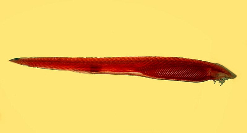
First up, is Amphioxus. This strange little creature is one of those borderline beasties; it is one of the most primitive of vertebrates. If you look carefully, you can see the notochord running down the length of its back.
Transitional forms between classes, orders, families, phyla, and kingdoms are always of great interest and importance. (Just consider Henry the Eighth!)
Nature seems indifferent to our attempts to classify and categorize and it’s at this boundary point that we find Urochordates (“primitive chordates”), Hemichordates, and Cephalochordates. The hemichordates, such as, Balanoglossus and Saccoglossus, the “acorn worms”, are thought to be the closest living invertebrate relatives of the chordates. Ascidians also have a notochord, but only in the larval stage as it disappears in the adult forms. If we start looking carefully and critically at the morphology of the organism, we quickly discover that we need to know much more, and recent advances in molecular biology can help in advancing our understanding while, at the same time, making everything enormously more complicated, so it’s serendipitous that we have computers or we would be totally lost.
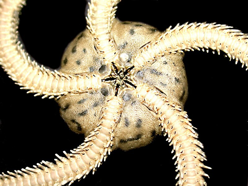
The next image is a closeup of the underside of an ophiuroid or brittle star. These are among the speediest and most prolific of the echinoderms. There are areas of the sea bottom which are carpeted with these organisms. They have very flexible arms and when grabbed by one can disconnect it at the point of impact and whisk away to safety (sometimes). This is a process known as autotomy and there are other echinoderms that are capable of it as well. If you look at the arms, you quickly realize how many small joints there are.
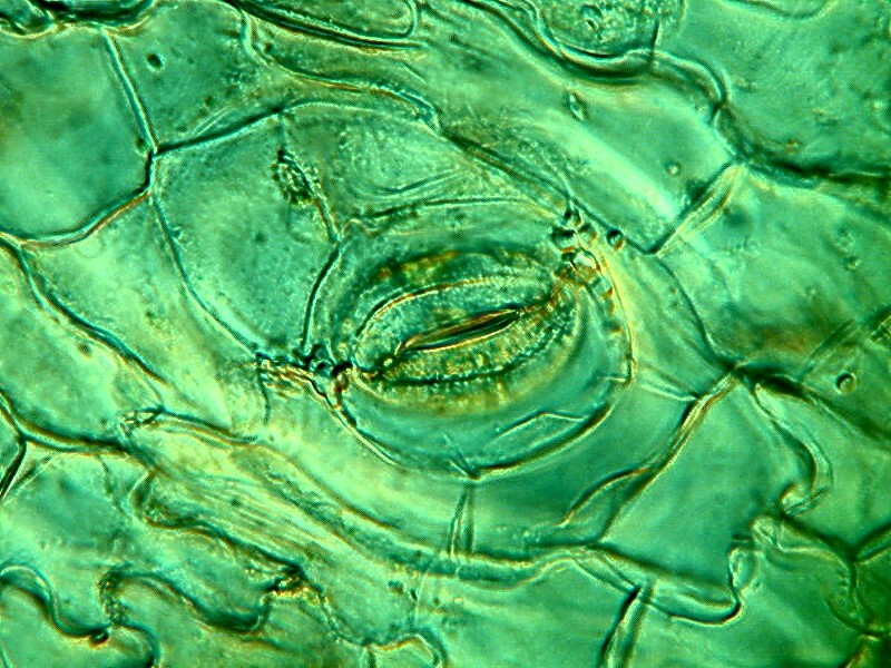
Now, we’ll shift kingdoms and jump over to a couple of closeup images of parts of a cactus. The first one shows the cells in a bit of tissue surrounding a stomata which is involved with respiration. The second image is a spine from that same cactus as viewed with polarized light.

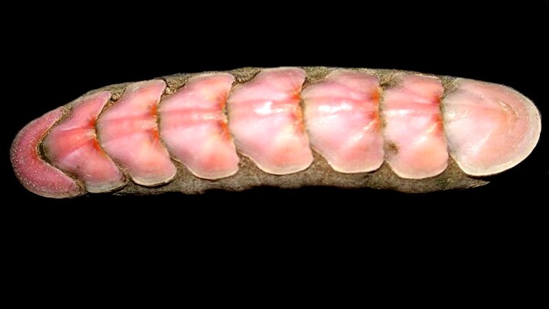
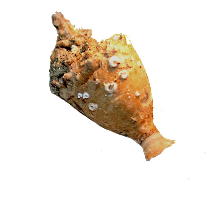
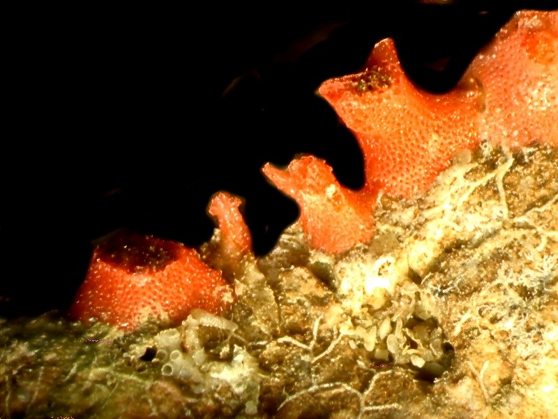
While I was procrastinating regarding doing the nitty-gritty work for Part 2 of the chiton article, I happened across this image below of a splendid chiton specimen which I took 9 years ago and then forgot! A clear indication that I need a keeper or, at the very least, a cataloger/secretary.
This is one of the veriform species and has lovely pastel color.
I mentioned earlier that I am absent-minded, well, here’s what happens to the brain of an absent-minded individual.
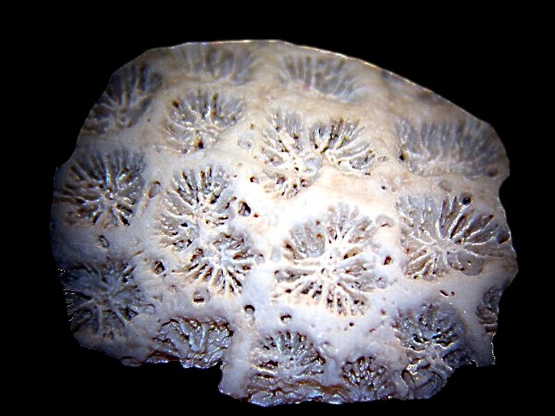
This is a fascinating piece of coral and, of course, the polyps are long gone as this is a fossil specimen.
When my wife and I got up this morning we were very grumpy, in fact, positively crabby. I’m the one at the top.
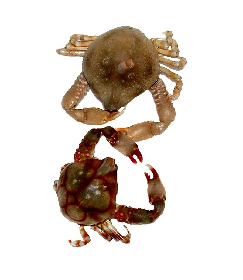
These delightful little creatures are popularly known as Porcelain crabs.
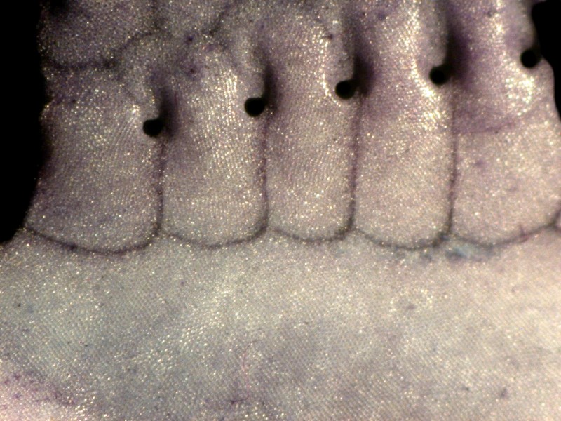
Sometimes, one finds something quite beautiful in unexpected places. Who would think of finding a delicate, lacy disk in the rubbery foot of a sea urchin?
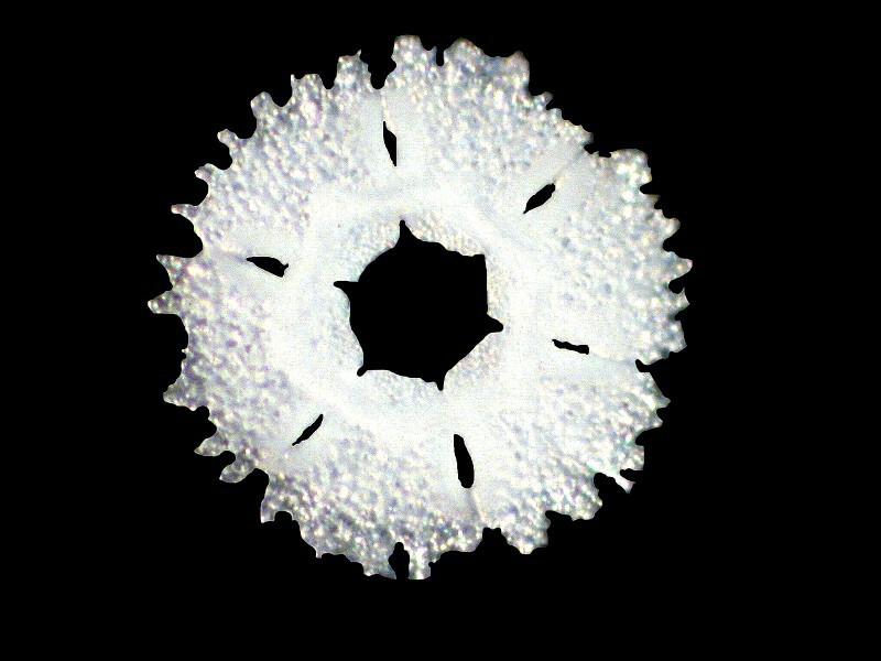
This elegant structure is from the echinoid Strongylocentrotus purpuratus. It is composed of a series of 6 fused calcareous plates each of which is composed of sub-plates each of which is composed of hundreds of tiny crystals.
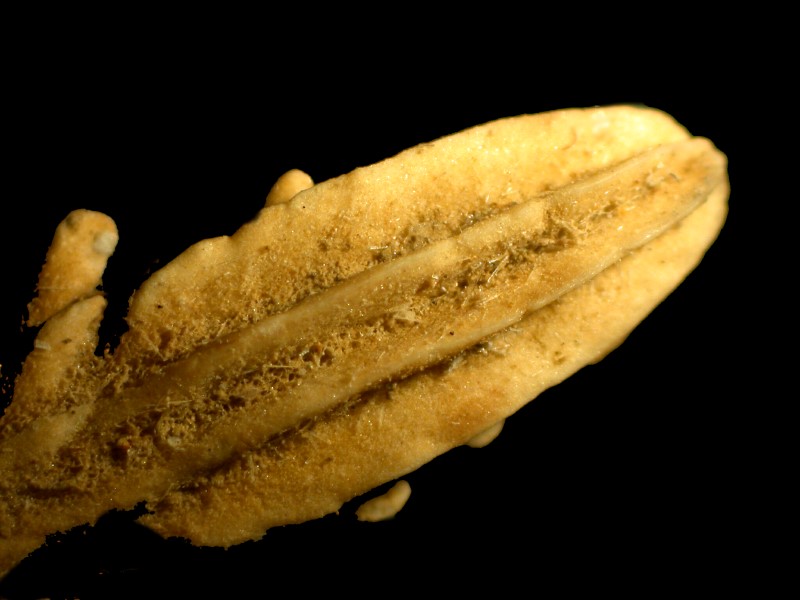
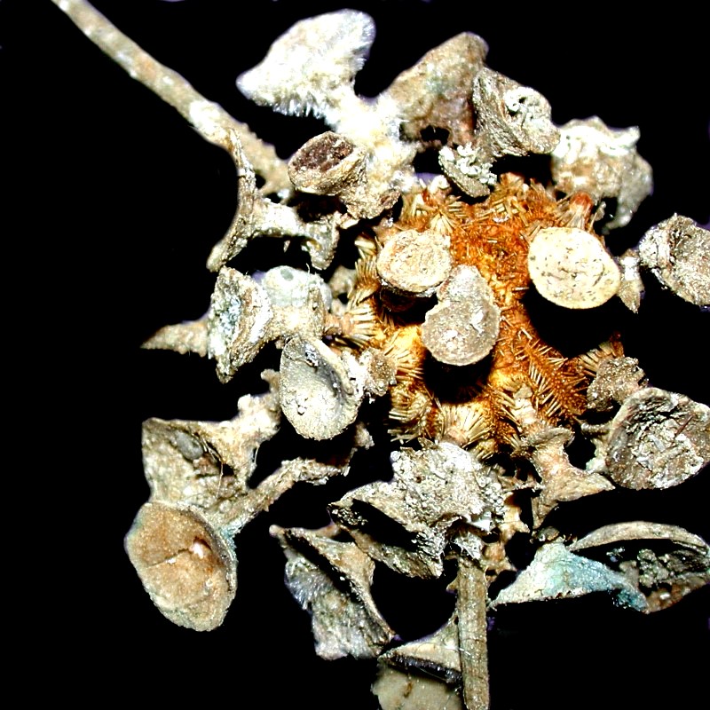
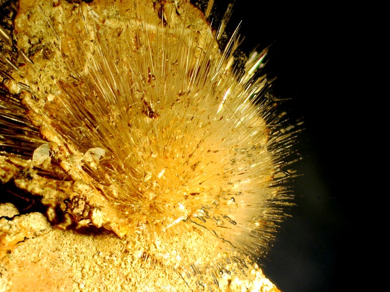
This next object is quite extraordinary; it is an ancient Greek pottery vessel and, as you can see, it has been excavated from an area with iron-rich brownish-red soil. Oh, Plato and I keep telling you that philosophers and poets are liars. What this actually is, is even more surprising. It is the spine of a sea urchin with the odd name of Psychocidaris ohshimai. I would tell you that the generic name means “crazy, psychotic little urchin,” but then I’d be lying again. First, I’ll show you the whole urchin and then an image of a bizarre spine.
The variation in sea urchin spines is truly astonishing and we’ll look at a couple of other examples a bit later on.
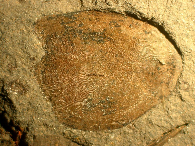
Here’s another fossil; this time an imprint of a fish scale. The rings on the scale can be used to determine the age of the fish and the first suggestion that this was so came from Aristotle.
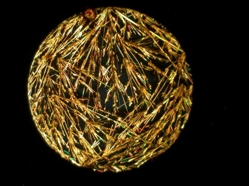
Several years ago, I came across a box of slides which had 10 small circles printed on them. I bought them because I thought it would be interesting to try a different combination of crystals in each circle and then examine them under polarized light. Well, here’s one example; it’s sodium bicarbonate.
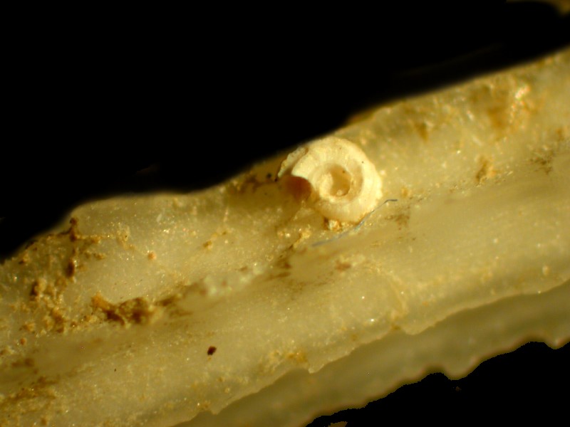
If you go back up and look at the strange urchin spine of Psychocidaris again, you’ll notice several small spiral white structures on the surface. These are tubes of the worm of the genus Spirorbis. They show up rather frequently on urchin spines and we’ll look at some examples here.
Here is an image of a portion of a bladed spine of a tropical urchin. And immediately following it, on a different part of that spine, we see a specimen of Spirorbis.
However, Spirorbis are equal opportunity hitchhikers; that is, they will take what ever opportunity is offered to attach, and, in the image below, we see one on a section of a plate from the test of a sea urchin.
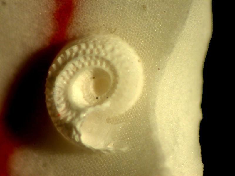
If you look closely, you can see the packed crystalline structure of the calcareous plate.
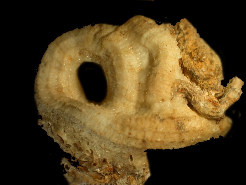
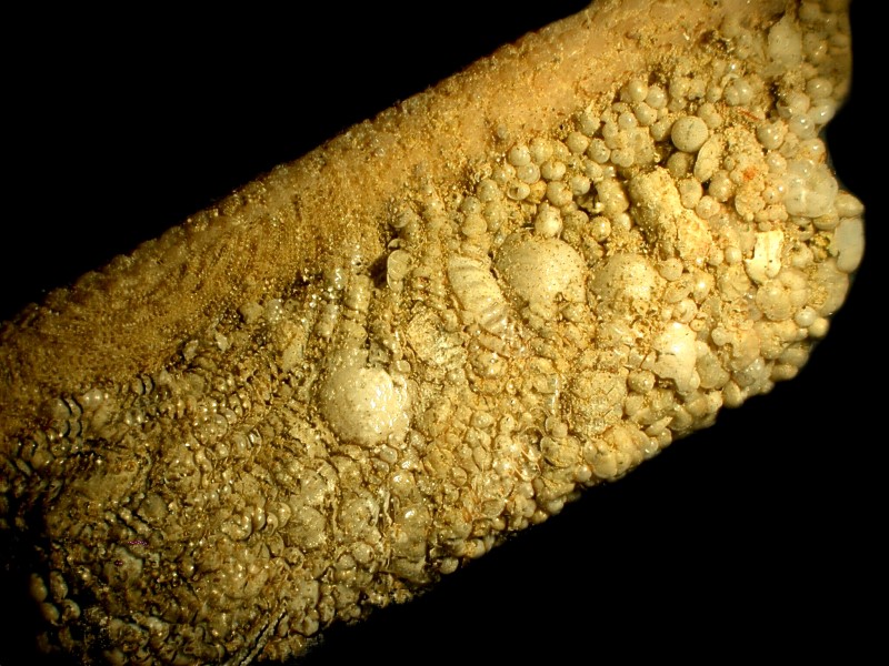
Tube worms are fairly common and can pose a problem both for other organisms and for humans and we have had to invent ways of covering and protecting undersea cables from damage by tube worms, boring sponges, and a variety of other creatures. Here are 2 other kinds of tube worms.
In that last photograph, you may have noticed something bright red in the upper right hand corner. That’s another amazing and anomalous organism.
Years ago, Brian Darnton kindly sent me a sample he collected from a coastal shore in Malta. It was full of wonderful forams, bryozoa, tiny mollusk shells, and some strange pink and/or reddish items which I had never seen before. I think that Brian was playing a subtle little game with me and I wrote and asked him if these were bits of red and pink coral. He wrote back promptly telling me that these were actually forams. FORAMS! I would never have guessed. They are in fact Miniacina miniacea. Talk about shaking up one’s categories.
And while we’re on the subject of forams, sea urchin spines, and tube worm tubes, here’s an image that combines all three.
Running along the top is the spine, then a tube worm somehow collected, glued together in a dense and compact form, an arrangement of forams to create a protective tube. At first, I didn’t realize what it was. The tube extends further down the spine past what we can see in this image and down there I found an opening and could see a mucous lining on the inside. Quite amazing!
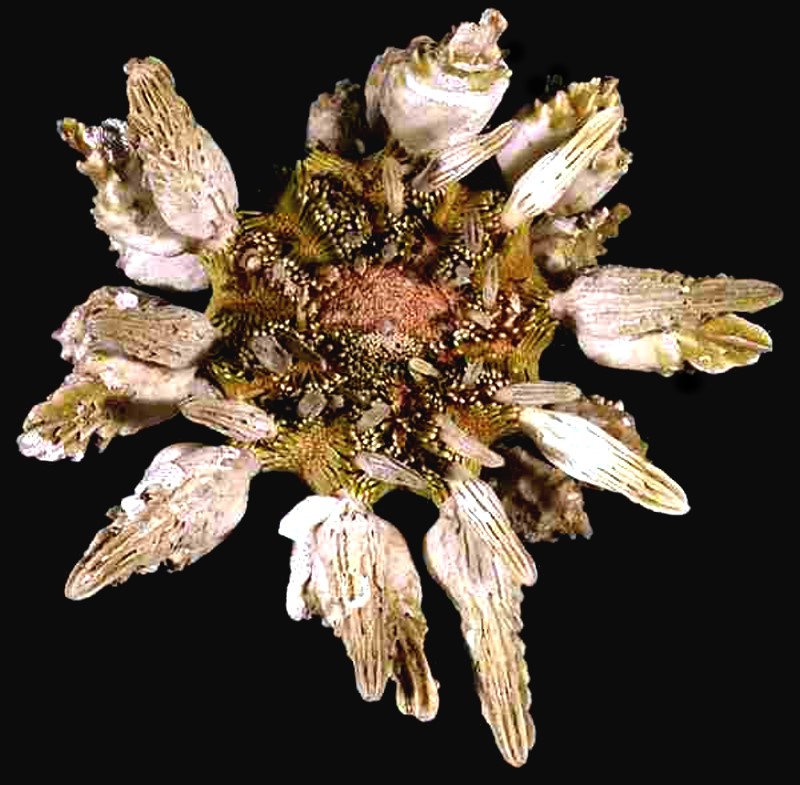
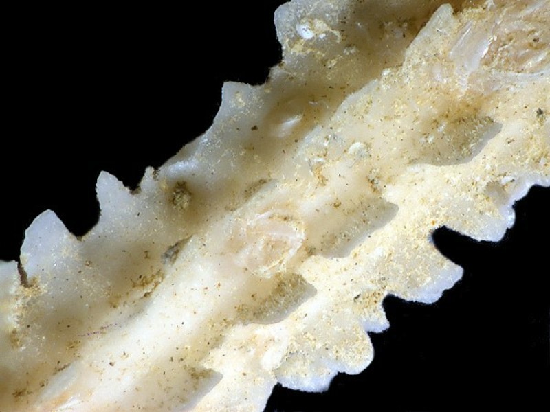
I mentioned earlier that we would look at some additional types of spines of sea urchins. An urchin that has adapted to living in coastal areas where there is heavy surf is Colobocentrotus atratus also known as the “helmet urchin” or the “cliff clinger”. Here you can see some splendid images of this intriguing organism.
I’ll show you a closeup of some of the smaller flattened spines which in this view rather remind me of foot pedals on a pipe organ.
Yes, I am often rather pre-occupied with sea urchins. They are in many ways both alien and beautiful and full of interesting puzzles and mysteries. Previously, we looked at the plate in a tube foot of Strongylocentrotus purpuratus. Now, I am going to show you a cross section of one of the spines; the color is natural and the image is brightfield. I produced this section using micro-tools and cut some spine sections until I got a few that were thin and relatively flat. I then used sandpaper and emery boards to make a section yet thinner and flatter.
Earlier, we also looked at a very eccentric spine of Psychocidaris ohshimai. It is fascinating that one can find considerable variation in the shape of spines in the same species and on the same specimen. Here is another rather differently eccentric spine from that same specimen.
And yet another species with highly unusual spines. There are a couple of “usual” “toothpick” spines, but most of these are little cups or inverted umbrellas (there are strong currents down there on the bottom). As you can see various sorts of extremely tiny things have taken up housing on the cups.
However, larger organisms also seek refuge here. In this case, a small sponge has settled comfortably into one of the cups.
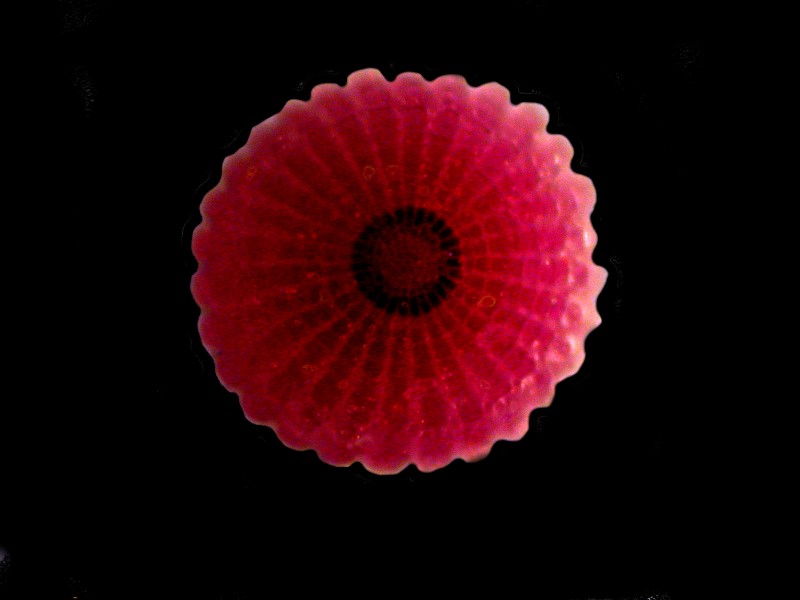
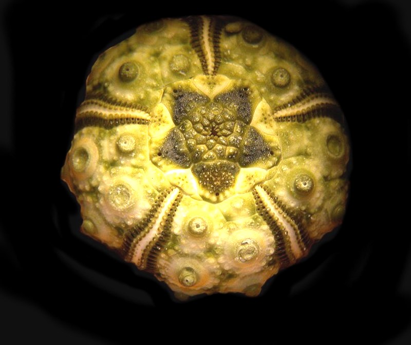
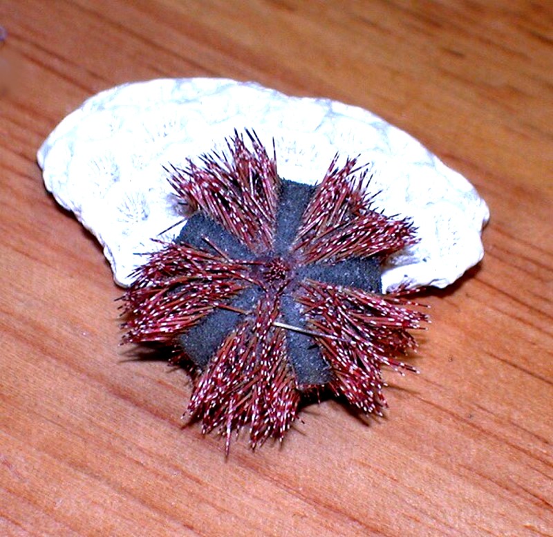
Next a look at 2 small urchins, the first is the test with the spines removed and the second is a specimen of Mespilia with spines intact, resting against a small piece of bleached coral.
These are beautiful objects in quite different ways and Nature is so full of such resplendent productions that we can easily be overwhelmed and reduced to silence. Just take a look at some of the varieties of Mespilia.
None of my images can approach this extraordinary display by Mother Nature and tomorrow (May 8, 2016) is Mother’s Day, so I’ll end here.
All comments to the author Richard Howey are welcomed.
Editor's note: Visit Richard Howey's new website at http://rhowey.googlepages.com/home where he plans to share aspects of his wide interests.
Microscopy UK Front
Page
Micscape
Magazine
Article
Library
Published in the May 2016 edition of Micscape Magazine.
Please report any Web problems or offer general comments to the Micscape Editor .
Micscape is the on-line monthly magazine of the Microscopy UK website at Microscopy-UK .
©
Onview.net Ltd, Microscopy-UK, and all contributors 1995
onwards. All rights reserved.
Main site is at
www.microscopy-uk.org.uk .