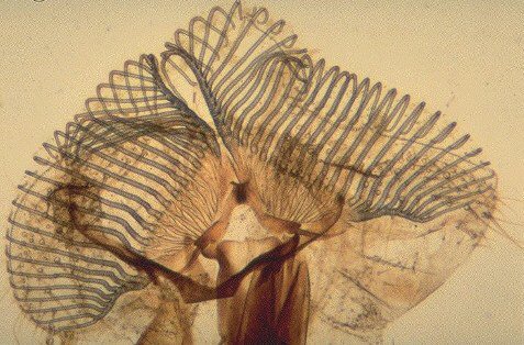
The main part of interest is of course the tongue, or more correctly the ligula. It is not really anything like a tongue in most insects, being quite indistinct, except in the crickets, bees and in flies. The ligula is extremely well developed in flies, forming what is commonly called the proboscis. This represents a curious modification of the ordinary tracheal structure where instead of the usual spiral structure the fibres are broken down into rings.
These rings do not surround the whole tube but are terminated in a set of arches that pass from one to another. These are the so-called pseudotrachea, suctorial organs which can take in liquid both at the extremities and throughout the whole length due to the interruptions of the rings.They really are quite wonderful muscular lips in effect, enabling the insect to employ the tongue as a prehensile organ. They are covered with rows of minute setae, directed a little backwards and arranged closely together.
There are very many rows of these minute hairs employed by the insect in scraping or tearing delicate surfaces. These hairs are what tickle somewhat when a fly alights on your skin in summer, to sip the sweat! It is this detail that makes the object useful as a test object of microscope objectives. The spiral form should be viewed under medium to high powers where it will be seen that the spiral structure doesn't exist, as mentioned above.
It is well worth considering here the depth of study carried out in the distant past. For instance, Mr Lowne's treatise on 'The anatomy and physiology of the blow-fly' published in 1869 and running to many pages. Also, the preparation of such objects. The mounting of the tongue must be done wth a considerable amount of care to show the structure. The most commonly used method is to slit it open, flatten it out and mount it in balsam. The famous mounter C.M. Topping started work in around 1839 and quickly acquired great skills in mounting. He refined a technique for preparing the proboscis of the blow-fly, in 1842, in a manner whuch became a favourite preparation. Others followed, and there are many of these Victorian slides still in existence, often in good condition. A number of other species were prepared and it is worth looking at these in comparison.
Finally, a quote from Richard Kerr, author of 'Nature through Microscope and Camera' 1905.;
"Perhaps no object in insect life has been photographed more frequently, yet it may be said with truthfulness that no microscopist has ever succeeded in either exhausting or in depicting all the detail. The proboscis of the blow-fly or the house-fly defies the highest amplification and still reserves to itself many mysteries never seen by the microscopist. It is more complicated than a locomotive, and more highly finished than a costly gold watch. It is a contrivance that commands our admiration although we are acquainted with but a few of its actions and uses. Its exquisite beauty, the minuteness of its thousands of springs, and the finish of its mechanisms have led many a man to reflect on his own impotence, and have suggested to his mind something of the sublime skill that must be behind all that we are pleased to call 'Nature.'"
Please report any Web problems
or offer general comments to the Micscape Editor,
via the contact on current Micscape Index.
Micscape is the on-line monthly
magazine of the Microscopy UK web
site at Microscopy-UK