Dileptus at Dinnertime

by Rosemarie Arbur, Oregon, US


In August, I had taken some soil from the edge of an irrigation reservoir—some merely damp, some barely covered by water at the shoreline—and, just when I thought I'd become familiar with its inhabitants, a Dileptus showed up on my slide.  My original idea for this article was simply to share what I saw.
Here: 
 
Dileptus and Litonotus A fair-sized Dileptus (650 µm long).  It's carnivorous. It has smooth-looking ciliation, an exquisitely tapered tail, and a pellicle that in dark field looks like beaten silver. Its neck, in which the myonemes are visible, is flat and, seen from the side, quite delicate. Dileptus waves it around a lot.


Litonotus is a carnivore, too; this is a big one. If Dileptus looks frantic, waving its neck, Litonotus swims elegantly and with poise.

 
My second idea for this article, after discussing it with Dave Walker, was to include some words about Dileptus and Litonotus, responding to "what's a 'Dileptus'?"

Dileptus: Phylum Ciliaphora, Class Gymnostomea, Order Haptorida, Family Trachelidae.
Litonotus: Phylum Ciliaphora, Class Gymnostomea, Order Pleurostomatida, Family Amphileptidae.

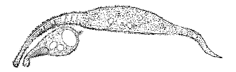
Both ciliates belong to the class Gymnostomea, which have an anterior mouth at or near the surface; it's supported but not constrained by trichites—internal rod-like structures—and can stretch greatly to accommodate large prey. The nucleus of both is polyploid, but the Haptorida (Dileptus) have toxicysts around the mouth, and the Pleurostomidae (Litonotus) do not. Both have long, flattened necks and relatively large bodies.

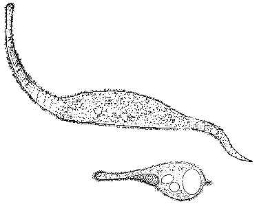
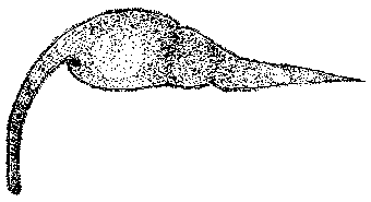
Dileptus' cytostome is round, at the base of the neck; its neck is usually in motion; water-expelling vesicles are small and numerous; and Dileptus is usually larger than 250 µm. Here's a site with more information and photomicrographs of different species of Dileptus. (And, while some ciliates reproduce every few hours,  Dileptus normally requires a day or more between fissions.)

Litonotus' cytostome is a long slit; it has a prominent posterior water-expelling vesicle, a smooth pellicle, and it's typically 75-150 µm long.

Dileptus and Litonotus can be confused with each other and with Lacrymaria olor. The position of the mouth is distinctive: Lacrymaria's is at the end of its very elastic neck, Litonotus' is on the side of its neck, and Dileptus' is where the neck and body join. Motion of the neck differs, too: Lacrymaria's is whipped out and around, noticeably changing length,  Litonotus' is usually carried at a slight angle to the body, and Dileptus' sweeps side to side in arcs exceeding 135°.  Litonotus is much smaller than the other two. Once you've seen all three, confusion vanishes. 

 
Now, back to what I saw.  First . . .
 
Dileptus and Litonotus


Then . . .


Just as  Litonotus draws even, Dileptus' neck reaches out. It conforms itself to the curve of Litonotus' body. The contact between the organisms is delicate as a caress,
Dileptus and Litonotus
but deadly.  Litonotus' pellicle ruptures, and cytoplasm begins to leak out. Dileptus' strong, flexible neck gathers and sweeps the dead Litonotus toward the elastic (now huge) mouth at the base of its neck. Very little of the Litonotus is lost; a food vesicle forms instantly, and the digestion of Litonotus is almost fast enough to see.


 And then . . .
 

Dileptus

Dileptus contracts itself slightly and performs metaboly (creates a "waist" that, moving from anterior to posterior and back, molds the body into temporary, round "waves"). The contraction and metaboly cause Dileptus' body to become less transparent because thicker. The vesicle containing Litonotus begins to grow indistinct.


My third idea for this article—actually, the very first motivation—is the appearance of the living organism vs. the usual drawing of it. Dileptus is usually drawn with its mouth open, a semicircular cutout at the base of its neck, and with long, shaggy cilia. But when Dileptus opens its mouth—an action that occurs at intervals; it doesn't swim about with its cytosome gaping—the mouth doesn't appear in silhouette but as a (relatively) huge, dark entry into the organism's body. As for the cilia, even my illustrations on-screen seem to me to emphasize it too much. Adjusting the focus and illumination carefully, one can see the cilia—on the body and around its edges—but, seeing a live Dileptus for the first time (and quite a few times thereafter), one is struck by its size, its beautiful "muscularity," and its fascinating behavior.
 
 
Comments to the author Rosemarie Arbur are welcomed.


Microscopy UK Front Page
Micscape Magazine
Article Library


© Microscopy UK or their contributors.

Published in the November 2000 edition of Micscape Magazine.

Please report any Web problems or offer general comments to the Micscape Editor,
via the contact on current Micscape Index.

Micscape is the on-line monthly magazine of the Microscopy UK web
site at Microscopy-UK

WIDTH=1


© Onview.net Ltd, Microscopy-UK, and all contributors 1995 onwards. All rights reserved. Main site is at www.microscopy-uk.org.uk with full mirror at www.microscopy-uk.net.