Well, there's the migraine-inducing Schoenberg and the Ueber-stentorian Wagner and Telemann (whose 800,000 works sound virtually identical) and there's the egregiously verbose Stephen King, the semi-literate Hemingway, and the ever-tedious Faulkner and then there's Rothko, whose mother must have deprived him of a paint set as a child, and Mondrian, the linoleum designer, and Jackson Pollack, the dribbleroops, I meant to write an essay on debris in cultures and samples and got off on the wrong track. Let me start again.
Whenever I'm looking at living pond cultures or preserved samples or dried up samples or debris attached to dried marine specimens, I can't resist going in and removing some samples for microscopic examination. I'm sure you all have nice, neat, orderly laboratories with everything properly arranged and you also, no doubt, know exactly where everything is. Imagine the antithesis of that and you will have a general idea of what my lab is likecluttered, messy, disorganized with room only to accommodate one and a half persons. As a consequence, I frequently come across dried up cultures and samples, so it's no wonder that I am so often distracted.
I remember some samples I took from a very fertile little pond and the samples eventually dried up. When I "re-discovered" them, I was fascinated by the debris which had accumulated on the surface of the mud. Of particular interest were dozens of minute snail shells, from, at least, three different species and the pink dried remains of several small freshwater "shrimp" (Gammarus). These intriguing creatures are of a grayish-green (or greenish-gray) color when they are alive and are acrobatically active, which makes them quite fun to observe in small aquaria. They are possessed of a special pigment such that when they manage to climb out of a culture dish (or when they dry up in one), they change to a distinctive pink color. They are a favorite food of trout and when they are abundant, they account for the delicate pink and very succulent meat of freshwater trout.
Strings of dried filamentous algae were also to be found and when mud samples were scraped, moistened, and examined on a slide, diatoms and desmids were also present. However, the thing which impressed me most was the realization that I was observing an early stage in the process of one type of fossilization. A few more layers of mud, some intense pressure, a few million years and some life form, hopefully much more intelligent than we are, might be observing the very same types of specimens which I was looking at.
It is not difficult to create a pond environment or two or three in your lab or in a basement window. A large jar or small aquarium serves well. If there is proper light, a few aquatic plants, and a cool room temperature, these pond environments can be self-sustaining for periods of several years. Too much light and you may find the sides of the jar coated with algae, which is fine if you want a supply of algae to study and so I usually keep one or two small jars just for that purpose. You have to keep the snails out however, or they'll make breakfast, lunch, and dinner out of your algae crop. From time to time, it's worthwhile examining the debris and sediment in such a jar and you may well find not only the remains of diatoms and desmids, but living flagellates and small amoebae as well.
In the case of the non-algae jars, you may wish, from time to time, to add parts of new samples you have collected or you may even wish to select specific organisms to isolate and introduce into your jarsDaphnia (or other cladocerans), copepods, Hydra, Stentor coeruleus, tube-building rotifers, such as Floscularia, or dragonfly nymphs or hellegramites. However, if you introduce either of these latter two into your tanks (Yes, by now, you won't be satisfied with jarsyou'll have aquaria all over the place and a tangle of tubing hooked to airpumps and filters) but, as I was saying, dragonfly nymphs and hellegramites are voracious, even vicious, feeders and virtually anything else of any size will quickly disappear from your tanks, including other dragonfly nymphs and other hellegramites. During all this feeding and breeding, all kinds of things are accumulating on the bottom, some of which are, as they decay, providing food for other organisms which like less oxygen than they would find in the upper layers of your pond.
In fact, there is a significant number of organisms which are anaerobic, that is, to them oxygen is a poison, just as methane or hydrogen sulfide are to us. A substantial number of evolutionary theorists have proposed that originally life evolved on Earth in anaerobic conditions and that, only as a consequence of a series of particular and peculiar biophysical and biochemical events, some organisms began producing oxygen on a large scale and over an enormous period of time produced this "poisonous" atmosphere which is essential for us, but spelt death to many of those primordial organismsexcept for those which were able to find new anaerobic niches or were able to adapt to conditions which included oxygen at relatively low levels of concentration. Think about it this way: human beings have adapted to living in a context which includes complex hydrocarbons, arsenic, lead, radon, methane, carbon monoxide, etc. So, we should not be surprised that less complex and much more adaptable organisms have adapted to all kinds of environments which would be lethal to us. Consider the example of Naeglaria gruberi. Taxonomists are still arguing about its name and taxonomic position. It is a quite small amoeboid protist (10-20 microns) which has a flagellated stage and lives in very low oxygen conditions, down in those environs where all the debris collects. As humans have polluted ponds and increased the thickness of the oxygen-deficient layer, Naeglaria have migrated upwards and in some ponds which have traditionally been used as swimming holes, have created a nightmarish situation. Naeglaria can invade the human body through the nose, into the sinus cavities, and end up in the brain where it produces a usually fatal kind of meningitis. It makes me rather glad that I was never very good at swimming as a consequence of respiratory difficulties and thus rarely swam. Brain sections from autopsies have revealed brain tissue riddled with Naeglaria. Some modern young people use their noses to sniff all kinds of strange and toxic substances but, my advice, as an amateur aquatic biologist, is don't sniff pond water from your lab samples.
Over the years, I have accumulated many preserved marine samples and the detritus on the bottom almost always yields up something of interest. One can almost always find some diatoms and some of the smaller ones are well worth tracking down and you may find diatoms in all kinds of intriguing places. They are, of course, likely to be abundant in filamentous algal mats and sometimes by taking a piece of the mat in a pair of forceps and giving it a vigorous shaking in a shallow dish of water, you can dislodge a number of interesting forms. Also look on other algae, on hydroid stalks, on sessile tunicates and spongesall of these can prove to be sources of diatoms. Larger animals, such as, sea urchins and sea cucumbers, can provide diatoms and other small algal forms in abundance. Cut open the stomach with a fine pair of scissors and place the contents in a small dish of water. Agitate the contents with a Pasteur pipet to help break up the clumps and then use the pipet to place small samples on slides for macroscopical examination. I found some wonderful specimens of Biddulphia in a sea-urchin stomach.
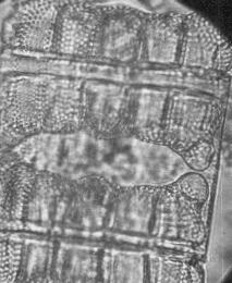
Biddulphia: A diatom from the
stomach of a sea urchin.
In addition to the very small remains in the debris which can be brought up using the Pasteur pipet, there is another range of somewhat larger creatures that cannot be removed in that fashion. For those, I use a plastic turkey baster and the large bulb allows one to produce considerable suction. My recommendation is to buy 4 turkey basters:
1) one for preserved samples,
2) one for collecting bottom sand or mud samples along the edges of ponds, lakes or streams,
3) one for collecting bottom sand or mud samples from marine environments, and
4) one for the turkey.
Among the things you may find in preserved marine samples are: ostracods, copepods, delightful little caprellid shrimp, larvae of all sorts, bits of echinoid spines, fragments of sponges, "heads" of hydroids with the tentacles, pieces of bryozoans, very small gastropod and bivalve shells, nematodes, small annelids, fish scales, and the list goes on and on.
I have already written about all the lagniappe one can find on the tunicate Styela plicata. However, at the moment, I am looking at debris from a very different source, namely, the deep sea glass sponge, Euplectella apergillum which has a holdfast to anchor itself on the bottom. When these sponges are collected, dried, and cleaned, they, fortunately, are not cleaned of the debris surrounding the holdfast which has many long glass "threads," creating a sort of network which retains all sorts of interesting things.
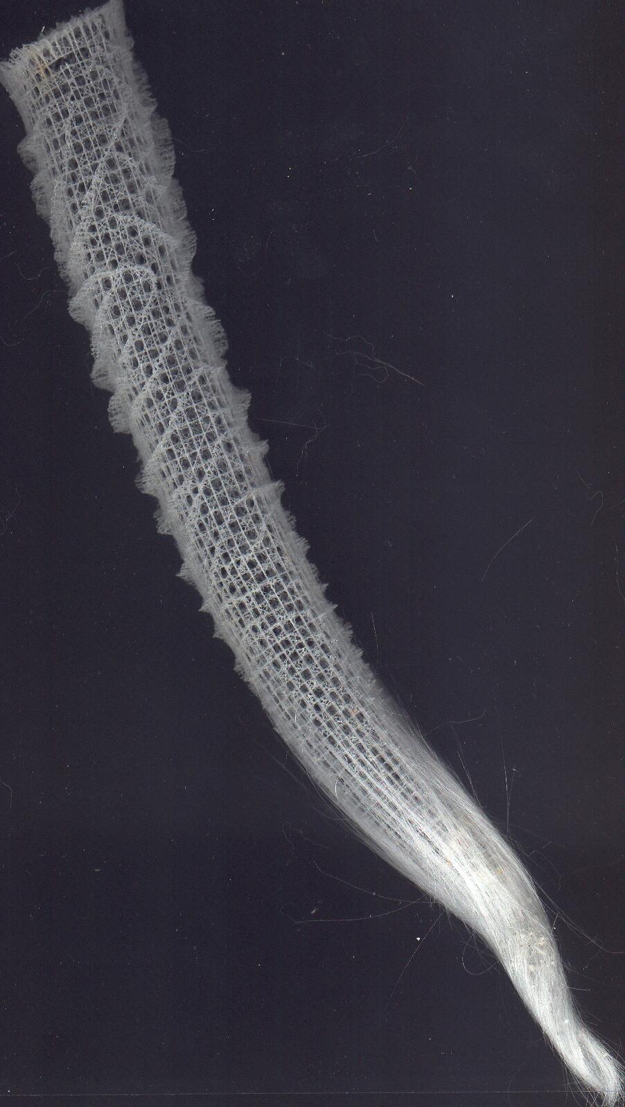
Euplectella: Image of whole sponge.
Click to view larger image.
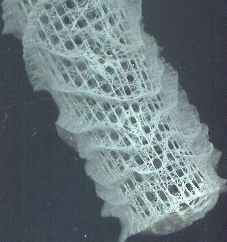
Detail of Euplectella. The sieve
plate over the osculum.
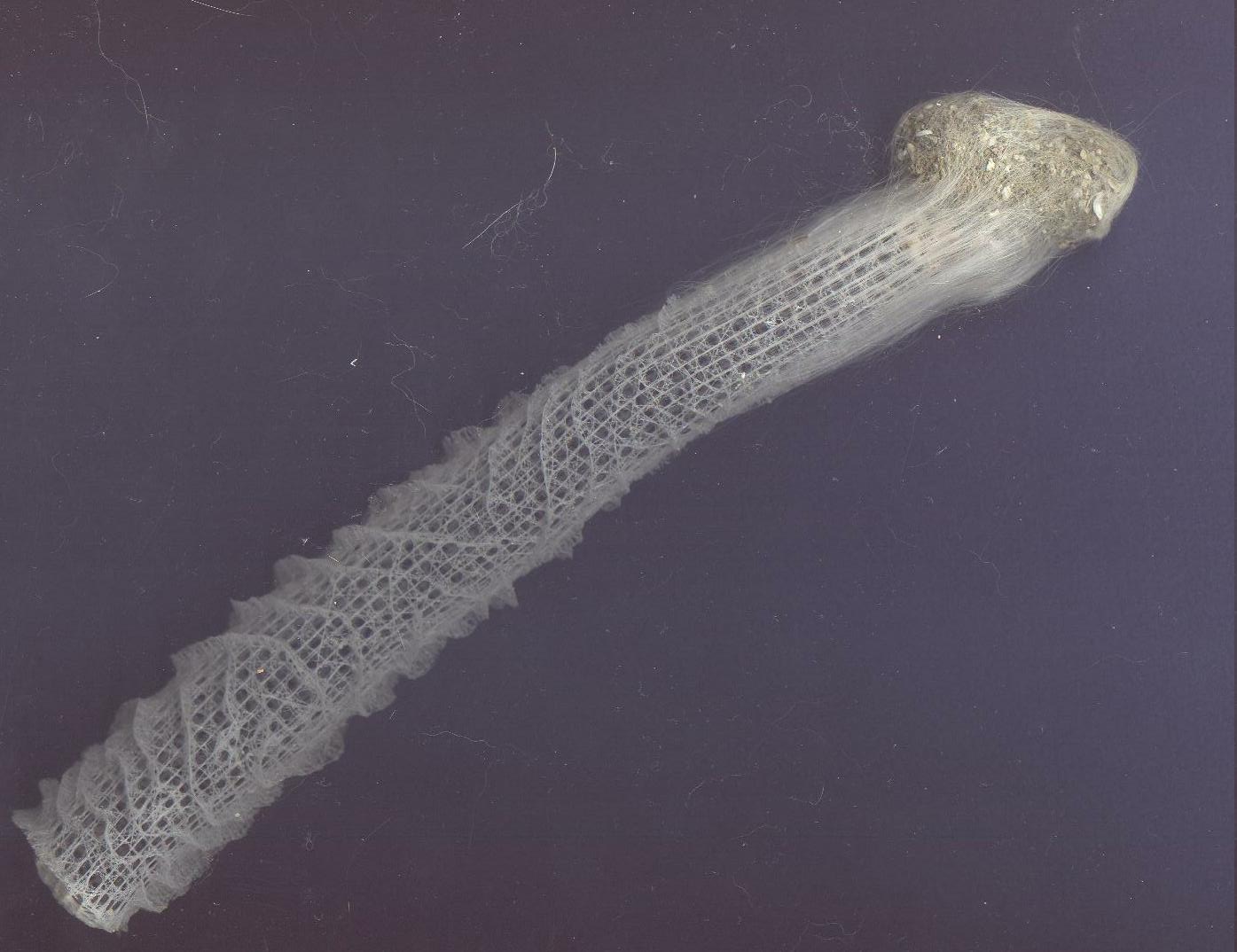
Euplectella: Image of whole sponge.
Another
specimen. Click to view larger
image.
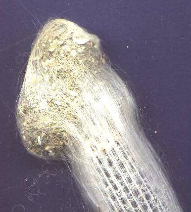
Detail of above, showing the
mass at the posterior end
from which the author extracted
the forams.
I took a micro-scalpel and began scraping off the detritus into a small plastic jar for storage. This needs to be done with some care, although the way the sponge is constructed makes it surprisingly strong despite its delicate appearance. But a second concern is that you are working with many tiny glass fragments and really should wear rubber gloves, a dust mask, and goggles. The first time I worked on my specimen, I didn't and I noticed afterwards that my hands were slightly irritated from the glass fragments. If you notice such irritation, the first thing to do is to rinse your hands thoroughly in running water. If you start off with soap directly, you are likely to increase the irritation by rubbing the particles into your skin.
The other night I fell upon (as it turns out, literallyI tripped on a rug) a wonderful old book buried at the bottom of one of my bookshelves. It is a 1935 textbook on invertebrate paleontology by Twenhofel and Shrock and as I browsed through it, I found a fascinating drawing of a glass sponge depicting what has been theorized to be its position when living on the sea bottom. The long glass threads of the holdfast extend from the posterior of the sponge down into the muddy substrate and then, remarkably, the sponge itself is shown at a 90 degree angle to the holdfast or, in other words, parallel to the sea bottom. This means that the currents would readily flow through the body of the sponge where the choanocytes are able to filter out the nanoplankton for food. This certainly seems a reasonable conjecture, and as, yet, I have not had time to check and see if there are other more recent alternative data.
I have now stripped away most of the debris and much of the holdfast and am approaching the point where I can begin to do a close examination of the body of the skeleton itself and that will be the subject for a completely separate essay.. Yesterday afternoon as I removed most of the final detritus, I was surprised to discover that the bottom of the sponge appears to be quite like the end of a pipe. I do not yet know if this is its natural condition or if it has been cut in the process of collecting. I'll keep you updated, as the television newspersons say.
However, for the moment, I would like to go back to the debris and give you an idea of what all the fuss is about. I am by no means an expert on forams and I shall just mention a few of the genera which I am reasonably confident are represented in this material: Globigerina and Globigerinoides, both of which look like small aggregates of spheres with textured surfaces, are fairly common. There are marvelous specimens of Globorotalia with prominent surface sculpturing and a slightly iridescent sheen. Bulimina and Uvigerina are among the most abundant and have somewhat the appearance of an upside-down pear with distinct ribbing running from top to bottom. Then there are a few Orbulina which are almost perfect spheres with a tiny round aperture and the surface of the shell looks rather as though it had been made of milk glass and then sprinkled with a fine coating of sand. Quinqueloculina (and related genera, such as, Sigmoilina and Massilina) are also present. Their shells have a distinctly white, chalk appearance. I love the name Quinqueloculina and I inflict it on my students, much to their annoyance. There is a species, Quinqueloculina semulina, which sounds like it should be the title of some popular Italian song.
For spicule fans, there's a treat. There are indeed some fairly large curved, bumpy needle-like spicules, which most likely are from Alcyonarians (soft corals), but just might be from a sponge, since there are some sponge spicules which appear very similar.
As one would expect, there are many little gastropod shells and a few bivalve shells as well. I found some fragments of sand dollars and a few spines, a few small sea urchin spines and one, slightly larger, which was quite remarkable and of a sort I had not previously seen. If you imagine a white spine in-laid with tiny raised, rectangular boxes along its length, you'll get some idea of its appearance. In addition, there were two lovely Dentalium or "tooth" shells.
However, what I did not expect and found in some numbers, were substantial fragments of a least two types of pteropod shells, and in three instances, the shells were nearly complete. (I have since found another 8 or 10 nearly complete ones in the debris around another specimen.) Pteropods are pelagic sea snails, the so-called "sea butterflies" and are indeed remarkable creatures, but from contemplating the shells alone, one would be hard put to get much of a sense of their diaphanous elegance. The first type of shell is a thin, white, fragile tube which comes to a point at the end and has two barbs giving it the appearance of an arrow. The other type is much more complex and has the appearance of a "rolled" trilobite and what looks like a distinct "cephalon" with several distinctive ridges running across its front.
It is an extraordinary sensation to be examining material from the ocean depths where only a very, very fortunate few have ever been able to venture. What is the depth from which this material comes? The consensus seems to be that these sponges are usually found in a range from 1,500 feet to 15,000 feet. The shallowest estimate I was able to find was 1,000 feet and the greatest depth23,000 feet! It used to be thought that the ocean depths were a barren landscape devoid of life, but in the last few decades, scientists have learned that there is an extraordinary diversity in the abyssal environments, populated by creatures stranger than anything in science fiction and much more interesting, because they can actually be investigated and examined. Furthermore, there are estimates that there may well be hundreds of thousands, perhaps even millions, of creatures that we have not yet encountered in this realm of dark, cold, and enormous pressure. That creatures can not only survive, but thrive in such environments, once again underscores the incredible tenacity of life.
Which bring me to two types of unusual clumps of mud which I found around the Euplectella holdfast. The first type looked like a miniature burrow in lumps of mud. After examining several such burrows, I think that it is likely that these were constructed by small tube worms. The other type, I was much slower to catch on to. I started to see clumps of forams and other debris stuck together by what almost appeared to be oddly-shaped bits of cellophane tape, some of them slightly curved. Gradually it dawned on me that these might also be fragments of a worm tube. Years ago when collecting in Maine and Oregon, I had come across a similar sort of construction in certain coastal tube worms. So, for now, that's what I'm guessing.
So far, I have been examining the debris at about 20x with my Olympus stereo zoom. This afternoon, I took a sample and started to examine it at 60x. Immediately, I began to find smaller forams of interesting new types that I hadn't noticed at 20x and noticed some spines on a larger foram that I hadn't noticed before and it turned out to be a very attractive species of Hantkia from which, unfortunately, most of the spines had been broken off. However, had I not taken the trouble to increase the magnification, I would have missed this completely. Even worse, I feel I have barely begun to look at this material. The next step will be to take part of this sample and run it through a series of washings to get rid of the mud particles and salt deposits. When this is done, then I can begin to take samples and explore them under the compound microscope under higher and higher magnifications. In ten or twelve years, I may have exhausted this sample.
So, once again, I would urge you not to ignore all the wonderful extras that you might find on a specimen, at the bottom of a culture, or on the bottoms of a preserved marine sample. There is a good chance that you will encounter some fascinating remains of creatures that you might never have noticed otherwise.
All comments to the author
Richard
Howey are welcomed.