|
M. Halit Umar Scientific Researcher, The Netherlands
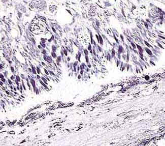
One characteristic of members of the Kingdom Animalia is their ability for movement. Animals achieve this property of movement by a specialized tissue called muscle. Muscle cells can contract and then go back to their initial state before contracting again. Such repetitive contraction of the muscle cells causes movement. We recognize three types of muscle cells which form specialized tissues: Skeletal muscle, heart muscle and smooth muscle. Skeletal and heart muscle cells are cross-striated, whereas smooth muscle possess no cross striations, contract rather slowly and cause a standing wave motion (of the stomach, intestines and large bowels, for example). Slugs have no articulate skeletal system so they have to crawl. They achieve this by using muscles especially in their foot. They are equally successful in climbing on vertical surfaces like walls. However, they have neither toes nor fingers to hold fast. Their ventral surface is rather flat and triangular with the apex towards the distal end (tail). Although a slug is not well equipped for quick movements, like those of a jumping cat or human athlete, they nevertheless wander steadily to find food. How do they accomplish this? Well, there is a substance called 'slime' of which all animals may benefit according to their physiological needs. Slugs are masters in secreting such slimy substances all over their body, especially on the ventral side. The aim of this short article is therefore to show microscopical aspects of the slime secretion of a slug. I collected a few small specimens of slug, probably of the genus Arion, in the forest stretching behind the laboratory where I worked. A sample measuring not more than 12 mm was fixed in a mixture of formaldehyde and glutaraldehyde in phosphate buffer pH 7.4. Twenty-four hours were enough for a complete chemical fixation at room temperature. Then, it was embedded in glycolmethacrylate and a tissue block was made. (See footnote 1). From this block, I obtained several hundred consecutive tissue sections which were mainly stained using crystal violet, toluidine blue, and a few of them with hematoxylin. After a short time in the stain, excess was drained off and washed in distilled water, and glass slides with stained sections were placed on a hot plate to dry. They were each sealed with a cover glass using a transparent synthetic resin. Here follows some microscopical images made through a Sony CCD camera attached to a Zeiss Axioscope microscope. They illustrate the ventral side of a slug body.
The lower part of the image shows foodstuff remnants containing lots of bacilli (bacteria). The upper half shows the ventral side of the tail. Note that there is an acellular space between the slug and the surface on which it crawls. This is not a completely empty place. The following images will show that some transparent slimy material is also present there (obj. 10x, see footnote 2). 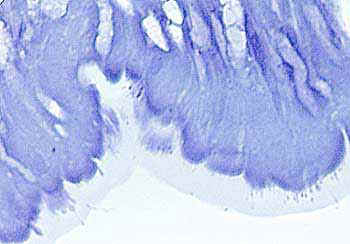 This image shows that there exists a transparent, slimy substance on the foot and it functions as a cushion between the animal and external surfaces. Note that this zone of secreted slime contains some small and oval dots in close proximity to the cell border (obj. 25x). 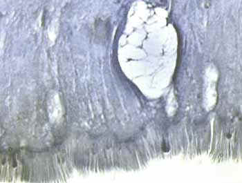 In sections stained with hematoxylin, we clearly see that the slimy zone has a structure with a brush-like border (obj. 25x). 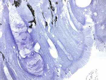 A detailed image which indicates the presence of some pigments (aggregation of small black dots in the upper part) and empty cells (actually a false impression) lying perpendicular to the brush-like border of the slime. On the left, there is a huge cell with many incomplete compartments. Such large and pear-shaped cells are called 'caliciform', 'mucigenetic' or 'goblet' cells which are the source of mucilaginous (slimy) substances for use anywhere in the body but mainly in mucous membranes (obj. 40x). 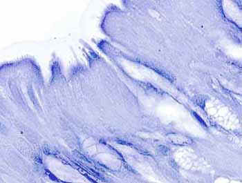 Such goblet cells have a foamy appearance. There are examples of them here at the lower part and at the right hand side of the image (obj. 40x). 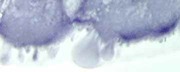 In this highly magnified microscopic field, we observe balloon- shaped, drop-like excretions within the slimy mass of the brush-like border on the ventral side (obj. 40x, intermediary 1.2). 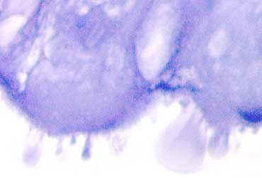 A still more magnified field reveals that such forms are closely related to foamy cells (obj. 40x, intermediary 1.6). 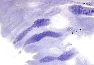 Actually, such drops are formed in the Golgi apparatus of the goblet cells and excreted like amorphous grains (obj. 40x). 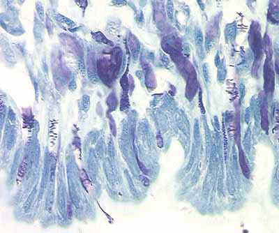 Note the abundance of slime producing goblet cells which are darkly stained and sometimes contain fine granules of the product to be discharged on the surface (obj. 40x). 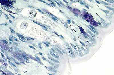 Finally, we see here a detailed illustration of the ventral side; a broad slime zone (at the right hand side), two goblet cells with emptied cytoplasmic contents (at the center) and other ones still containing mucilaginous substances seen as dark blue / purple globules (obj. 40x, intermediary 1.6). |