This short paper describes an astonishing biological mechanism which can readily be observed by anyone who has access to a modest microscope. Some plants and animals can easily enter a state of suspended animation, during which the metabolic activity of their cells can effectively be stopped, and the delicate architecture of the plasma membranes and intracellular organelles protected against extremes of cold, heat, radiation and other insults.
Examples which we can easily observe are dried yeast, brine shrimp eggs, tardigrades and certain metazoa such as bdelloid rotifers, which I have chosen to illustrate the process.
In my front garden is a birdbath, completely ignored by birds, but which has for many years harboured a profuse population of bdelloid rotifers. These quite complex organisms are very active, and,. incidentally, almost exclusively female. When their habitat dries up, they contract into ovoid cysts, in which state they can survive for extended periods of time. When rehydrated, they gradually reconstitute, and return to full activity within a couple of hours. Not all survive, and a proportion rupture during rehydration, but usually, the process works pretty well.
The bdelloids in fig.1 are encysted - left to dry out on a slide, and they have been in this state for several weeks, lying on a slide on my desk. They are contained within a thick tough case, probably derived from the syncytial membrane which is the skin of the active animal.
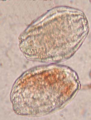
Fig. 1 Dried cysts of bdelloid rotifer.Some wrinkles in the case are visible, as are some internal structures such as the trophi. There is absolutely no movement - unsurprising considering the contents are now effectively solid!
When water is added, slow swelling occurs, and a progressive line of hydration can be seen to extend slowly across the cyst. (Fig. 2).
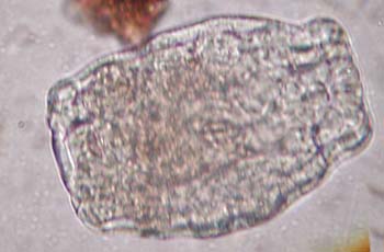
Fig. 2. Cysts 30 mins after adding water.
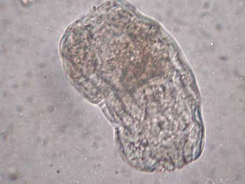
Fig. 3. Cysts 60 mins after adding water.
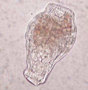
Fig. 4. Cysts 90 mins after adding water.After some further time, a very slow, intermittent peristaltic churning of the cyst contents starts, at the caudal pole of the cyst. In this region, the cyst contents have become sufficiently hydrated to return to a physiological condition, and the churning may help to distribute water towards the cranial pole. This process continues for some time, and it is at this stage that the rotifer is most at risk of rupture. The next sign of life is in the anterior third of the animal, where tiny flickering structures can be seen, beating with a frequency of about 5 beats/sec. These are probably flame cells, part of the excretory apparatus of the organism, and their early activity suggests that they may be an important priority for life support. Eventually, the organism as a whole starts to move - not yet driven by its wheel organs, but a rotatory drifting suggesting that the outer integument may be changing from a rigid case into a more flexible form, possibly secreting mucous material in the process. The contractions now start to involve the cranial part of the animal, and become quite forceful, forcing the ovoid shape first into a cylinder, and then in to a recognisable contracted version of the active animal. Finally, with more muscular activity, the metamorphosis into the final worm form is completed, the last phase being the extension of the wheel organs and resumption of feeding and full locomotion.
Transformation from an inert, dry cyst into an active swimming, feeding rotifer, takes less than two hours.
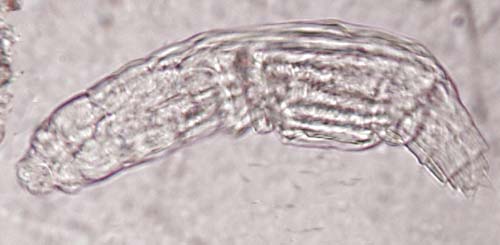
Fig. 5. An active bdelloid rotifer emerges from the cyst.The process becomes even more astonishing when you consider what is happening at the cellular level. Cells depend for their continued existence on a complex and delicate architecture, which is very vulnerable to damage and degradation. Cell membranes and organelles, and intracellular proteins are all very easily damaged by dehydration. They generally do not reconstitute when water is added. My houseplants have certainly not mastered desiccation resistance.
So how does the rotifer, the brine shrimp, and, best of all, the tardigrade, manage to pull off the trick?
The answer seems to lie with a remarkable, and remarkably simple molecule called trehalose. This is basically 2 glucose molecules linked together in a special configuration, to form a slightly more complex 1-alpha sugar. It appears that this molecule, which is both non-toxic and a potential energy source, concentrates as the cell dehydrates, and forms a very special complex state with protein and other molecules, effectively splinting them in place and preventing disruption or shape changes which would otherwise be lethal.This is a bit like an archaeologist encasing a fragile specimen in plaster of paris before transporting it. The master of desiccation, the tardigrade (see previous Micscape articles) can survive freezing in liquid nitrogen, and boiling point extremes of temperature, and still survive. Dried yeast is resistant to huge doses of ionising radiation, presumably because the nuclear material is protected from ionisation and free radical damage in a similar way.
When rehydration occurs, the sugar/protein complexes dissociate, and because membranes, proteins, and enzyme systems are in their rightful place, metabolism, biochemistry, and life processes can resume.
Desiccation resistance would seem to be a very valuable attribute for simple organisms in periodically hostile environments. This mechanism can apparently make some organisms virtually indestructible by dehydration, and highly resistant to cold and radiation, and to a lesser extent, to extremes of heat. It is a wonder to behold, and there are some rotifers near you who will be glad to demonstrate this almost miraculous feat.