I am sure that many of you reading this article will have spent time gazing into ponds, perhaps even dipping the odd jam jar into those special ones that are rich in organic matter and high in biodiversity, a sample of which can keep the amateur microscopist occupied for hours. But on the way to get your sample, you might just have passed an ecosystem that is positively bursting with protozoa, one that is almost constantly on the move, a perfectly ordinary sight in most countries yet rather inaccessible to most amateurs on a rainy afternoon. I am referring to domestic ruminants like sheep, goat, cows, bison, and oxen; wherever you are in the world there will be a ruminant nearby. And I'll tell you a little secret: it is highly likely to be stuffed full of ciliates; not parasites but endosymbionts living in mutual and (reasonably happy) coexistence.
Take, for example, the average cow. The rumen of a cow is about 100 L in volume. Inside, there may be half to one million ciliates per mL. That means a population more than ten times the world's human population. And, if you are persistent (or mad?) enough to get a sample, you are guaranteed many different and quite unique species.
I studied rumen ciliates for my PhD but I actually started as an undergraduate. I had always been fascinated by protozoa and I really wanted to try out an electron microscope. So I approached a possible supervisor about looking at Amoeba or Paramecium (something obvious) but he pointed me in a different direction. “Try rumen ciliates” he said, “no one has done much on them. Ameoba and Paramecium, they've all been done” (or words to that effect). So I set about obtaining a sample. Fortunately, one of our lecturers was a parasitologist who had links with the local abattoirs. He arranged for me to go with a large black bin bag and collect a sheep's rumen. A friendly technician drove me up in the departmental van. I met a meat inspector who disappeared into the bowels of the abattoir and sure enough, a few minutes later my black bag returned with a rather heavy load. It turned out that this one sample would keep me going for years (I still have material from it), although I did subsequently collect others.
The pictures I am showing are mostly from the first sample, and were all taken at Manchester University with equipment in what was the Zoology department. I used a Vickers light microscope with Nomarski optics, a Cambridge S150 scanning electron microscope and a Phillips 400T transmission electron microscope. All images were acquired onto black and white negative film from Ilford, so any colours used here are for demonstration purposes only and have been added digitally using Adobe Photoshop™.
And here are some examples of what I found. Basically, a rumen contains a very large number of bacteria, fragments of semi-digested plants and large numbers of entodiniomorphid ciliates. The name comes from the type species Entodinium, which is actually rather uninspiring (Fig. 1). Entodinium is rather a simple ciliate in some ways: it is small (about 20 micrometers long), it has a macronucleus and micronucleus, a single contractile vacuole that cannot always be seen, a zone of oral cilia grouped into membranelles (but they are not true membranelles like more advanced ciliates) and a cytoproct (bottom to everyone else).
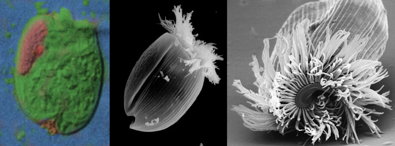
Figure 1. Panel of images showing Entodinium (typically 20 - 40 um). In the false-colour Nomarski image on the left, the ciliate can be seen to contain a macroncleus (pink), micronucleus (red) and at the front two lips that close the cell and hide the cilia when disturbed. Middle panel, a scanning electron micrograph of a similar example, this time with the lips open and the cilia extended out. Right panel, scanning EM of a front view showing the extruded cilia.
But entodiniomorphs actually get a bit more interesting than Entodinium. It seems that Entodinium, or more likely a similar ancestor, was probably the original source of the stock.
From these humble beginnings, a plethora of forms has evolved within the rumen, in almost perfect isolation from the rest of the world. During the course of this evolution, they have, like other creatures evolving across the world, diversified and become increasingly complex. The first sign of this increasing complexity is the acquisition of two new features and an increase in size. The new features, elegantly demonstrated by a beautiful species called Eudiplodinium neglectum, are a second zone of membranelles, independent of the mouth, and a structure called the skeletal plate (Fig 2).
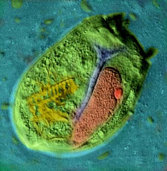
Figure 2. False colour Nomarski view of Eudiplodinium negelectum (typically 80 um). The macronucleus (pink) and micronucleus (red) are visible, along with two extruded zones of cilia, beating so they appear blurred, the skeletal plate (blue) and ingested plant fragments (yellow).
So what is the skeletal plate? In the electron microscope it resembles starch grains from plant cells, so the chances are it is some form of food storage device. In Eu. neglectum and its larger relative, Eudiplodinium maggii (Fig. 3) it is a long rod with a short fork at the top. But Eu. maggii differs from Eu. neglectum in having an unusual hook-shaped nucleus and its larger size, more contractile vacuoles, and bigger zones of cilia all argue for a process called allometry taking place along this line of ciliates (a gradual change in size and complexity during evolution).
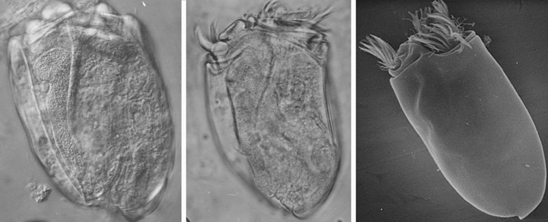
Figure 3. Panel of images showing Eudiplodinium maggii (typically 100-200 um). In the left Nomarski image the skeletal plate, hook shaped macronucleus and tiny micronucleus are easily visible. In the middle Nomarski view, the ciliate is swimming and its two zones of cilia are extruded. A similar view can be seen in the scanning EM on the right.
Other rumen ciliates demonstrate this principle too. Polyplastron (Fig. 4) has two skeletal plates, and is as big as, or even bigger than, Eu Maggii. Epidinium caudatum (Fig. 5) (probably my favourite) has three skeletal plates, all fused at the lower end into one large one. Epidinium has a tail, too, and a beautiful stripy pellicle best seen by scanning electron microscopy. Finally Ophryoscolex (Fig. 6) has many spines and at least three skeletal plates. It is probably the most unusual in appearance of all the entodiniomorphs. The tail seems to appear in several forms, even in some versions of Entodinium, and seems to be a plastic feature that appears in a population of tailless forms in the presence of predatory ciliates - presumably it makes them harder to swallow!
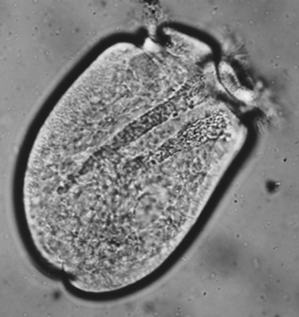
Figure 4. A phase contrast view of Polyplastron showing its two skeletal plates.
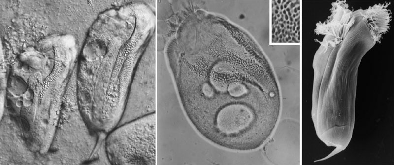
Figure 5. Panel of images showing Epidinium caudatum (typically 100 um). In the left panel, the Nomarski optics reveal part of the beautiful skeletal plate, the tail and contractile vacuoles. The middle image is a phase contrast view of a swollen cell which nicely reveals the three skeletal plates in full (the inset in this picture shows an enlarged region of the middle plate). The right scanning EM shows the cell with its cilia extruded, its stripy pellicle and the tail.
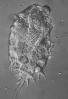
Figure 6. Nomarski view of Ophryoscolex (typically 100 um). These cells have a complex shape with multiple spines, but contain skeletal plates and structures similar to other entodiniomorphs.
I have many thousands of pictures of rumen ciliates, and I largely concentrated my work for my PhD on Epidinium, Eu. maggii and Entodinium. This work has been published in academic journals with my supervisor should you be interested in looking it up, or feel free to contact me if you would like more information. But I thought I would finish the article with two more pictures, of the cilia and basal bodies of Eu maggii, taken using a transmission electron microscope (Fig. 7). Most microscopists will know what cilia look like, so the pictures may not be that informative, but for me they sum up the beauty of the ultrastructural world of ciliates, of which rumen ciliates are a unique, almost secret, example.
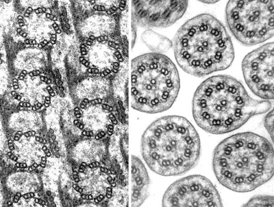
Figure 7. Transmission EMs of the infraciliature (basal bodies) and cilia of Eudiplodinium maggii. The cytoskeleton of these cells is a highly intricate structure built up from fibres, filaments and mictrotubules.