 |
The Heine Condenser ( Part 2) The operational aspects and imagery. By Paul James (uk)
|
Click here for part 1 of this article.
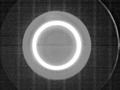
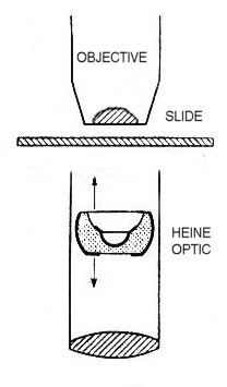
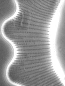
The variation of width of the annulus using the Heine condenser can be seen either down the draw tube or more ideally through a phase telescope or built-in Bertrand lens. A 0.95na X40 objective will allow the observer to see the full range of the annulus's diameter from its narrowest, when its optic is at its lowest setting to a point where it strikes the boundary of the back lens of this objective when raised. Here's a typical view through a phase telescope of the annulus about 3/4 of the maximum aperture of the x40 achromat :-
|
Objectives of any aperture can therefore be illuminated with its variable annulus up to and including the theoretical limit of 1.0 na without oil immersion assistance.
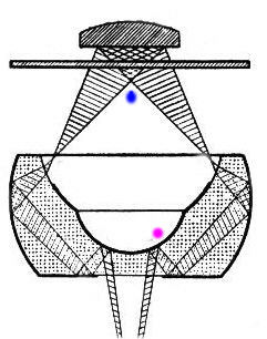
LAYOUT AND RAY DIAGRAMS FOR THE HEINE CONDENSER
 |
 |
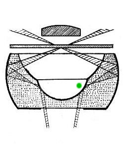 |
The diagrams above give some indication of the general design of the Heine condenser. Note how the subtle change in angle at the purple and green points of the ascending light from the substage bring about the annulus ring or darkfield illumination respectively. Note also the blue area just beneath the most oblique rays of the annulus where the maximum resolution occurs in the 'transition' zone cited below.
IN USE
The most interesting and rewarding imagery occurs when the annulus is near the maximum aperture of the objective in use. When the Heine's optic is raised a little further the effects rapidly change in the 'transition' zone between COL and DF. Here the maximum resolution of the objective can be realised. Lower settings of the condenser optic often bring about increased contrast, and so the beauty of the Heine condenser is that it can be set to optimize either resolution or contrast or a compromise of both in COL.
The following images demonstrate the effects on the imagery of butterfly scales using a x10 achromatic objective using subtle changes of elevation of the condenser optic :-
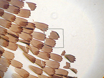 |
Typical appearance of full field with Heine set near x 10's maximum aperture value of 0.25na. Note the bright central section which is less noticeable in live view. |
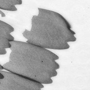 |
Detail of greyscaled crop from above to show crisp imagery of good contrastand resolution from first class achromat (Hund). |
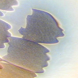 |
Heine set near 'transition' zone between COL and DF. Even in this imperfect CCD image the slightly higher resolution is just in evidence, though the colour is not reproduced accurately. |
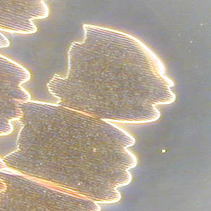 |
A little higher, the Heine's optic starts to bathe the specimen in DF, but the specimen is not a suitable one in DF and therefore not at its best. |
|
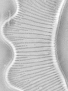
The specimen here is an Eutonia Diatom, a nice test for x40 achromat with Heine set at about 80% of the objective's aperture. (Empty mag of x 1200 to show more clearly detail on screen ). |
 |
As above but Heine set in 'transition' zone revealing better resolution from max obliquity of illumination and shorter wavelength of blue light. |
|
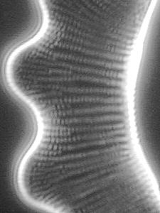
Optic elevated again producing more detail expected with darkfield lighting. |
|
|
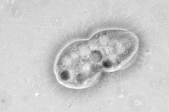
| Unidentified protozoan x1200 Wild Achromat 0.65na with 95% annulus setting of Heine Optic. Again empty magnification to minimise the effects of CCD noise. |
PHASE CONTRAST USE WITH THE HEINE CONDENSER
The prime purpose of the Heine, that of utilising the variable annulus for phase contrast allows any phase objective to be used. Leitz made a series of objectives suited specifically for the Heine, with the designated Pv symbol. I have no experience with these objectives, but would imagine they convey a rather unique phase contrast image.
| All comments welcome to the author Paul James |
Microscopy
UK Front Page
Micscape
Magazine
Article
Library
Please report any Web problems or offer general comments to the Micscape Editor.
Micscape is the on-line monthly magazine of the Microscopy
UK web
site at Microscopy-UK