|
|||
| Blepharisma | |||
by Jannette Hanna
All images and video footage copyright Jannette
Hanna 2004
Blepharisma americanum
Kingdom Protista
Phylum Ciliophora
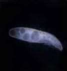
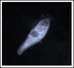
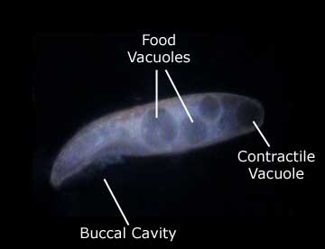
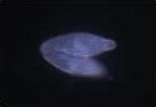
Blepharismas are protists that are commonly viewed under microscopes in science classes. They are readily available from scientific supply stores. Blepharisma is a microphagus filter feeder and a ciliate, it is classified in the phylum Ciliophora. Ciliates are generally considered the most evolved and complex of the protozoans. They are single celled organisms with organelles that are used for movement and food gathering.(1) Cilia are short hairlike organelles used for motion, as well as to sweep food into its buccal cavity (mouth). Blepharismas generally eat bacteria from decomposing vegetation, which is drawn into the buccal cavity and then passes to the food vacuoles at the posterior end of the cell. (2)
Blepharismas are particularly notable because of their unusual color. Unlike most protists, they are a faint shade of pink. This is not visible under brightfield illumination, so these images were shot using darkfield illumination set up on a Nikon microscope. The objective magnifications used were 10x and 20x.
Despite many still images available about their structure, there is not as much information easily accessible on the Blepharisma's behavior and movement. Still images can provide excellent views of their internal organelles, but video footage is necessary to view their movement. As with any small, transparent, moving organism there are challenges involved in video recording.
The specimens were placed onto a slide for microscope viewing, and a ring of petroleum jelly was used to seal in the water and give the Blepharismas room to move. This provided a water tight seal that kept the protists alive for over a week after the slide was prepared. If kept at room temperature the Blepharismas move very quickly making it difficult to keep them in view by moving the microscope stage. After being placed into a refrigerator for several hours, they slow down notably making them easier to follow. Various substances such as glycerin can be used to suspend or greatly deter motion, however chilling the specimen was sufficient for viewing their normal movement.
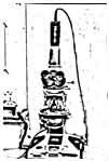 A Nikon microscope was used, with a darkfield
condensor to illuminate the ciliates. A peanut microscope camera was
placed where the photo reticle would ordinarily be, and it was hooked
up to its control unit. An S-video cable out of the control unit was
hooked up to a Canon GL-1 digital video camera for recording.
A Nikon microscope was used, with a darkfield
condensor to illuminate the ciliates. A peanut microscope camera was
placed where the photo reticle would ordinarily be, and it was hooked
up to its control unit. An S-video cable out of the control unit was
hooked up to a Canon GL-1 digital video camera for recording.
As you can see, because of its cilia on all sides, the blepharisma can turn in any direction and often spins around. It spirals as well as spins in a circular motion. It can change direction very easily, which makes it somewhat difficult to keep in view on a microscope. Also, because it is suspended in a 3-dimensional water bath, it moves up and down in and out of the focus of the microscope. I obtained the best results by using a maximum of the 10x and 20x objectives, and by persevering in capturing enough footage that it could be edited to include only segments that were in good focus. I used Adobe Premiere for the video editing, and exported it as a QuickTime file, compressed for web viewing.
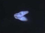
Some ciliates reproduce by fission, and some reproduce by conjugation - including Blepharismas. Conjugation is an exchange of genetic information to reproduce that occurs when two organisms of the same species fuse for a period of time.(3) They exchange nuclear products that results in the reshuffling of hereditary characteristics, as in sexual reproduction. Conjugation does not always result in immediate increase in numbers, often it is immediately followed by binary fission.(1)
 As you can see the pair of ciliates is less
agile than the individuals. They still spin, and in general they move
together, but at times they appear to conflict or to attempt to pull in
different directions. There is a slower rotation with the pair, but
they do move up and down in and out of the focus of the microscope.
Finding conjugating pairs is more difficult than finding individuals.
When the slides were initially prepared all of the specimens on them
were individuals. The slides were then left to sit for several days, at
which point there were multiple sets of conjugating pairs present.
As you can see the pair of ciliates is less
agile than the individuals. They still spin, and in general they move
together, but at times they appear to conflict or to attempt to pull in
different directions. There is a slower rotation with the pair, but
they do move up and down in and out of the focus of the microscope.
Finding conjugating pairs is more difficult than finding individuals.
When the slides were initially prepared all of the specimens on them
were individuals. The slides were then left to sit for several days, at
which point there were multiple sets of conjugating pairs present.
The still images shown above were taken from the video
footage. Larger still images of the Blepharismas can be seen at some of
the sites listed below.
Bibliography
(1)Encyclopedia Britanica, 15th edition, 2002
Volume 3 "Ciliate," volume 14 "Conjugation"
(2)Department of Zoology, University of
British Columbia
http://www.zoology.ubc.ca/courses/bio332/Labs/CiliateProject/blepharisma/bleph1web.html
(3)"Conjugation," Wikipedia
http://en.wikipedia.org/wiki/Conjugation
"Blepharisma americanum," Protist Information Server
http://protist.i.hosei.ac.jp/PDB/images/ciliophora/blepharisma/americanum/americanum_4.html
The music in the video clips is Mozart - Concerto in F for Three
Pianos and Orchestra. It is a MIDI file, a computer generated music
file. Examples of MIDI files can be found at:
http://www.classicalmidiconnection.com/cmc/mozart.html
ALL IMAGES AND VIDEO FOOTAGE COPYRIGHT JANNETTE HANNA 2004
I am currently an undergraduate student studying Biomedical Photographic Communications at the Rochester Institute of Technology in Rochester, New York, USA. I have completed my photographic concentration in high magnification photography as well as a magazine publishing project, and will be graduating with a Bachelor's of Science in May 2005.
This article was designed as a final project for a Photomacrography class, and I would like to thank Micscape for giving us this opportunity. If you have any questions or comments please contact me at j.hanna@gmail.com
-Jannette Hanna
Return to index of articles
written by students on the 'Principles and techniques of photomacrography'
course, November 2004,
Biomedical Photographic Communications (BPC)
program at the Rochester Institute of Technology (RIT).
Article hosted on Micscape
Magazine (Microscopy-UK).