Hair Through The Microscope
Tim Crandall
All the images below were captured with a Nikon D-70 Camera attached to a Nikon Optiphot microscope. I used a Nikon 20x primary objective and a Nikon 4x photo eyepiece. The exception is the comparison picture which was captured on a Leitz Wetzlar comparison microscope with a 10x Leitz primary objective and a 4x Olympus photo eyepiece housed in a modified Minolta mechanical tube. The camera was steadied with a copy stand.
Getting a decent image out of the Leitz proved to be beyond my grasp and resources. The electronics in one of the scopes were no longer working, but I made use of a fiber optic illumination system in place of the integrated light bulb. The result was an uneven and unmatched color between the two images. I would like to utilize a modern comparison microscope system in the future.
Specimens were collected from those who would hold still in my vicinity as well as from myself. Immersion oil matching the refractive index of glass was used as a mounting medium on a slide under a cover slip.
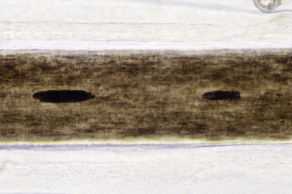
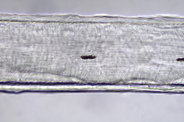
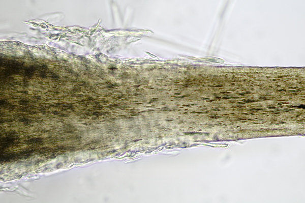
Below you will find photomicrographs of hair samples that illustrate different sections of hair that may be compared to determine matches. One of the examples is a brown and a white hair, both from my head that possess very different characteristics. The technicians that analyze hair samples for forensic evidence are highly skilled professionals. Differences and similarities are so slight that it takes years of familiarity to become competent in the field.
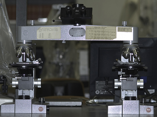
In forensic applications, hair samples can be closely examined through the use of the light microscope. Comparison microscopes similar to the now antiquated Leitz Wetzlar model shown below are commonly used in investigatory procedures. Through the use of a quartz beam splitter joining two identical imaging systems, technicians can analyze specimens alongside one another. Images of samples can also be superimposed. These images can then be recorded to a light sensitive material.
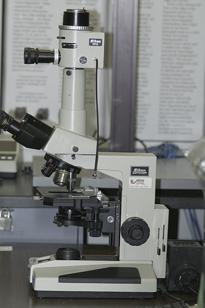
Nikon Optiphot microscope
Leitz Wetzlar beam splitter microscope
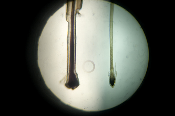
Example of a Leitz beam splitter image. Root tips from two subjects
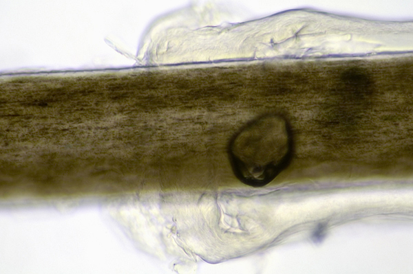
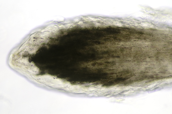
Brown hair with root sheath from a different subject
I am currently a student at the Rochester Institute of Technology in Rochester, New York, USA, studying in the Biomedical Photographic Communications department. My interests include photographic comparison in technical applications as well as macro and close-up photography.
Contact: Tpcrandall@yahoo.com