|
|
A Gallery of Palmitic Acid Photomicrographs (using a variety of illumination
techniques) |
|
|
A Gallery of Palmitic Acid Photomicrographs (using a variety of illumination
techniques) |
Palmitic
acid is an extremely common substance. The name correctly implies
that it is a major constituent of palm oil (from palm trees), but it is
also present in many of the foods we eat like meat, milk, butter and
cheese.
Industry makes use of this chemical
in the manufacture of products as diverse as soap and food
additives. Many shaving creams use palmitic acid as an emulsifier
to prevent separation of the oil and water constituents.
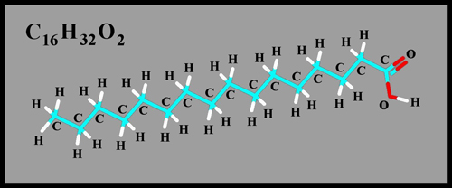
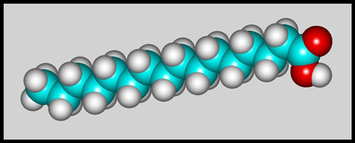
The structural formula and
molecular shape are shown below. (HyperChem software was used to
prepare both illustrations.)
The white crystals have a very low
melting temperature of about 63 degrees Celsius and thus make an ideal
candidate for a melt specimen. Only very gentle heating is
required to melt a thin layer between slide and cover-glass. Note
that palmitic acid is considered a skin, eye, and respiratory
irritant. (One wonders then, why it is present in so many of the
products that we use in everyday life!) If the acid is heated to
decomposition, carbon dioxide and poisonous carbon monoxide are
produced.
The images in the article were
photographed using a Nikon Coolpix 4500 camera attached to a Leitz
SM-Pol polarizing microscope. Images were produced using several
illumination techniques: transmitted light, dark-ground
illumination, phase contrast and polarized light. Crossed polars
were used in all polarized light images. Compensators, ( lambda
and lambda/4 plates ), were utilized to alter the appearance in some
cases. A 2.5x, 6.3x, 16x or 25x flat-field objective formed the
original image and a 10x Periplan eyepiece projected the image to the
camera lens.
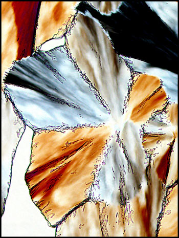
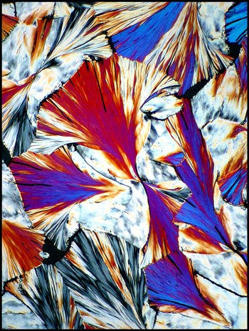
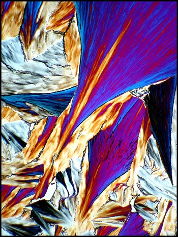
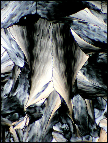
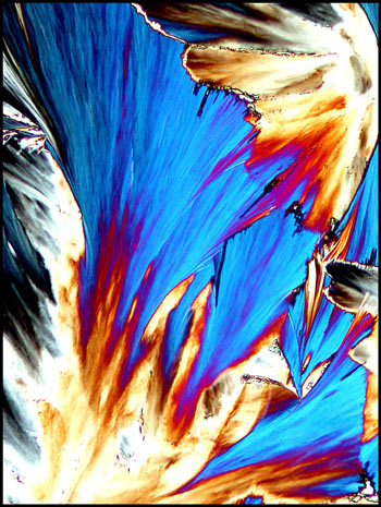
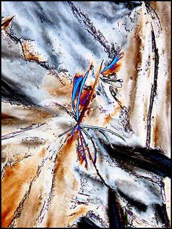
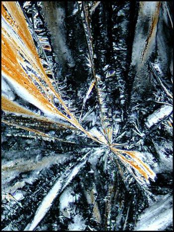
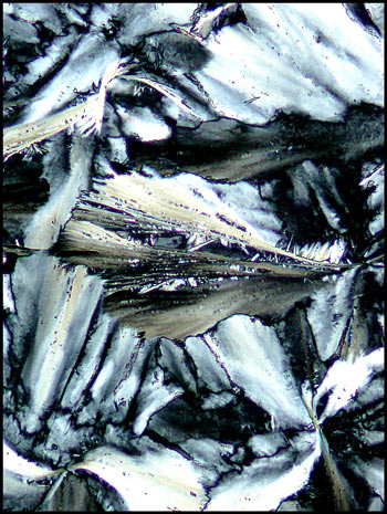
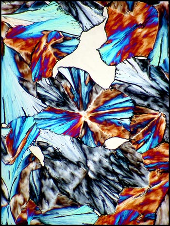
Most melt specimens contain double
fan-shaped structures. Two examples are shown below, with the
left image using dark-ground illumination, and the right polarized
light.
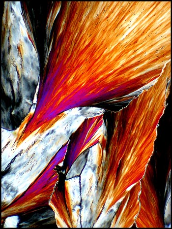
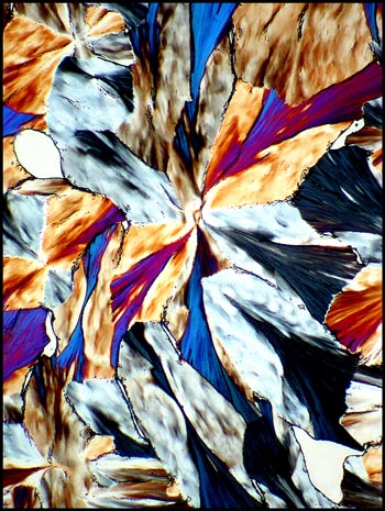
Higher magnification gives a
different view of the base of the fans.
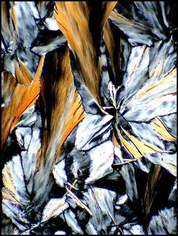
If the layer of crystals being
studied is thinner than in the two examples above, the colours are more
muted and tend towards gray.
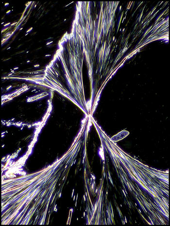
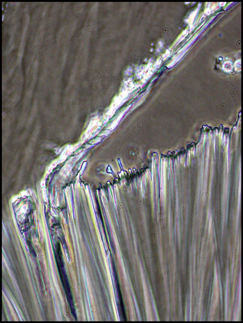
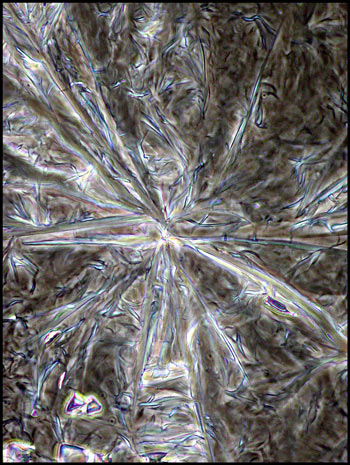
When the top edge of a fan is
highly magnified, and phase-contrast illumination is utilized, details
are revealed. Notice that the fan seems to be composed of long
crystal fibers that project out from the growth front during
crystallization.
Many double fan structures have
complex detail at the joining point, where growth started. Notice
the coloured radial spikes in the high magnification image to the right.
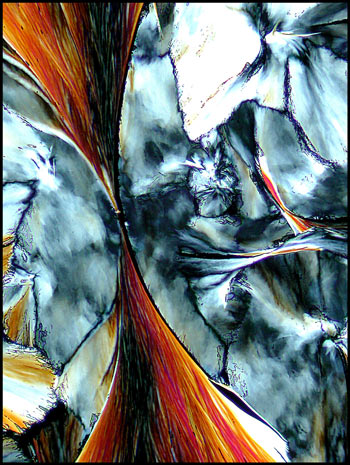
Two other examples follow.
The low magnification image on the left uses polarized light, while the
much higher magnification image on the right uses phase-contrast
illumination.
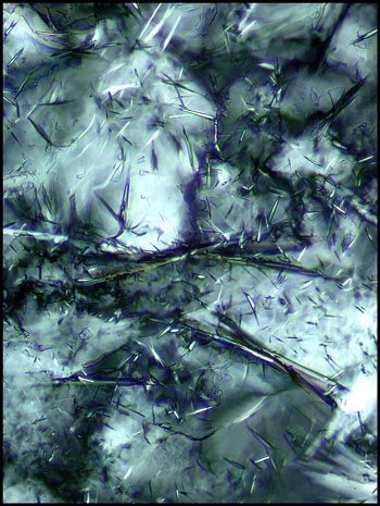
The three images below of a melt
specimen are in order of increasing magnification. Notice the
large number of randomly oriented needle-like structures in the last
image.
Both of the low magnification
images of specimen fields that are shown below use crossed polars, with
the addition of two lambda/4 compensators, (one below and one above the
specimen). This combination produces white bubble areas.
The first image in the article utilizes the same illumination.
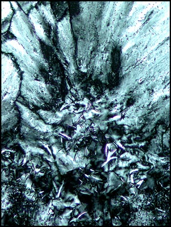
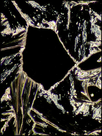
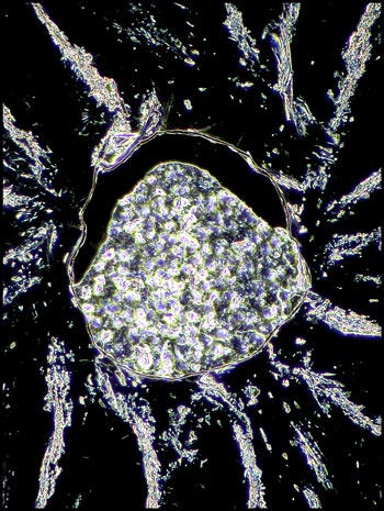
When these bubble areas are
studied with dark-ground illumination, they may contain no crystal
material (like that on the left) or an amorphous crystal mush (like
that on the right).
Palmitic acid would make a good
starting substance for the investigation of melt specimens under the
microscope. It is relatively harmless if treated carefully, and
has a melting temperature lower than that of boiling water! The
distinctive, colourful fan shapes make for a rewarding viewing
experience.
Published in the
November
2005 edition of Micscape.
Please report any Web problems or
offer general comments to the Micscape
Editor.
Micscape is the on-line monthly magazine
of the Microscopy UK web
site at Microscopy-UK
© Onview.net Ltd, Microscopy-UK, and all contributors 1995 onwards. All rights reserved. Main site is at www.microscopy-uk.org.uk with full mirror at www.microscopy-uk.net .