 The 3D-effect in
photomicrography –
The 3D-effect in
photomicrography –
Anaglyphs of recent tropical gastropod shells
Robert Sturm, Austria
Introduction
|
T |
he
investigation of gastropod shells represents a thrilling feature of biological
work due to the enormously high variability of shapes, patterns and colours.
Since Aristoteles, human fascination on mollusc biology and ecology has
continuously increased, and it was Georges Cuvier
(1769-1832), who for the first time described a comparative anatomy among the
whole phylum of the mollusca, thereby dividing the mollusc body into its
well-known parts (head, foot, and visceral lump). Besides the research of
mollusc biology itself, there has also been established a specific field of
scientific investigation solely dealing with the morphology of gastropod and
bivalve shells. This science is called conchyliology and has also aroused the
interest of non-scientists during the last decades. Unfortunately some people
have concentrated their holiday activities on sampling as many shells as possible,
and in some cases also living animals have been killed for obtaining their
shells. It has to be clearly mentioned that most gastropods and bivalves are
highly protected (most of them are included in the national Red Lists of
endangered animals), so that only a moderate sampling of empty shells on the
beach is justified.
Due
to their variable shape, which may be also characterized by the formation of
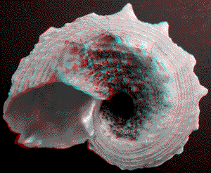
bizarre extensions (an example for that is exhibited on plate 3), gastropod shells are preferred objects for
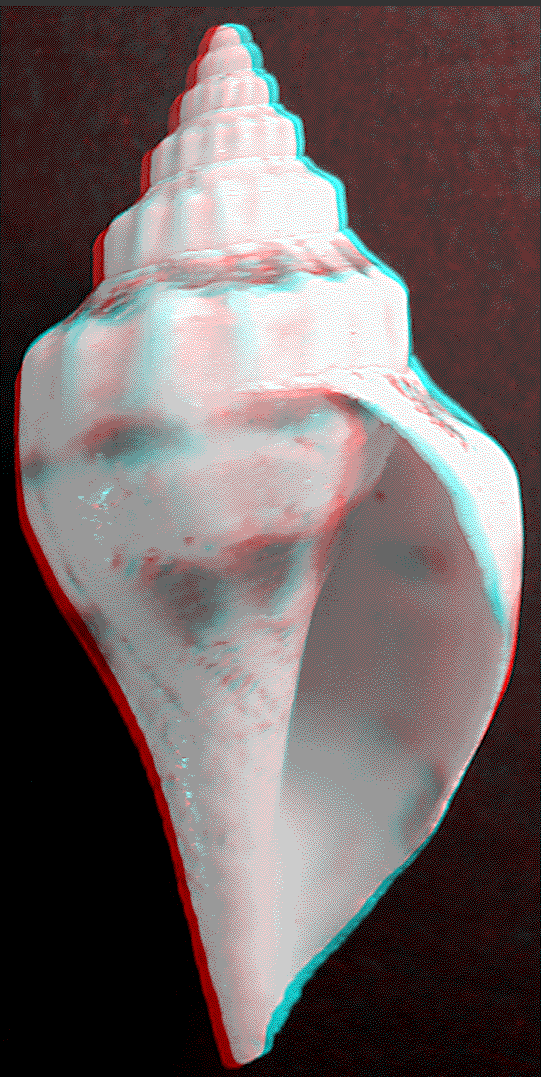
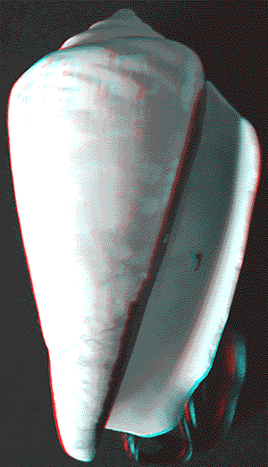
stereophotography. This photographic technique, where the object of desire is
photographed from two closely related positions, allows us to
create three dimensional images, whereby the spatial impression can be
only percepted by using stereographic glasses. In the present contribution,
four plates showing stereograms or anaglyphs of diverse tropical gastropod
shells are presented. In some cases, it is necessary to fix the object for some
seconds, because the eyes have to adapt to the stereographic effect. Spatial
impression can be also obtained from prints of the images.
Selected anaglyphs of tropical gastropod shells
 Plate 1
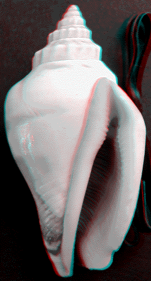
Left: Strombus (Canarium) urceus Linnaeus 1758,
“White mouth”,
height: 45 millimetres.
Middle: Lyria mitraeformis (Lamarck 1811), height: 40 millimetres.
Right: Mauritia histrio (Gmelin 1791), length: 56 millimetres.
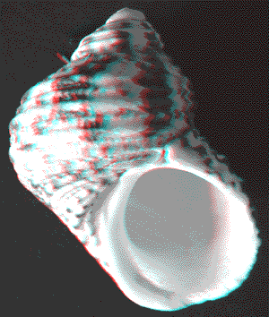
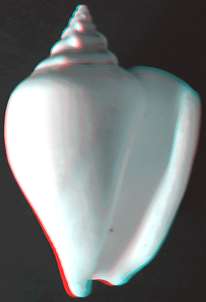
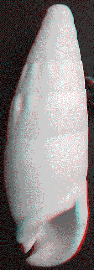
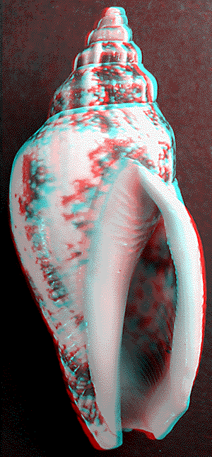
Plate 2
Left: Rhinoclavis vertagus (Linnaeus
1758), height: 64
millimetres.
Middle: Strombus (Laevistrombus) canarium Linnaeus
1758,
height: 50 millimetres.
Right: Turbo (Marmarostoma) chrysostomus Linnaeus 1758,
“Gold-mouthed Turban Shell”, height: 52 millimetres.
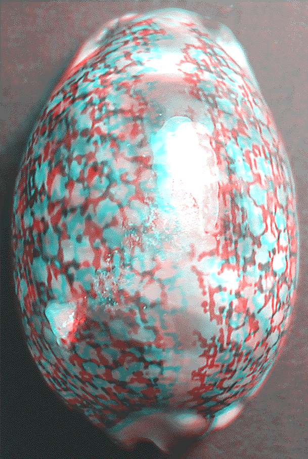
 Plate 3
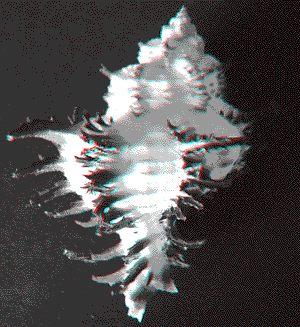
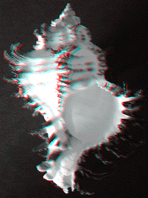
Hexaplex
cichoreus (Gmelin
1791), “Endive Murex”, back and front view, height: 95 millimetres.
 Plate 4

Left: Angaria delphinus
(Linnaeus 1758), “Dolpin
Shell”,
diameter: 50 millimetres (bottom
view).
Middle: Strombus (Conomurex) luhuanus Linnaeus
1758,
height: 60 millimetres.
Right: Turbo (Batillus) cornutus Lightfoot 1786,
“Horned
Turban”, height: 50 millimetres.
The
author highly acknowledges any response or questions concerning the shells or
the production of the stereograms. You can contact the author under the
following E-mail address.