|
|
A Gallery of Benzoic Acid Photomicrographs (using
polarized light illumination) |
|
|
A Gallery of Benzoic Acid Photomicrographs (using
polarized light illumination) |
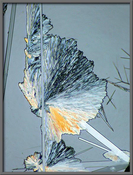
This article is actually a second look
at benzoic acid. In an earlier
article, I used phase-contrast illumination exclusively to produce
a series of photomicrographs of the compound. In the present
article, I have used the slides prepared earlier to provide an
alternative gallery of images using polarized light. If you take
a look at the earlier article, it should be noted that the same
magnification was used for all images, whereas here, several
magnifications were used.
Benzoic acid is a white crystalline
solid with a melting temperature of about 122 degrees Celsius.
This low melting point makes it easy to produce a melt specimen by
placing a few crystals on a slide, covering with a cover-glass, and
heating gently over an alcohol lamp until the solid melts. Slides
prepared in this way cool to room temperature in about a minute.
It should be kept in mind that the MSDS safety document for the
compound states: May be harmful if
swallowed. May act as an eye or respiratory irritant. May cause
allergic respiratory or skin reaction.
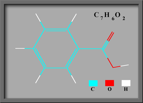
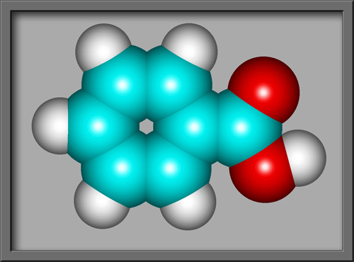
Benzoic acid C6H5COOH is the simplest aromatic (based
upon a benzene ring) carboxylic acid (containing the
COOH group). The structural formula and molecular shape,
(produced using HyperChem Pro),
can be seen below.
This compound is often used as an
anti-microbial agent in products like cosmetics, toothpastes,
mouthwashes and deodorants. Fruit products, beverages and
condiments may use benzoic acid as a preservative. In such
applications the quantity used, is of course very small, in order to
reduce the harmful effects mentioned above.
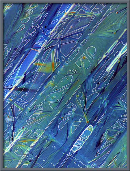
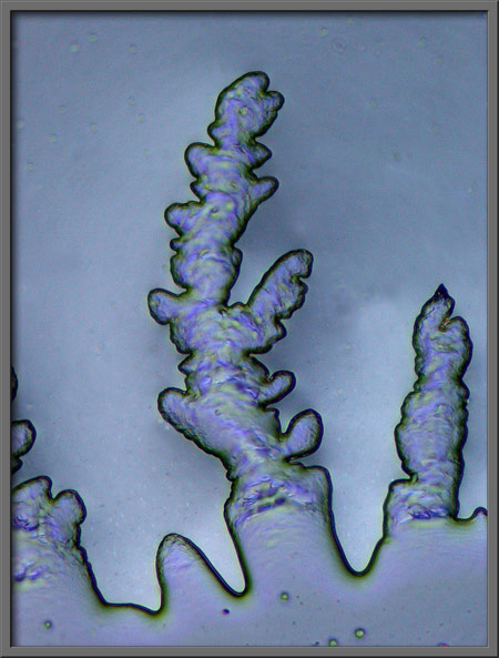
In addition to crossed polars, a
quarter wave plate and whole wave plate
were used to change the
colouration of the crystals.
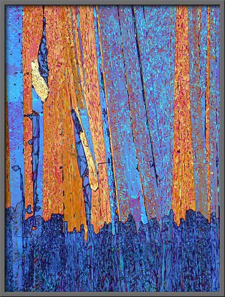
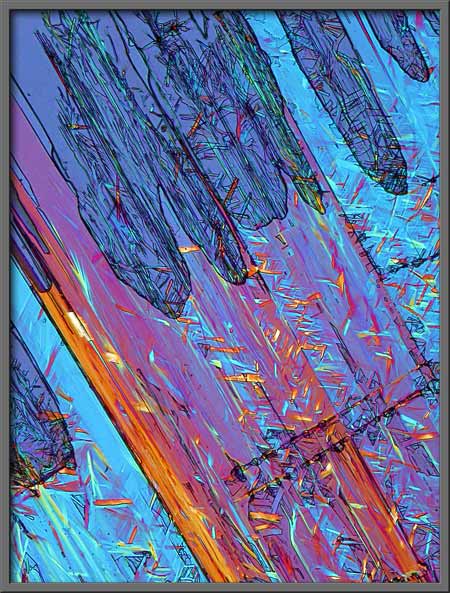
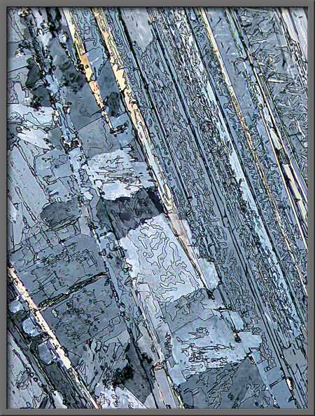
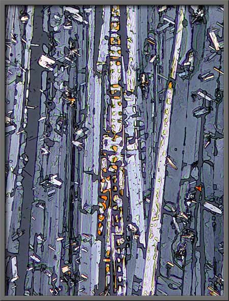
Notice the parallel lines and bands
that often occur in melt specimens of the compound. (quarter-wave
plate & whole-wave plate)
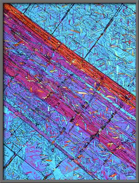
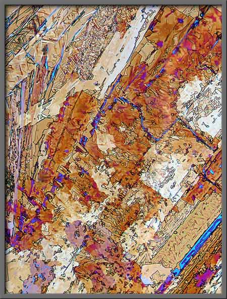
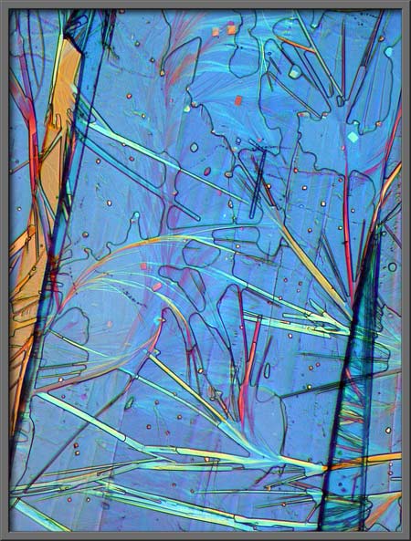
The different colouration shown in
the image below resulted from the use of two quarter-wave plates.
This combination produces elliptically polarized light rather than the
plane polarized light resulting from crossed polars alone.
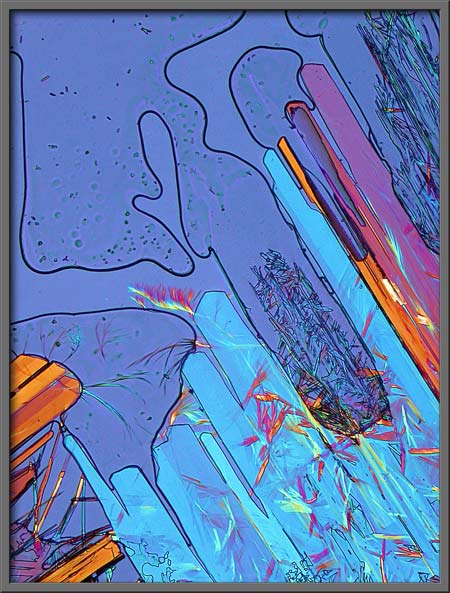
In the two images that follow, note
the small random crystal inclusions that have formed within the larger
crystal structures. (quarter-wave plate
& whole-wave plate)
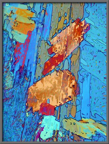
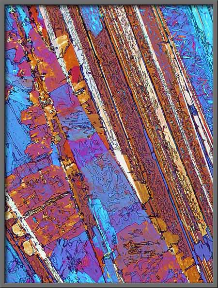
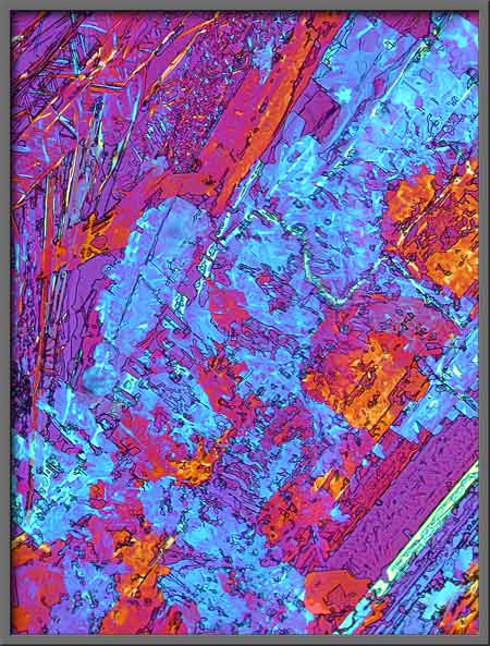
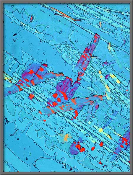
In the photomicrograph that
follows, tiny colourful crystals seem to bridge the gap between larger
blue formations. (quarter-wave
plate & whole-wave plate)
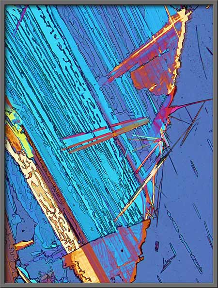
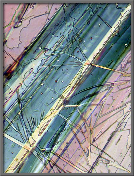
It takes a moment of study to
determine that the two images below are of exactly the same field of
view. The use of plates, (sometimes called compensators), allows
the photomicrographer to accentuate different details of a crystal
field. (quarter-wave
plate & whole-wave plate on the left and two quarter-wave plates on
the right)
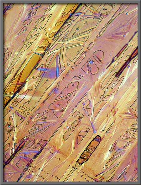
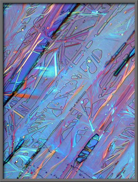
Another example follows.
Exactly the same field is shown in both images! (two quarter-wave
plates on the left and a quarter-wave plate & whole-wave
plate on the right)
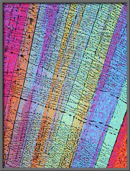
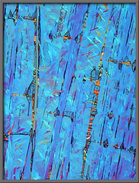
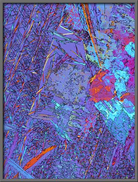
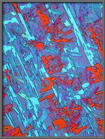
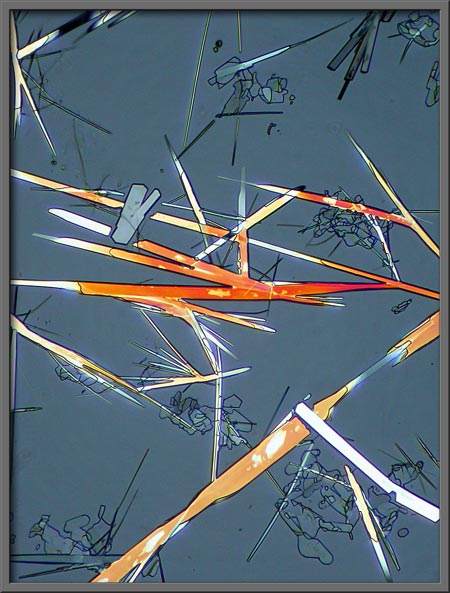
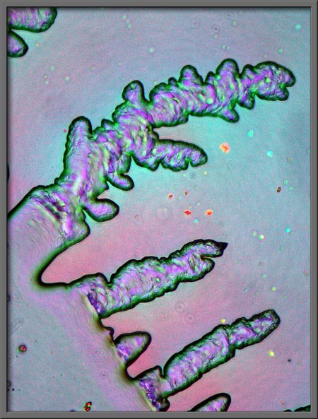
Many of the fields on the slides
were rather chaotic. (quarter-wave plate
& whole-wave plate)
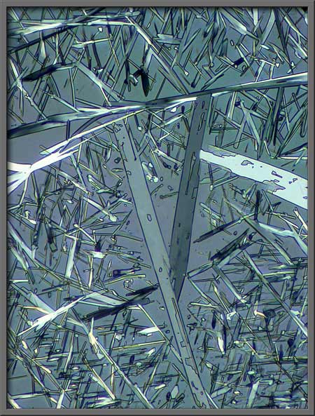
Elliptically polarized light tends
to give gray backgrounds. (two quarter-wave plates)
Just to the right of center in the
photomicrograph below, several straight streamers radiate out from the
interface between two crystal structures. (quarter-wave plate
& whole-wave plate)
Other similar formations are shown
below. (quarter-wave
plate & whole-wave plate on the left and two quarter-wave plates on
the right)
The two images of the same field
that are shown below are my favourite polarized light benzoic acid
photomicrographs. (quarter-wave plate
& whole-wave plate) The
whole-wave plate was rotated to produce the difference in
colouration. (Note: The
first image in the article was produced by using Adobe Photoshops Invert (image) command on the image
on the left below.)
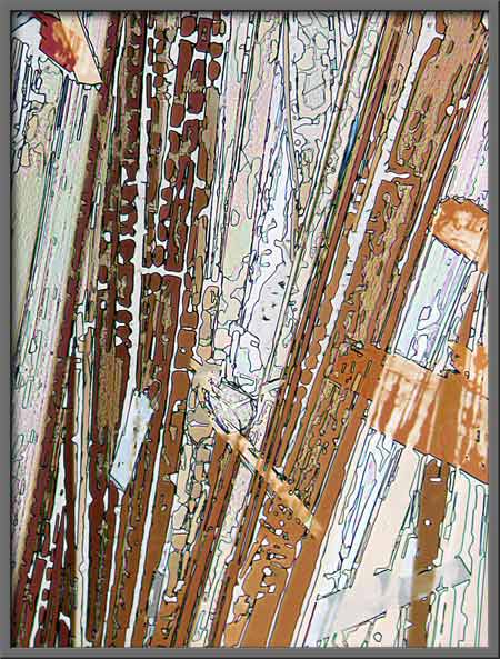
These strange flow patterns
occurred at the edge of the cover-glass in one melt specimen.
Photoshops Auto-level command was used to increase the contrast in
both images.
Most chemical compounds do not
allow the use of phase-contrast illumination to produce
photomicrographs. Benzoic acid is a major exception to this, and
I must confess that I prefer phase-contrast to polarized light images
of this compound.
Photomicrographic
Equipment
The images in the article were
photographed using a Nikon Coolpix 4500 camera attached to a Leitz
SM-Pol polarizing microscope. Images were produced using a
polarizing condenser. Crossed polars were used in all
images. Compensators, ( lambda and lambda/4 plates ), were
utilized to alter the appearance in some cases. A 2.5x, 6.3x, 16x
or 25x flat-field objective formed the original image and a 10x
Periplan eyepiece projected the image to the camera lens.
All comments to the author Brian Johnston are welcomed.
Published in the
November 2006 edition of Micscape.
Please report any Web problems or
offer general comments to the Micscape
Editor.
Micscape is the on-line monthly magazine
of the Microscopy UK web
site at Microscopy-UK
© Onview.net Ltd, Microscopy-UK, and all contributors 1995 onwards. All rights reserved. Main site is at www.microscopy-uk.org.uk with full mirror at www.microscopy-uk.net .