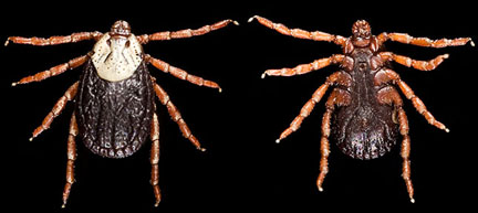
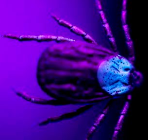
Dermacentor andersoni: Female
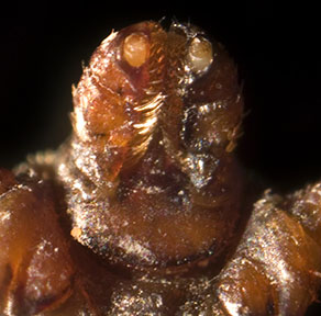
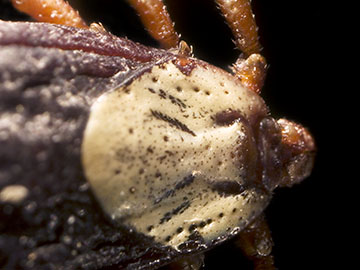
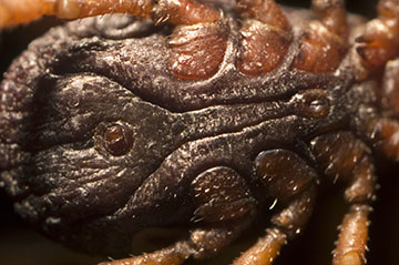
The Rocky Mountain wood tick ( Dermacentor andersoni ) is usually found in the most arid areas of the Rocky Mountains, but can be found in varying areas of North America . Adult and nymph ticks have eight legs while larval and seed ticks only have six. Females also have a distinctive white shield near the head and front of the body; males have no distinctive shield but are covered in a mottled gray pattern.
Rocky Mountain wood ticks are the primary carriers of Rocky Mountain spotted fever, which is the worse of tick-borne diseases. Rocky Mountain spotted fever is characterized by fever, nausea, emesis, severe headache, muscle pain, and a lack of appetite occurring within one to fourteen days after the bite. If left untreated, the disease progress into skin rashes, abdominal pain, and joint pain; the worse symptoms include partial paralysis of the extremities, gangrene, hearing loss, and movement and language disorders.
Photographing a Rocky Mountain wood tick requires all of the following equipment: a Nikon D70 camera, copy stand, Nikor 60mm Macro lens, 38mm thimble lens, 20mm thimble lens, bellows, lab jack, fiber optic lights, microscope slides, forceps, and a black light.
To work with a potentially infectious insect, the best course of action is to acquire preserved specimens from Ward's Scientific to photograph. For small subjects, it is frequently beneficial to mount the subject on a microscope slide to prevent it from being damaged. Forceps can be used to position the specimen and adjust the subject during high magnification photography. Lighting the subject is best done using fiber optic lighting, the smaller lights create higher levels of contrast. Two lights were used
in this photograph, one was the primary source of illumination and the other served as a fill light to even the lighting. Unfortunately, the chemicals used in preservation can frequently introduce highlights that cannot be eliminated.
It is worthwhile to try a variety of lenses before taking a final image, including thimble lenses using bellows to achieve higher magnifications. This allows the smaller structures to be photographed more easily. Long exposure times work the best, as a high aperture such as F8 or F11 is required in order to have good depth of field. It may be necessary to take several images and use a tool such as Combine ZM to create a single composite image of the subject to have a perfectly focused image of the entire tick.
My name is Alycia Yee, I am currently a fourth year student majoring in the Biomedical Photographic Communications with a minor in Chinese at the Rochester Institute of Technology, New York . I have completed my photo concentration in ophthalmic photography and completing a second concentration in photo macrography and photo microscopy. Please feel free to contact me.
© Alycia Yee 2006
For more information on Rocky Mountain wood ticks:
http://en.wikipedia.org/wiki/Rocky_Mountain_spotted_fever
http://www.agr.gov.sk.ca/apps/insectPest/pests/rocktick.asp
Return to
index of articles
by students on the 'Principles and techniques of photomacrography'
course, November 2006,
Biomedical Photographic Communications (BPC)
program at the Rochester Institute of Technology (RIT).
Article hosted on Micscape
Magazine (Microscopy-UK).